. 2013 Jan 14:10:6.
doi: 10.1186/1742-2094-10-6.

Absence of IL-1β positively affects neurological outcome, lesion development and axonal plasticity after spinal cord injury

Affiliations

Absence of IL-1β positively affects neurological outcome, lesion development and axonal plasticity after spinal cord injury

Francesco Boato et al. J Neuroinflammation. .

Abstract

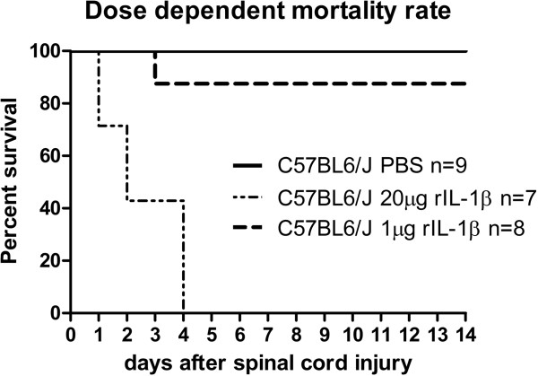
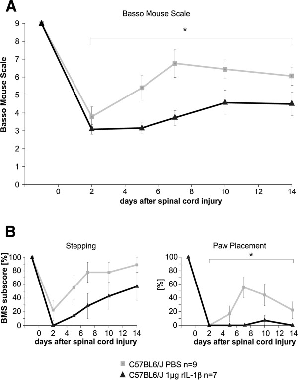
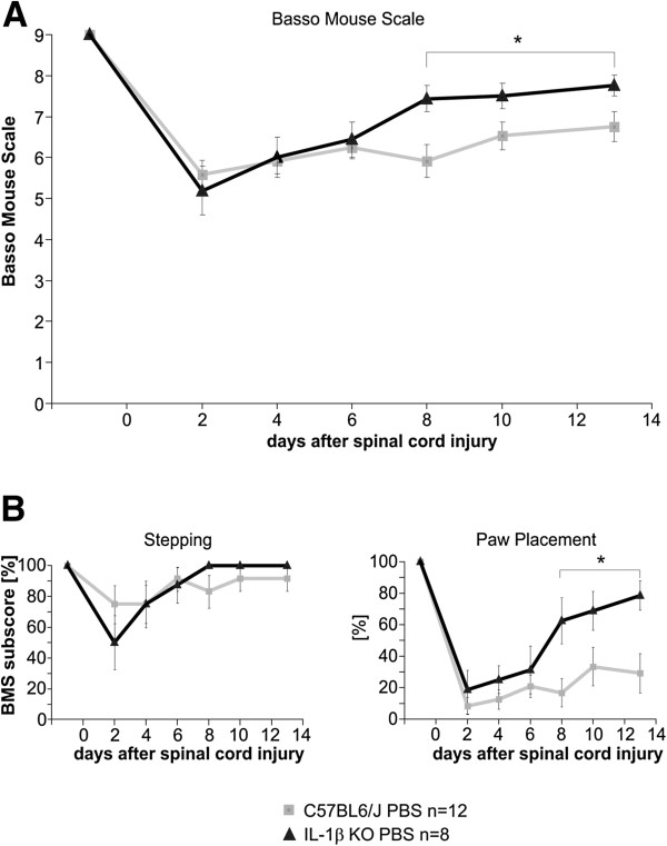
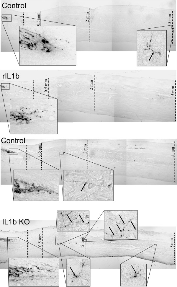
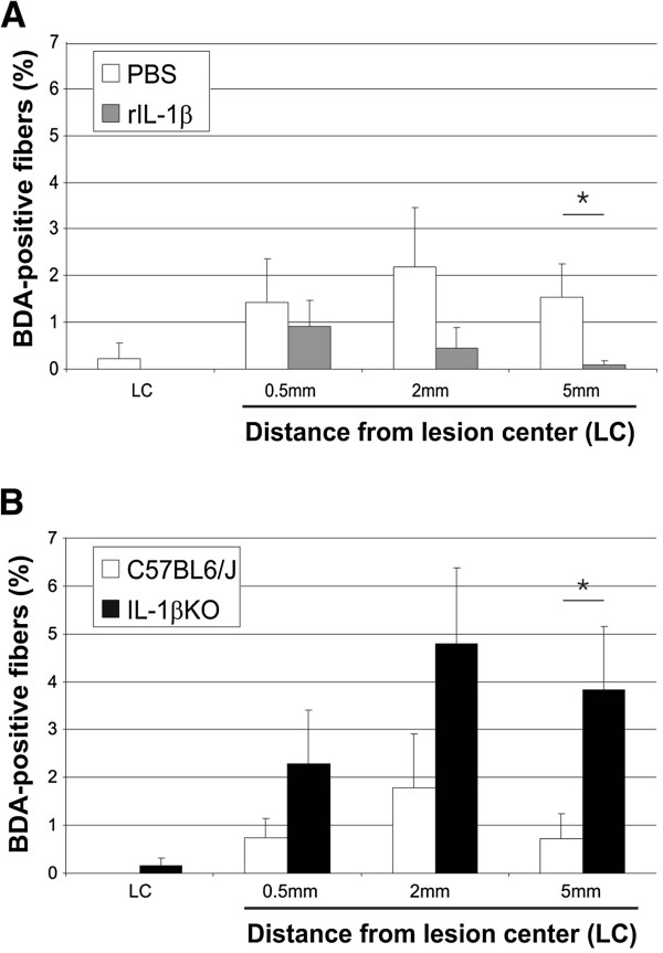
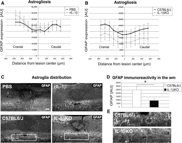
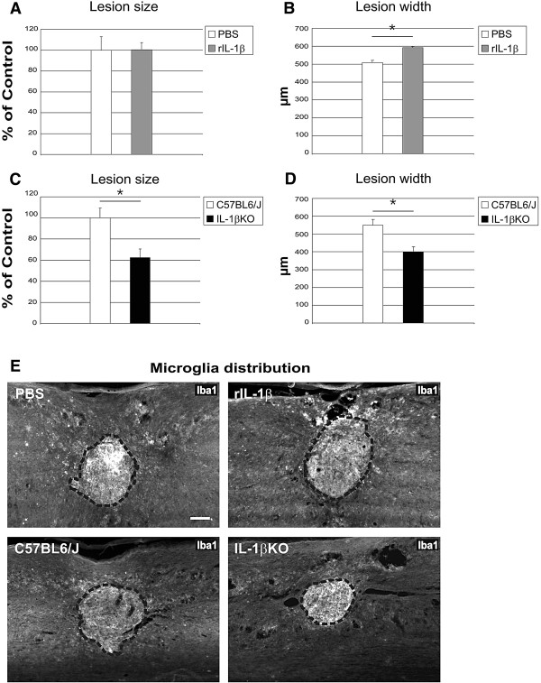
Precise crosstalk between the nervous and immune systems is important for neuroprotection and axon plasticity after injury. Recently, we demonstrated that IL-1β acts as a potent inducer of neurite outgrowth from organotypic brain slices in vitro, suggesting a potential function of IL-1β in axonal plasticity. Here, we have investigated the effects of IL-1β on axon plasticity during glial scar formation and on functional recovery in a mouse model of spinal cord compression injury (SCI). We used an IL-1β deficiency model (IL-1βKO mice) and administered recombinant IL-1β. In contrast to our hypothesis, the histological analysis revealed a significantly increased lesion width and a reduced number of corticospinal tract fibers caudal to the lesion center after local application of recombinant IL-1β. Consistently, the treatment significantly worsened the neurological outcome after SCI in mice compared with PBS controls. In contrast, the absence of IL-1β in IL-1βKO mice significantly improved recovery from SCI compared with wildtype mice. Histological analysis revealed a smaller lesion size, reduced lesion width and greatly decreased astrogliosis in the white matter, while the number of corticospinal tract fibers increased significantly 5 mm caudal to the lesion in IL-1βKO mice relative to controls. Our study for the first time characterizes the detrimental effects of IL-1β not only on lesion development (in terms of size and glia activation), but also on the plasticity of central nervous system axons after injury.


Figures

Figure 1
Figure 1
Dose-dependent mortality after spinal cord compression injury. The mortality rate in mice with local administration of recombinant IL-1β (rIL-1β) in Gelfoam directly after spinal cord compression injury (SCI) was 100% in mice treated with 20 μg rIL-1β, and was significantly higher compared with mice treated with 1 μg rIL-1β or only with PBS. P <0.0001 using the log-rank test.
Figure 2
Figure 2
Application of recombinant IL-1β impairs neurological outcome after spinal cord compression injury. (A) Locomotion analysis using the Basso Mouse Scale (BMS) showed significant worsening of neurological outcome after spinal cord compression injury (SCI) and local administration of recombinant IL-1β (rIL-1β) in Gelfoam directly after SCI. The rIL-1β-treated mice scored more than 1 point of the BMS less than control mice (scoring respectively 4.5 and 6), most probably reflecting lack of coordination and consistent plantar stepping in the treated mice. (B) The paw positioning subscore differed significantly between the two groups, while the stepping subscore did not. *P <0.05, two-way analysis of variance.
Figure 3
Figure 3
Systemic application of recombinant IL-1β impairs neurological outcome after spinal cord compression injury. Locomotion analysis using the Basso Mouse Scale (BMS) showed a significant early worsening of neurological outcome after spinal cord compression injury (SCI) and systemic administration of recombinant IL-1β (rIL-1β) compared with control mice. At days 1 and 2 after SCI, the rIL-1β-treated mice scored more than 2 points of the BMS lower than the control mice, but the significant difference was progressively lost and they scored in the same range of values as control mice at day 7 after lesion. *P <0.05, two-way analysis of variance.
Figure 4
Figure 4
Absence of IL-1β in IL-1βKO mice promotes functional outcome after spinal cord injury. (A) Locomotion analysis using the Basso Mouse Scale (BMS) showed a significant increase of neurological outcome after spinal cord compression injury (SCI) in IL-1βKO mice, as evidenced by a difference of1 point of the BMS between treatment groups (6.8 for wildtype (WT) mice and 7.8 for IL-1βKO mice). (B) The paw positioning subscore was significantly different between the two groups. Conversely, the stepping subscore was almost identical. *P <0.05, two-way analysis of variance.
Figure 5
Figure 5
Administration of recombinant IL-1β or absence of IL-1β alters numbers of corticospinal tract fibers. (A), (B) Representative micrographs of the area of the spinal cord between the corticospinal tract (CST) end and 5 mm caudal to the lesion center (LC). Higher magnification panels highlight the area between the end of the CST and the LC and one selected area (recombinant IL-1β (rIL-1β)) to four selected areas (IL-1βKO) caudal to the LC, where diaminobenzidine-positive fibers could be detected. Arrows indicate CST fibers caudal to the LC.
Figure 6
Figure 6
Quantification of biotinylated dextran amine-positive corticospinal tract fibers in recombinant IL-1β-treated or IL-1βKO mice. (A) The quantity of corticospinal tract (CST) fibers (shown as a percentage of the total number of biotinylated dextran amine (BDA)-positive fibers at C4 level in a standardized, 20 μm wide area [18]) was significantly decreased 5 mm caudal to the lesion in recombinant IL-1β (rIL-1β)-treated mice compared with controls. (B) Conversely, the percentage of CST fibers of IL-1βKO mice increased about fivefold compared with controls. Bars represent the percentage of CST fibers at the lesion center (LC) and in the area 0.5 mm, 2 mm and 5 mm distal to the LC. *P <0.05; n = 7 mice (PBS), n = 6 mice (rIL-1β), n = 9 mice (C57BL6/J), n = 6 mice (IL-1βKO). Values throughout are represented as mean ± standard error of the mean and P values were determined using the Mann–Whitney U-test.
Figure 7
Figure 7
Reduced glial fibrillary acidic protein expression in IL-1βKO white matter after spinal cord compression injury. (A), (B) Quantification of the intensity of glial fibrillary acidic protein (GFAP) immunoreactivity in the entire dorso-ventral axis of the spinal cord from 600 μm cranial to 600 μm caudal to the lesion center (LC) in a standardized, 100 μm wide, area showed no significant difference in astrocytic reactions between PBS-treated and recombinant IL-1β (rIL-1β)-treated animals (A) or between wildtype controls and IL-1βKO animals (B). (C) Representative micrographs of spinal cord sections stained with GFAP showing the perilesional astroglia distribution for the four different study conditions. Upper panels: comparison of PBS-treated and rIL-1β-treated spinal cord. Lower panels: comparison of wildtype control with IL-1βKO spinal cord. (D) Quantification of GFAP intensity in a standardized area limited to the white matter (wm) shows a significant difference of more than 60% in immunoreactivity and astroglia expression when using IL-1βKO mice compared with controls. (E) Higher magnification of the boxes in (C) representing GFAP expression in the white matter of control and KO mice. *P <0.05; n = 5 mice (PBS), n = 5 mice (rIL-1β), n = 7 mice (C57BL6/J), n = 5 mice (IL-1βKO). Scale bar = 100 μm.
Figure 8
Figure 8
Application of recombinant IL-1β and its deficiency influence lesion size after spinal cord compression injury. (A) to (D) Quantification of the lesion size and lesion width based on a clearly distinguishable Iba1-positive area. Lesion size measurement in the central sections of recombinant IL-1β (rIL-1β)-treated mice indicated no difference compared with controls (A), while the lesion width was about 20% greater (B). Both the lesion size (C) and the lesion width (D) were reduced by 40% and 25%, respectively, in IL-1βKO mice compared with controls. (E) Representative micrographs of Iba1 immunoreactive microglia distribution around the compression injury site in spinal cord sections. Upper panels: comparison of PBS-treated and IL-1β-treated spinal cord. Lower panels: comparison of wildtype control with IL-1βKO spinal cord. Dashed line, area of the lesion. Iba1 intensity did not differ significantly between groups (data not shown). *P <0.05; n = 5 mice (PBS), n = 5 mice (rIL-1β), n = 7 mice (C57BL6/J), n = 5 mice (IL-1βKO). Scale bar = 100 μm.

References

    1. Allan SM, Tyrrell PJ, Rothwell NJ. Interleukin-1 and neuronal injury. Nat Rev Immunol. 2005;5:629–640. doi: 10.1038/nri1664. - DOI - PubMed
    1. Bauer J, Berkenbosch F, Van Dam AM, Dijkstra CD. Demonstration of interleukin-1 beta in Lewis rat brain during experimental allergic encephalomyelitis by immunocytochemistry at the light and ultrastructural level. J Neuroimmunol. 1993;48:13–21. doi: 10.1016/0165-5728(93)90053-2. - DOI - PubMed
    1. Desson SE, Ferguson AV. Interleukin 1β modulates rat subfornical organ neurons as a result of activation of a non-selective cationic conductance. J Physiol. 2003;550(Pt 1):113–122. - PMC - PubMed
    1. Viviani B, Bartesaghi S, Gardoni F, Vezzani A, Behrens MM, Bartfai T, Binaglia M, Corsini E, Di Luca M, Galli CL, Marinovich M. Interleukin-1β enhances NMDA receptor-mediated intracellular calcium increase through activation of the Src family of kinases. J Neurosci. 2003;23:8692–8700. - PMC - PubMed
    1. Boato F, Hechler D, Rosenberger K, Lüdecke D, Peters EM, Nitsch R, Hendrix S. Interleukin-1 beta and neurotrophin-3 synergistically promote neurite growth in vitro. J Neuroinflammation. 2011;8:183. doi: 10.1186/1742-2094-8-183. - DOI - PMC - PubMed

Publication types

MeSH terms

LinkOut - more resources