. 2013 Feb;94(1):39-46.
doi: 10.1111/iep.12005.

Lymphangiogenesis and prognostic significance of vascular endothelial growth factor C in gastro-oesophageal junction adenocarcinoma

Affiliations

Lymphangiogenesis and prognostic significance of vascular endothelial growth factor C in gastro-oesophageal junction adenocarcinoma

Liang-Xi Xie et al. Int J Exp Pathol. 2013 Feb.

Abstract

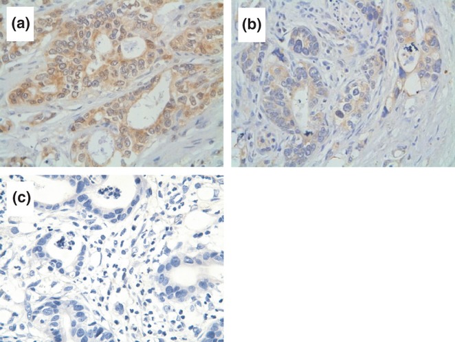
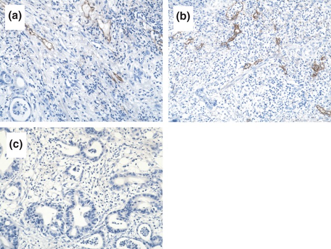
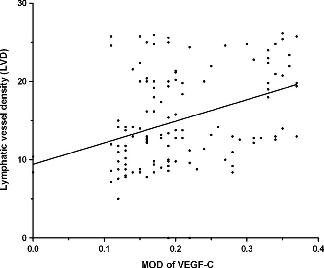
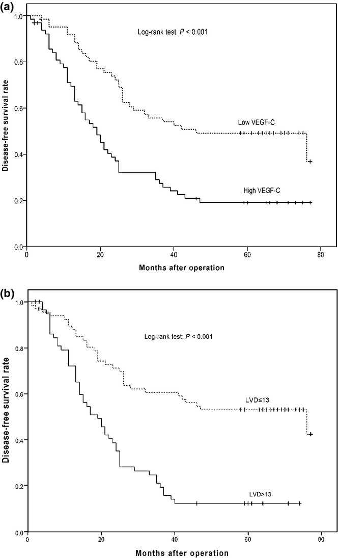
Vascular endothelial growth factor C (VEGF-C) is a crucial regulator of the development of lymphatic vessels and is involved in the lymph node metastasis of cancer. The levels of VEGF-C expression and lymphatic vessel density (LVD) in 128 gastro-oesophageal junction adenocarcinoma (GEJA) tissues were examined by immunohistochemistry and analysed for their association with clinicopathological features and disease-free survival. We found that 75.0% of tumour samples displayed strong immunoreactivity to VEGF-C. The levels of VEGF-C expression in the tumour tissues were associated with the stages of the clinical tumours and the lymph node metastasis status, but not with the age, gender and the size and type of tumours in the cohort. Similarly, LVD, as evaluated by anti-D2-40 staining, was also associated with the clinical stages of GEJA. The values of LVD were positively correlated with the levels of VEGF-C expression in these samples (r = 0.3760, P = 0.0001). High levels of VEGF-C expression and high values of LVD were associated with shorter periods of disease-free survival (DFS) in patients with GEJA (P < 0.001). In addition, GEJA at N1 and N2 stages, at T4 stage, chemotherapy after surgery, high levels of VEGF-C expression and lower marginal resection were independent factors for the prognosis of DFS in patients with GEJA. Our data indicate that VEGF-C may promote the lymphangiogenesis and lymphatic metastasis of GEJA and that VEGF-C may be a valuable biomarker for the diagnosis of lymphatic metastasis and a prognostic factor of the survival of patients with GEJA.


Figures

Figure 1
Figure 1
Immunohistochemical analysis of Vascular endothelial growth factor C (VEGF-C) expression in gastro-oesophageal junction adenocarcinoma (GEJA) tissues. The levels of VEGF-C expression in GEJA tissues were characterized by immunochemistry using anti-VEGF-c antibodies. Data shown are representative images (magnification ×400) of positive and negative GEJA tissue sections from 128 patients. (a) Strong immunoreactivity of anti-VEGF-C. (b) Moderate immunoreactivity of anti-VEGF-C. The yellow-brown staining was mainly identified in the cytoplasm of tumour cells. (c) Negative control.
Figure 2
Figure 2
Immunohistochemical analysis of D2-40 expression in GEJA tissues. The levels of D2-40 expression in gastro-oesophageal junction adenocarcinoma (GEJA) tissue sections were characterized by immunohistochemistry using an anti-D2-40 monoclonal antibody. Data shown are representative images (magnification ×200) of 128 samples. (a) Strong immunoreactivity of anti-D2-40 antibody. (b) Moderate immunoreactivity of anti-D2-40 antibody. The D2-40 was mainly expressed in the cytoplasm and membrane of lymphatic endothelial cells. (c) Negative staining with control IgG.
Figure 3
Figure 3
Correlation analysis of the levels of vascular endothelial growth factor C (VEGF-C) with the lymphatic vessel density (LVD) in GEJA tissues. Individual MOD and LVD values were analysed for the potential correlation by the Spearman rank correlation analysis. Data shown are individual values of 128 patients with GEJA. The levels of VEGF-C expression in GEJA were significantly correlated with the values of LVD (r = 0.3760, P < 0.0001).
Figure 4
Figure 4
Stratification analysis of disease-free survival (DFS) in patients with gastro-oesophageal junction adenocarcinoma (GEJA). The patients were stratified, according to the levels of VEGF-C expression or lymphatic vessel density (LVD) values, and the DFS curves were estimated using the Kaplan–Meier method, followed by log-rank analysis. (a) The levels of vascular endothelial growth factor C (VEGF-C) expression are associated with DFS. There were 65 patients with high levels of VEGF-C expression in their tumour tissues and 63 patients with low levels of VEGF-C expression in their tumour tissues. (b) The LVD-associated DFS. Patients with high levels of LVD (solid line, n = 61) had a DFS period significantly shorter than those with low LVD (dotted line, n = 67, P < 0.001).

Similar articles

Cited by

References

    1. Akagi K, Ikeda Y, Miyazaki M, et al. Vascular endothelial growth factor-C (VEGF-C) expression in human colorectal cancer tissues. Br. J. Cancer. 2000;83:887–891. - PMC - PubMed
    1. Amioka T, Kitadai Y, Tanaka S, et al. Vascular endothelial growth factor-C expression predicts lymph node metastasis of human gastric carcinomas invading the submucosa. Eur. J. Cancer. 2002;38:1413–1419. - PubMed
    1. Bunone G, Vigneri P, Mariani L, et al. Expression of angiogenesis stimulators and inhibitors in human thyroid tumors and correlation with clinical pathological features. Am. J. Pathol. 1999;155:1967–1976. - PMC - PubMed
    1. Cunnick GH, Jiang WG, Gomez KF, Mansel RE. Lymphangiogenesis and breast cancer metastasis. Histol. Histopathol. 2002;17:863–870. - PubMed
    1. Dadras SS, Paul T, Bertoncini J, et al. Tumor lymphangiogenesis: a novel prognostic indicator for cutaneous melanoma metastasis and survival. Am. J. Pathol. 2003;162:1951–1960. - PMC - PubMed

Publication types

MeSH terms

Substances

Supplementary concepts

LinkOut - more resources