. 2013 Feb;94(1):65-73.
doi: 10.1111/iep.12007.

Experimental development of bisphosphonate-related osteonecrosis of the jaws in rodents

Affiliations

Experimental development of bisphosphonate-related osteonecrosis of the jaws in rodents

Nicolau Conte Neto et al. Int J Exp Pathol. 2013 Feb.

Abstract

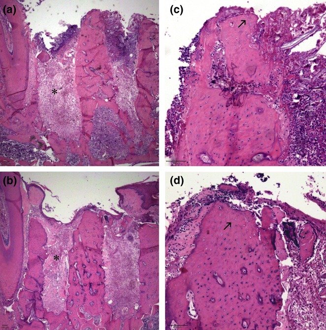
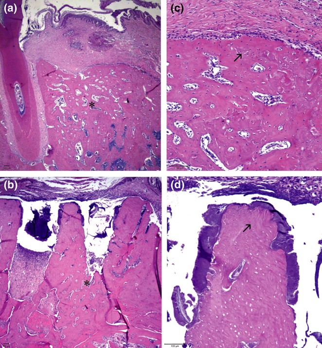
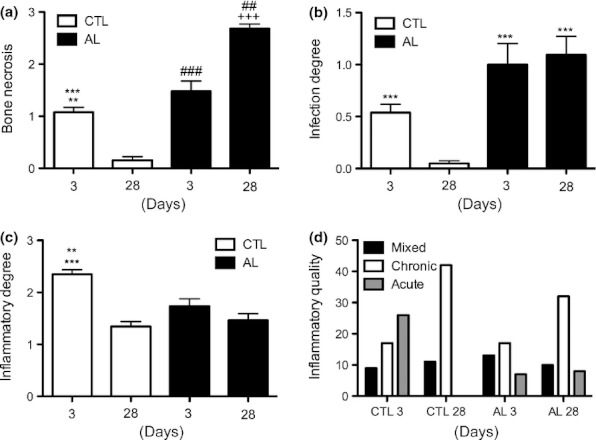
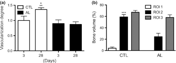
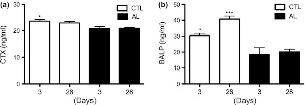
Osteonecrosis of the jaw (ONJ) following the use of bisphosphonates has become of increased interest in the scientific community, due in particular to its as-yet-unsolved pathogenesis. An experimental model of ONJ was induced in normal male rats [alendronate (ALN); 1 mg/Kg/day; n = 10] and matched controls (saline solution; n = 10). After 60 days of drug treatment, all animals were subjected to extractions of the left first lower molars and were euthanized at 3 and 28 days postsurgery. The following analyses were performed: (i) descriptive and quantitative (scores) histological evaluation, (ii) stereometry of distal sockets and (iii) biochemical measurement of C-telopeptide cross-linked collagen type I (CTX) and bone-specific alkaline phosphatase (BALP). The results showed that 28 days postsurgery the animals treated with ALN had areas of exposed and necrotic bone, associated with significant infection, especially in the interalveolar septum area and crestal regions, compared with controls. The levels of CTX, BALP and bone volume, as well as the degrees of inflammation and vascularization, were significantly reduced in these animals. Therefore, analysis of the data presented suggests that ALN therapy is associated with the development of osteonecrosis in the jaws of rodents after tooth extraction.


Figures

Figure 1
Figure 1
Schematic view of the regions of interest (ROIs) for stereometric analysis. The measurements were taken within the distal root of the left M1 at the ROIs that included three different areas inside the sockets. Initially, a standard ROI was determined by a quadrangular area extending from the level of the cemento-enamel junction (CEJ) of the left second mandibular (M2) to the apical end level of the M2 and between the mesial and distal alveolar bone surfaces. This quadrangular area was subsequently divided into three equal ROIs (1, 2 and 3).
Figure 2
Figure 2
Mean of body weight gain (g) of groups AL and CTL after surgical procedure. Baseline represents the mean of the body weight gain (g) on the day of teeth extractions. All animals tended to a decrease in body weight gain after 3 days of teeth extractions compared with baseline. At 7 and 14 days, both groups showed a tendency to an increase in the measures compared with 3-day period, and at 28 days after tooth extractions, all animals presented a slight decrease in the mean body weight gain compared with 14-day measures. No significant differences were found between CTL and AL groups in any periods.
Figure 3
Figure 3
Clinical aspects of a lower first molar (M1) alveolar socket 28 days after tooth extraction. Animals treated with sterile physiological saline (a) presented a full epithelial lining on the alveolar socket, with no signs of inflammation or infection (asterisk). In contrast, animals treated with alendronate (b) exhibited significant areas of exposed bone with a marked impairment of alveolar socket re-epithelialization (asterisk).
Figure 4
Figure 4
Clinical aspects of bone lesions around upper incisors of animals treated with alendronate (a, b). Some animals of the AL group presented, at euthanasia, areas of exposed bone surrounding the upper incisors (arrow). Of note, these lesion areas were associated, in all animals, with coronal fractures. Histological sections of these lesions (c) showing areas of bone necrosis and infection (arrow; original magnification × 100).
Figure 5
Figure 5
Histological sections of tooth sockets 3 days after left lower first molar extraction and stained with haematoxylin and eosin. Both the CTL (a) and AL (b) groups presented granulation tissue formation at this time (asterisk; original magnification × 25), as well as empty osteocyte lacunae at superior areas of the inter-radicular septum (arrow) (c, CTL; d, AL; original magnification × 100).
Figure 6
Figure 6
Histological sections of tooth sockets 28 days after left lower first molar extraction and stained with haematoxylin and eosin. (a) Animals in the CTL group presented complete alveolar bone regeneration, while animals in the AL group (b) exhibited only slight bone formation, restricted to the apical socket area (asterisk; original magnification × 25). The animals treated with alendronate (d) demonstrated retention of the inter-radicular septum, which was associated with bone necrosis and infection, while in the CTL (c) group, this area was completely remodelled, with signs of bone necrosis (arrow; original magnification × 100).
Figure 7
Figure 7
The effects of alendronate (ALN) therapy on bone necrotic levels (a), degrees of infection (b), degree (c) and quality (d) of inflammation at 3 and 28 days after tooth extraction. (a) Animals treated with ALN developed a higher proportion of bone necrosis compared with matched controls (**P < 0.01 in relation to CTL 28 days; ##P < 0.01 in relation to AL 3 days; ***P < 0.001 in relation to AL 28 days; ###P < 0.001 in relation to CTL 28 days and AL 28 days; +++P < 0.001 in relation to CTL 28 days). (b) Animals treated with ALN presented an increased degree of infection compared with matched controls (***P < 0.001 in relation to CTL 28 days). (c) At 3 days after tooth extraction, animals in the CTL group presented a higher degree of inflammatory response compared with all groups and periods (***P < 0.001 in relation to CTL 28 days and AL 3 days; **P < 0.01 in relation to AL 28 days).
Figure 8
Figure 8
Degree of vascularization (a) and bone volume (%) (b) in experimental groups. (a) At 28 days after tooth extraction, animals in the CTL group presented the highest levels of vascularization compared with all groups and periods (*P < 0.05 in relation to CTL 3 days and AL 3 days; **P < 0.01 in relation to AL 28 days). (b) The region of interest 2 (ROI 2) of CTL animals exhibited a statistically significant increase in bone volume compared with the ROI 2 of animals treated with alendronate (***P < 0.001 in relation to ROI 2 AL). Furthermore, no significant differences were found in other ROIs.
Figure 9
Figure 9
Levels of collagen type I (CTX) (a) and bone-specific alkaline phosphatase (BALP) (b) in experimental groups at 3 and 28 days after tooth extractions. In the CTX analysis (a), animals in the CTL group presented levels statistically significantly higher at 3 days compared with all periods for group AL (*P < 0.05 in relation to 3 and 28 days AL). At 28 days, no significant differences were found between groups and periods. In the BALP analysis (b), at 3 days, significant differences were found between the CTL and AL groups (*P < 0.05 in relation to 3 days AL), and at 28 days, animals in CTL exhibited increased levels of BALP compared with all periods for the AL group (***P < 0.001 in relation to AL periods).

Similar articles

Cited by

References

    1. Aghaloo TL, Kang B, Sung EC, et al. Periodontal disease and bisphosphonates induce osteonecrosis of the jaws in the rat. J. Bone Miner. Res. 2011;26:1871–1882. - PMC - PubMed
    1. Aguirre JI, Altman MK, Vanegas SM, Franz SE, Bassit AC, Wronski TJ. Effects of alendronate on bone healing after tooth extraction in rats. Oral Dis. 2010;16:674–685. - PubMed
    1. Allen MR, Burr DB. Mandible matrix necrosis in beagle dogs after 3 years of daily oral bisphosphonate treatment. J. Oral Maxillofac. Surg. 2008;66:987–994. - PMC - PubMed
    1. Allen MR, Follet H, Khurana M, Sato M, Burr DB. Antiremodeling agents influence osteoblast activity differently in modeling and remodeling sites of canine rib. Calcif. Tissue Int. 2006;79:255–261. - PubMed
    1. Bi Y, Gao Y, Ehirchiou D, et al. Bisphosphonates cause osteonecrosis of the jaw-like disease in mice. Am. J. Pathol. 2010;177:280–290. - PMC - PubMed

MeSH terms

LinkOut - more resources