

Cystic schwannoma of the pelvis
- PMID: 23317707
- PMCID: PMC3964667
- DOI: 10.1308/rcsann.2013.95.8.e1
Cystic schwannoma of the pelvis
Abstract
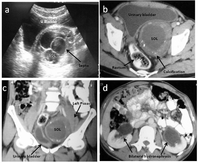
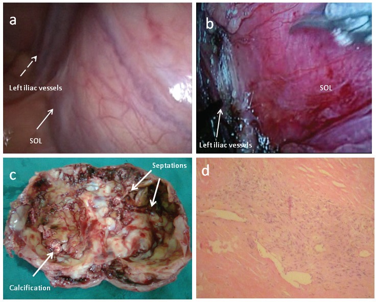
Schwannomas are benign tumours that arise from the Schwann cells of nerve fibres. They commonly occur in the head and neck, mediastinum and extremities. They are extremely rare in the pelvis. These are usually slow-growing tumours and are often detected incidentally. Preoperative diagnosis is extremely difficult as there are no definitive signs on imaging. Aspiration biopsy is often inconclusive or misleading. Surgical excision is both diagnostic and therapeutic. As these tumours are often large in size, open excision is most commonly performed. We describe a case of a large, cystic schwannoma of the pelvis causing bladder outlet obstruction and bilateral hydroureteronephrosis. Complete surgical excision was performed laparoscopically.
Figures
References
-
- Yi K, Wang YM, Chen J. Laparoscopic resection of an obturator schwannoma: a case report. Chin Med J 2010; 123: 1,804–1,806 - PubMed
-
- Andonian S, Karakiewicz PI, Herr HW. Presacral cystic schwannoma in a man. Urology 2003; 62: 551. - PubMed
-
- Konstantinidis K, Theodoropoulos GE, Sambalis Get al Laparoscopic resection of presacral schwannomas. Surg Laparosc Endosc Percutan Tech 2005; 15: 302–304 - PubMed
-
- Witherspoon P, Armitage J, Gatt M, Sagar PM. Laparoscopic excision of retrorectal schwannoma. Dis Colon Rectum 2010; 53: 101–103 - PubMed
-
- Ningshu L, Min Y, Xieqiao Yet al.Laparoscopic management of obturator nerve schwannomas: experiences with 6 cases and review of the literature. Surg Laparosc Endosc Percutan Tech 2012; 22: 143–147 - PubMed
Publication types
MeSH terms
LinkOut - more resources
Full Text Sources
Other Literature Sources
Research Materials