Review
. 2013:301:291-358.
doi: 10.1016/B978-0-12-407704-1.00006-3.

Regulation of blood-testis barrier (BTB) dynamics during spermatogenesis via the "Yin" and "Yang" effects of mammalian target of rapamycin complex 1 (mTORC1) and mTORC2

Affiliations
Review

Regulation of blood-testis barrier (BTB) dynamics during spermatogenesis via the "Yin" and "Yang" effects of mammalian target of rapamycin complex 1 (mTORC1) and mTORC2

Ka Wai Mok et al. Int Rev Cell Mol Biol. 2013.

Abstract

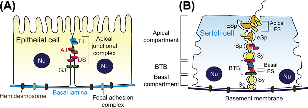
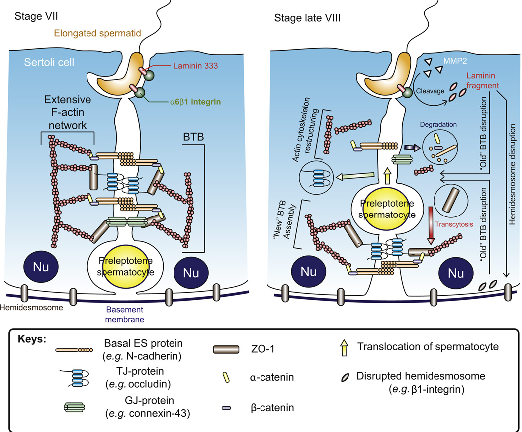
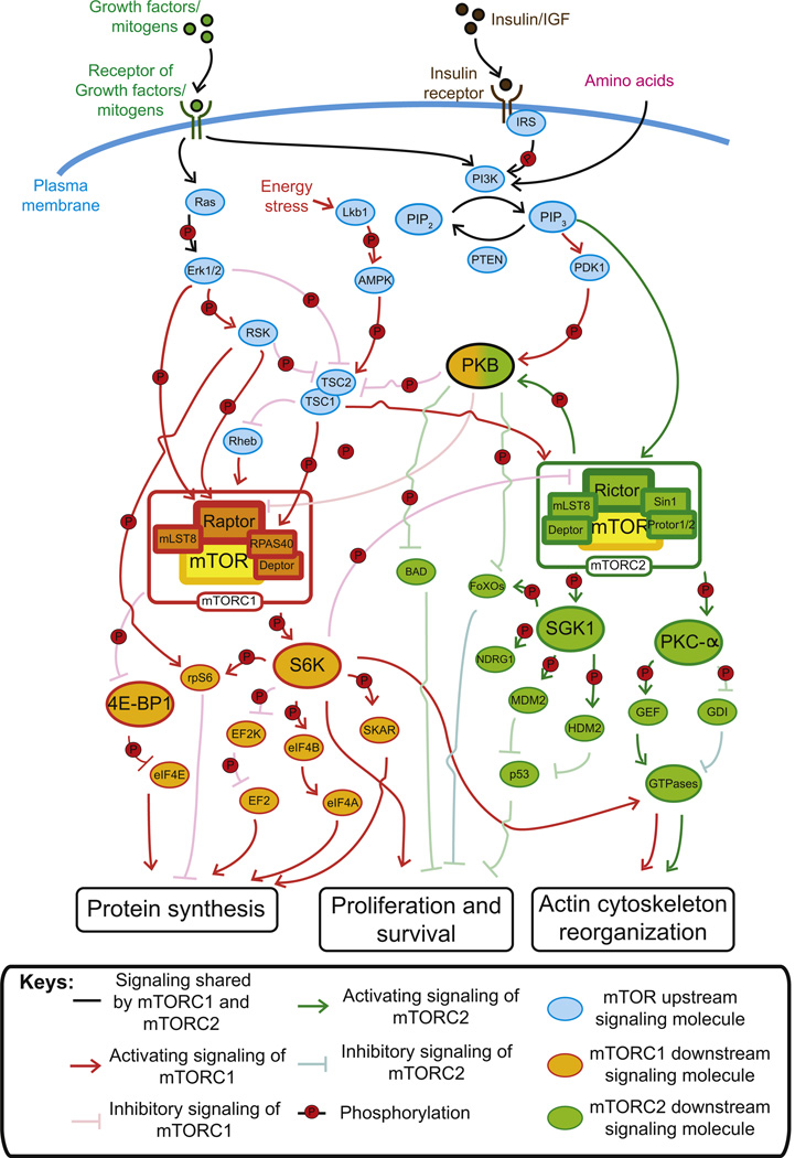
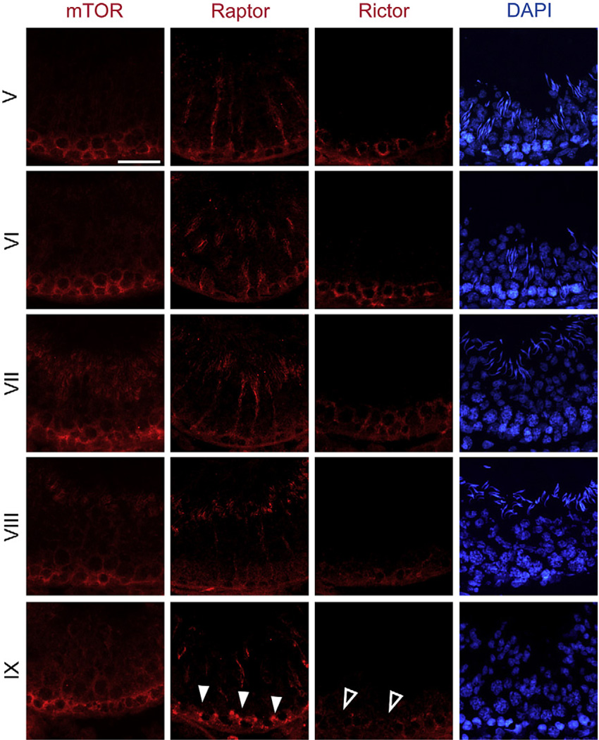
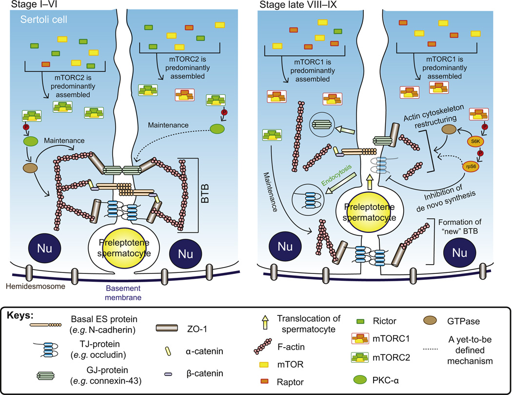
In mammalian testes, haploid spermatozoa are formed from diploid spermatogonia during spermatogenesis, which is a complicated cellular process. While these cellular events were reported in the 1960s and 1970s, the underlying molecular mechanism(s) that regulates these events remained unexplored until the past ∼10 years. For instance, adhesion proteins were shown to be integrated components at the Sertoli cell-cell interface and/or the Sertoli-spermatid interface in the late 1980s. But only until recently, studies have demonstrated that some of the adhesion proteins serve as the platform for signal transduction that regulates cell adhesion. In this chapter, a brief summary and critical discussion are provided on the latest findings regarding these cell-adhesion proteins in the testis and their relationship to spermatogenesis. Moreover, antagonistic effects of two mammalian target of rapamycin (mTOR) complexes, known as mTORC1 and mTORC2, on cell-adhesion function in the testis are discussed. Finally, a hypothetic model is presented to depict how these two mTOR-signaling complexes having the "yin" and "yang" antagonistic effects on the Sertoli cell tight junction (TJ)-permeability barrier can maintain the blood-testis barrier (BTB) integrity during the epithelial cycle while preleptotene spermatocytes are crossing the BTB.


Figures

Figure 6.1
Figure 6.1. Differences in the morphological layouts of junction types between a typical epithelium/endothelium and the seminiferous epithelium
(A) For the junctional complex in typical epithelia/endothelia, TJs, which are responsible for sealing the intercellular space to create the barrier function by regulating paracellular and transcellular transport, are located at the apical region of the lateral membrane between adjacent epithelial/endothelial cells. Underneath TJs, there are AJs that contribute to most of the adhesive force of the apical junctional complex by connecting to a dense F-actin network, creating the zonula adherens plaque, to be followed by desmosomes. Both TJ and AJ are actin-based cell–cell anchoring junctions, whereas DS is intermediate filament-based cell–cell anchoring junction. Other junctional molecules such as GJs, which are not part of the junctional complex, are localized basal to the junctional complex (constituted by TJ, AJ and DS). (B) Unlike the junctional complex in typical epithelia which are furthest away from the basal lamina, the BTB in seminiferous epithelium is located near the basement membrane (a modified form of extracellular matrix in the testis). Instead of being arranged as discrete structure as in other epithelia/ endothelia, TJs, basal ES (a testis-specific actin-rich AJ) and GJs are coexisting at the BTB, which together with DS are all involved in creating the BTB. The BTB physically separates the seminiferous epithelium into the basal and apical (adluminal) compartments. Spermatogonia and preleptotene spermatocytes reside at the basal compartment, and preleptotene spermatocytes that arise at stage VII-VIII of the epithelial cycle in the rat testis are the only germ cells that can traverse the BTB. After traversing the BTB, spermatocytes undergo meiosis and eventually differentiate into elongating/elongated spermatids, and spermatids (step 8–19 spermatids in the rat testis) anchored to the Sertoli cells by apical ES. Furthermore, hemidesmosomes (intermediate filament-based cell–matrix anchoring junction) and focal adhesion complexes (FAC, or known as focal contacts, an actin-based cell–matrix anchoring junction) are also found in most epithelia, but FAC is absent in the seminiferous epithelium. Abbreviations used: Sg, spermatogonium; Sy, spermatocyte; rSp, round spermatid; eSp, elongating spermatid; ESp, elongated spermatid; Nu, Sertoli cell nucleus; DS, desmosome; AJ, adherens junction; GJ, gap junction; TJ, tight junction; ES, ectoplasmic specialization. For color version of this figure, the reader is referred to the online version of this book.
Figure 6.2
Figure 6.2. Restructuring of the BTB to facilitate the transit of preleptotene spermatocytes at stage VIII of the epithelial cycle
Before BTB restructuring takes place, its integrity is maintained by coexisting TJs, basal ES and GJs which interact with each other and linked to actin cytoskeleton for structural support via adaptor proteins such as ZO-1. Besides, desmosome is also present at the Sertoli cell–cell interface at the BTB. On the other hand, elongated spermatids are also anchored to the Sertoli cell via a testis-specific apical ES protein complex in which laminin-333 residing at the elongating spermatid is linked to α6β1-integrin restricted to the Sertoli cell. At stage VIII of the epithelial cycle, when preleptotene spermatocytes are in transit at the BTB to enter the apical compartment for further development, the “old” BTB above the spermatocyte disassembles to “open” the BTB. This process is mediated by the apical ES–BTB–hemidesmosome functional axis, in which laminin 333 at the apical ES is cleaved by MMP2 to generate bioactive laminin fragments. The laminin fragments induce disruption of the “old” BTB and cause the loss of hemidesmosome function which also contributes to the “opening” of the “old” BTB. Besides, BTB restructuring is also facilitated by mTORC1 as well as by the reorganization of actin cytoskeleton mediated by actin-regulating proteins, such as the Arp2/3–N-WASP complex and Eps8 which induce a “branched/debundled” and “bundled” configuration of the actin filaments at the basal ES, respectively. Without the support from the dense F-actin network, BTB proteins are internalized through endocytosis and the internalized BTB proteins can either undergo degradation or being recycled for the assembly of “new” BTB via transcytosis at the base of the preleptotene spermatocytes. It is likely that molecules, such as testosterone, that promote BTB integrity may be working in concert with mTORC2 underneath the spermatocyte in transit to assemble a “new” BTB before the “old” BTB above the transiting spermatocyte is disassembled, so that the barrier function can remain intact during germ cell movement at the site. For color version of this figure, the reader is referred to the online version of this book.
Figure 6.3
Figure 6.3. The likely mTOR signaling pathways involving mTORC1 and mTORC2 and the corresponding interacting/regulatory proteins that regulate different cellular events including BTB function in the testis via the effects on F-actin organization
By assembling with different subunits, two mTOR complexes can be formed, namely, mTORC1 and mTORC2. Besides mTORC1 that is specifically regulated by the energy status of a cell, both mTOR complexes are activated by growth factors (e.g. insulin), mitogens and amino acids. Upon activation, except that upregulation of protein synthesis for cell growth is specifically mediated by mTORC1, the control of cell proliferation and survival as well as actin cytoskeleton organization is modulated by both complexes, despite the fact that they have their unique substrates and downstream signaling molecules. Moreover, mTORC1 and mTORC2 share several upstream signaling molecules. For example, PIP3 can activate both complexes while TSC1/2 complex inhibits mTORC1 but activates mTORC2. Furthermore, the signaling pathways of the two mTOR complexes are interconnected in which S6K1, the substrate of mTORC1, is able to phosphorylate rictor and thus inhibits mTORC2. As such, phosphorylation of PKB, which is the substrate of mTORC2, can be reduced. Since PKB phosphorylation is required for activating mTORC1, this leads to suppression of mTORC1 signaling and therefore, a negative feedback loop is established. For color version of this figure, the reader is referred to the online version of this book.
Figure 6.4
Figure 6.4. Stage-specific expression of raptor and rictor versus mTOR in the seminiferous epithelium of adult rat testes
Relative expression level and localization of mTOR, raptor and rictor (red) in the seminiferous epithelium from stage V–IX tubules were examined by immunofluorescence microscopy. Cell nuclei were stained with DAPI (blue) to show the stages of the tubules. This figure shows that from stage V to IX, mTOR was expressed at relatively similar level at the basal compartment where BTB was located. On the other hand, the expression of raptor, which is the key binding partner of mTORC1, was transiently induced at stage IX (indicated by white arrow head) that begin in late stage VIII, most notably at the BTB, whereas the expression of rictor, which is the key subunit of mTORC2, was found to decline gradually from stage VII and became barely detectable at late stage VIII through stage IX (indicated by open arrow head), even though it remained weakly expressed in these stages. Bar, 50 μm, which applies to all micrograph. For interpretation of the references to color in this figure legend, the reader is referred to the online version of this book.
Figure 6.5
Figure 6.5. mTORC1 and mTORC2 display antagonistic effects on the BTB and their combined effects can protect the immunological barrier integrity during the transit of preleptotene spermatocytes at the BTB
It is noted that rictor and raptor compete for mTOR for the formation of mTORC2 and mTORC1, respectively, to promote BTB and disrupt BTB integrity. At stages I–VI in which prior to BTB restructuring, the relative high expression of rictor favors the assembly of mTORC2, which is necessary for keeping the integrity of BTB by maintaining the dense F-actin network (namely the actin filament bundles at the basal ES), expression level of GJ proteins and GJ communication. On the other hand, in stage late VIII to stage IX that the BTB is transiently “open” to facilitate the transit of spermatocyte, the expression level of raptor is induced, whereas that of rictor is reduced. Thus, formation of mTORC2 is reduced and mTORC1 is favored. mTORC1 activates rpS6, which in turn disrupts the “old” BTB above the preleptotene spermatocytes in transit at the BTB by inhibiting de novo synthesis of BTB proteins. In addition, actin cytoskeleton reorganization during BTB restructuring is induced by (i) mTORC1 signaling via S6K1 and rpS6 and (ii) the decrease in phosphorylated PKC-α due to reduced mTORC2. The disorganized F-actin network leads to internalization of BTB proteins, which perturbs the “old” BTB. Furthermore, the decrease in GJ proteins and GJ communication caused by reduced mTORC2 also facilitates the disruption of the “old” BTB for the translocation of spermatocytes across the BTB. However, while mTORC2 expression is reduced, it remains to be robust enough to sustain the maintenance of the “new” BTB that is being assembled behind the preleptotene spermatocytes in transit. In short, utilizing the antagonistic effects of the mTORC1 and mTORC2 on the TJ-permeability barrier, the immunological barrier function can be maintained during the passage of preleptotene spermatocytes, which are connected in “clones” via intercellular bridges, at the BTB. For color version of this figure, the reader is referred to the online version of this book.

Similar articles

Cited by

References

    1. Abraham RT, Wiederrecht GJ. Immunopharmacology of rapamycin. Annu. Rev. Immunol. 1996;14:483–510. - PubMed
    1. Alessi DR, James SR, Downes CP, Holmes AB, Gaffney PR, Reese CB, Cohen P. Characterization of a 3-phosphoinositide-dependent protein kinase which phosphorylates and activates protein kinase Balpha. Curr. Biol. 1997;7:261–269. - PubMed
    1. Aliabadi AZ, Pohanka E, Seebacher G, Dunkler D, Kammerstatter D, Wolner E, Grimm M, Zuckermann AO. Development of proteinuria after switch to sirolimus- based immunosuppression in long-term cardiac transplant patients. Am J. Transplant. 2008;8:854–861. - PubMed
    1. Amato R, D’Antona L, Porciatti G, Agosti V, Menniti M, Rinaldo C, Costa N, Bellacchio E, Mattarocci S, Fuiano G, et al. Sgk1 activates MDM2-dependent p53 degradation and affects cell proliferation, survival, and differentiation. J. Mol. Med. (Berl) 2009;87:1221–1239. - PubMed
    1. Andjelkovic M, Alessi DR, Meier R, Fernandez A, Lamb NJ, Frech M, Cron P, Cohen P, Lucoq JM, Hemmings BA. Role of translocation in the activation and function of protein kinase. B.J. Biol. Chem. 1997;272:31515–31524. - PubMed

Publication types

Substances

LinkOut - more resources