Review
. 2013 Feb;46(1):1-132.
doi: 10.1017/S0033583512000157. Epub 2013 Jan 15.

Why nature really chose phosphate

Affiliations
Review

Why nature really chose phosphate

Shina C L Kamerlin et al. Q Rev Biophys. 2013 Feb.

Abstract

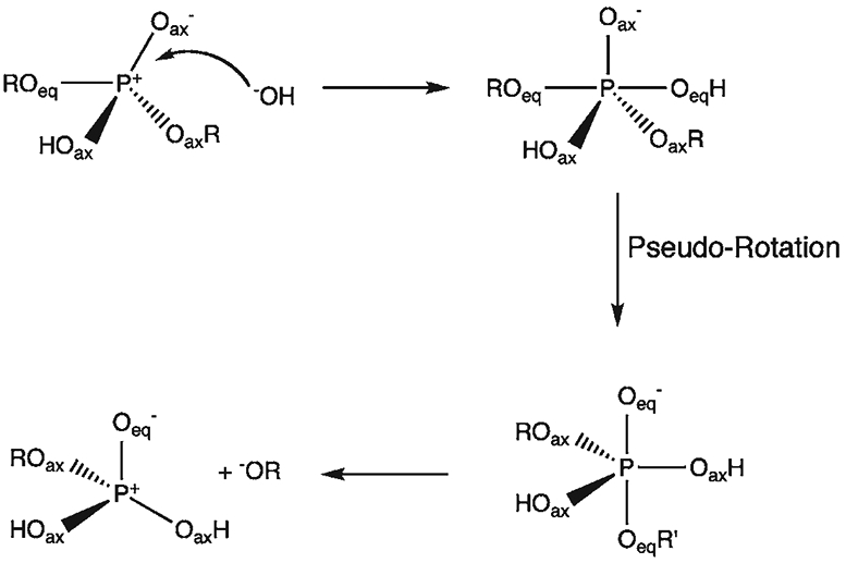
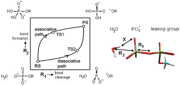
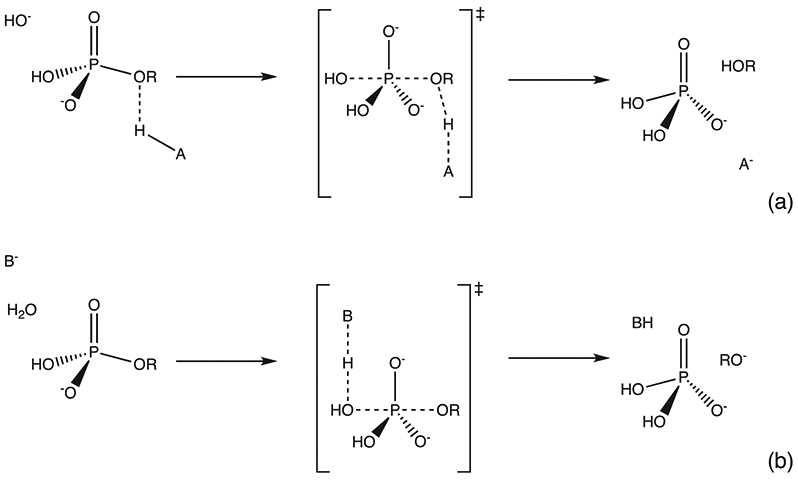
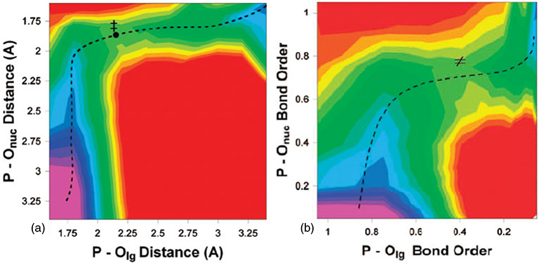
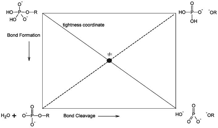
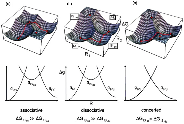
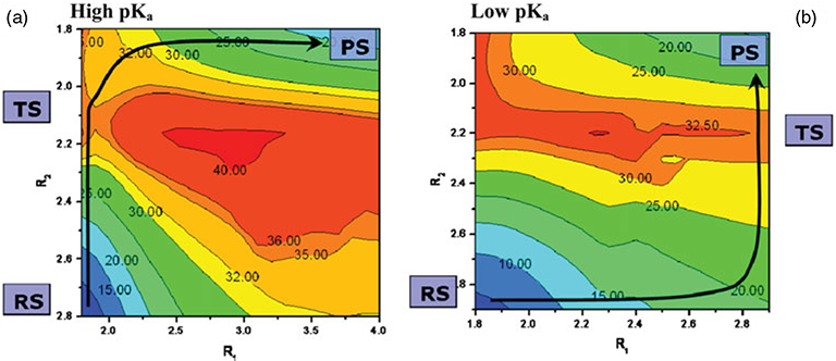
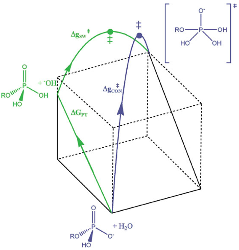
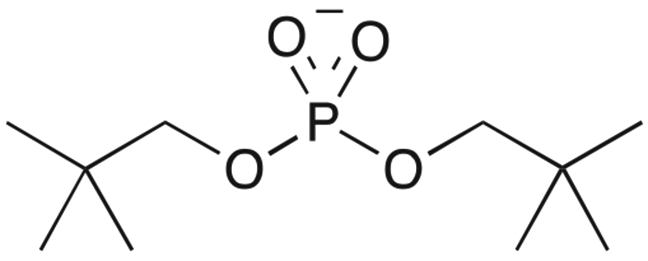
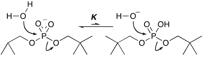
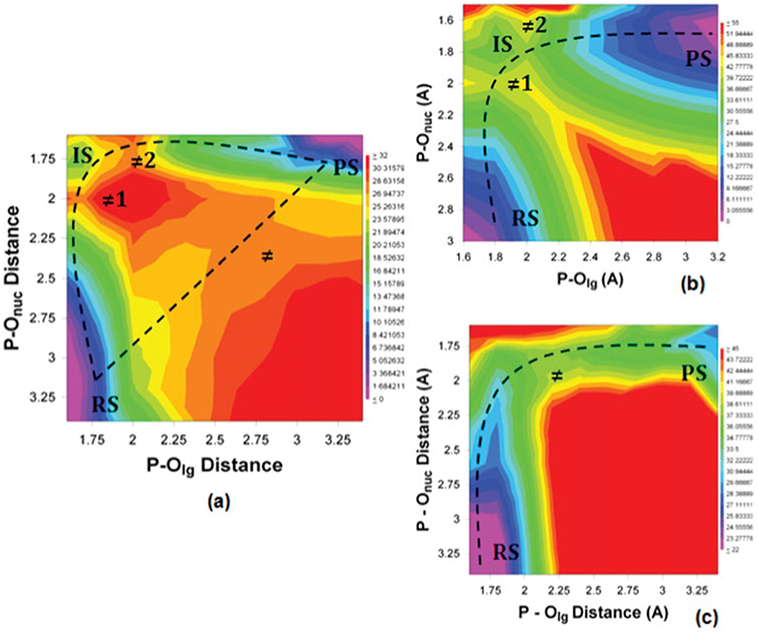
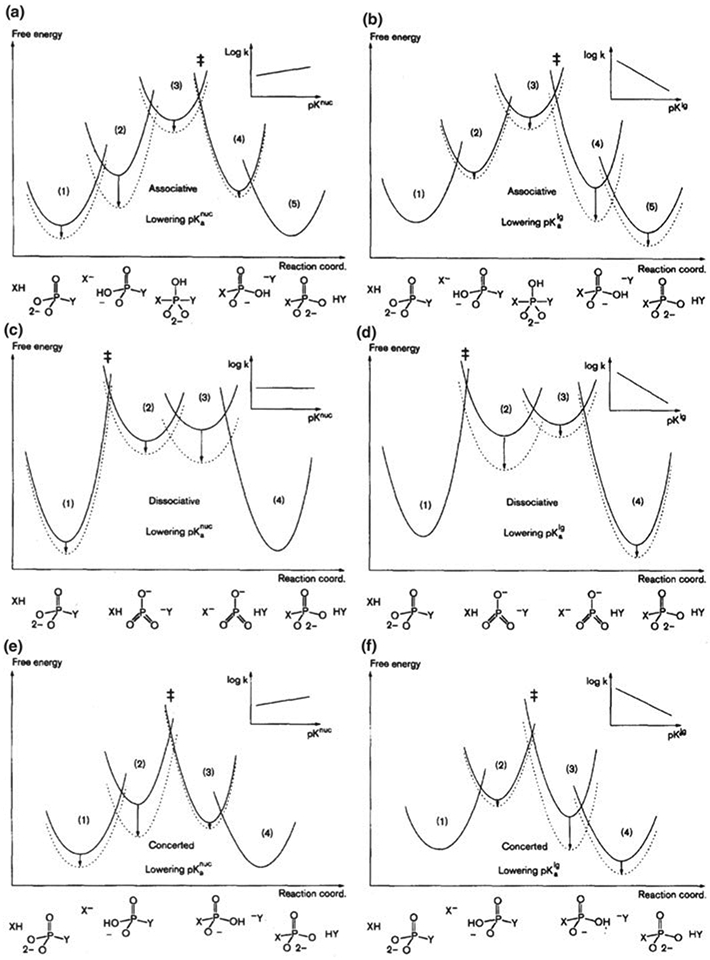
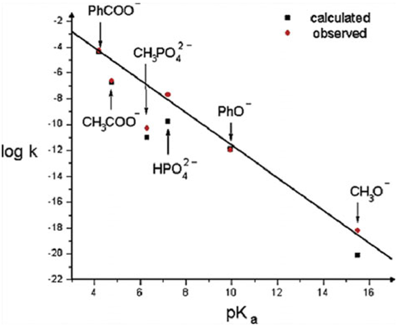
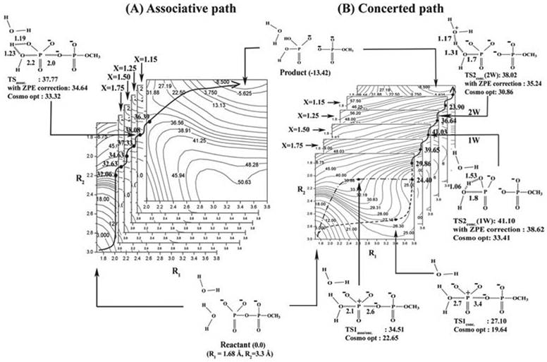
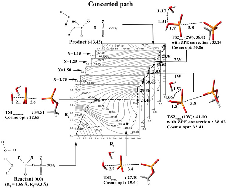
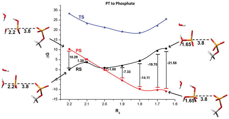
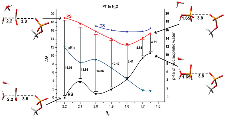
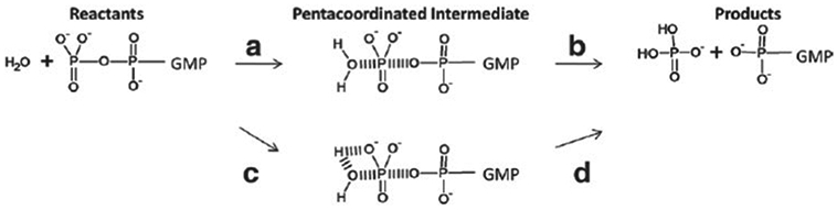
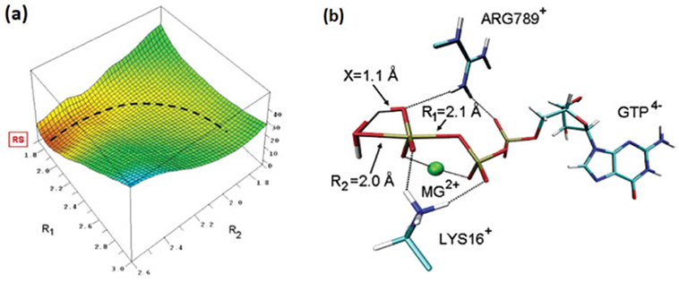
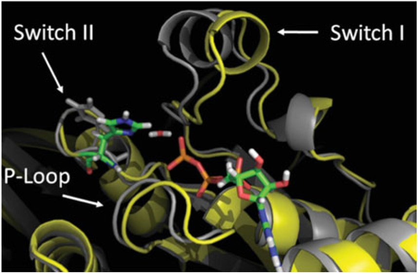
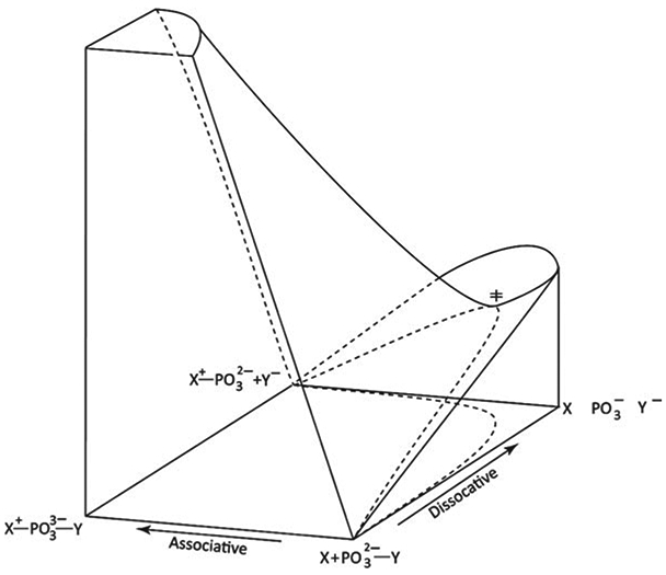
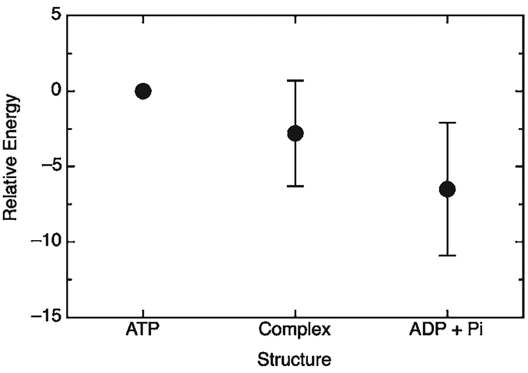
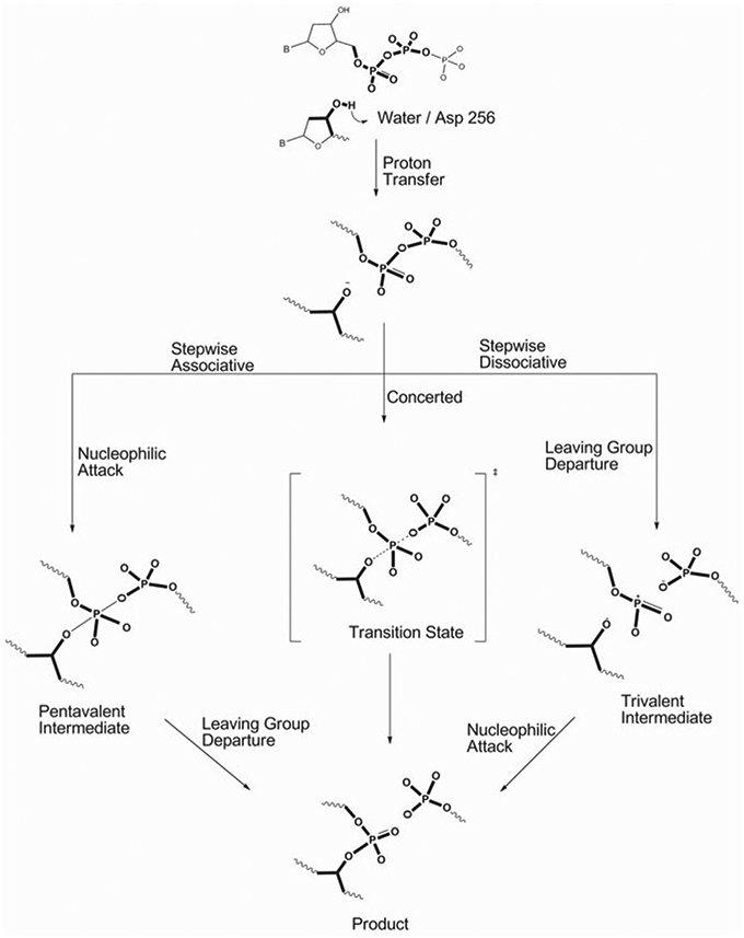
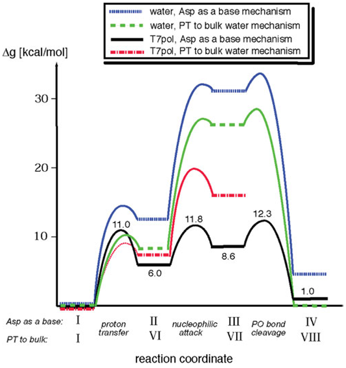
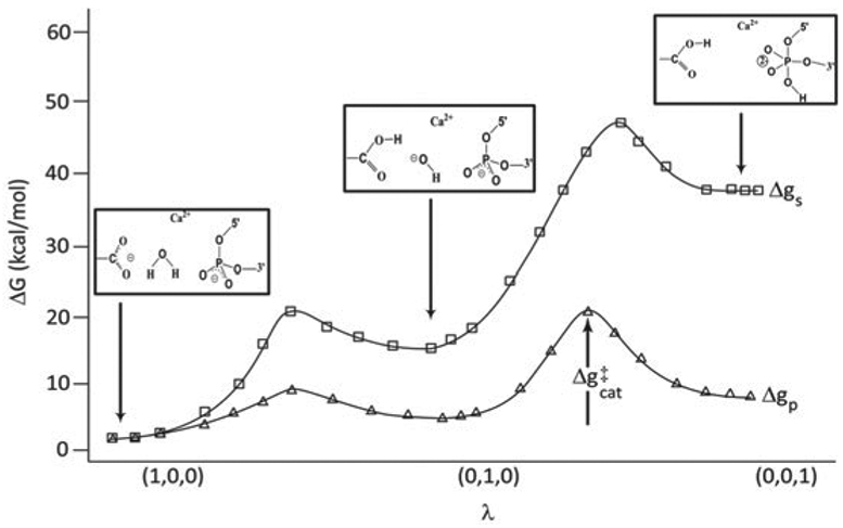
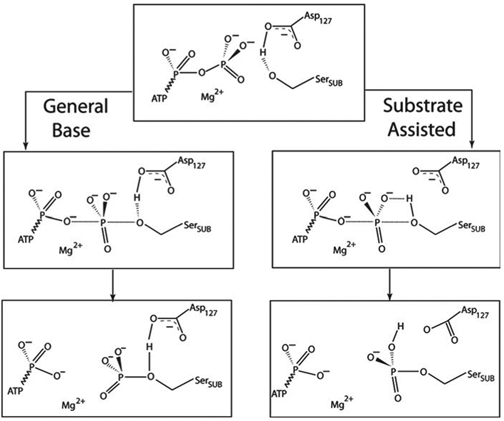
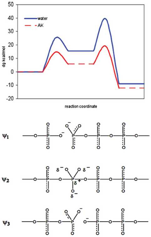
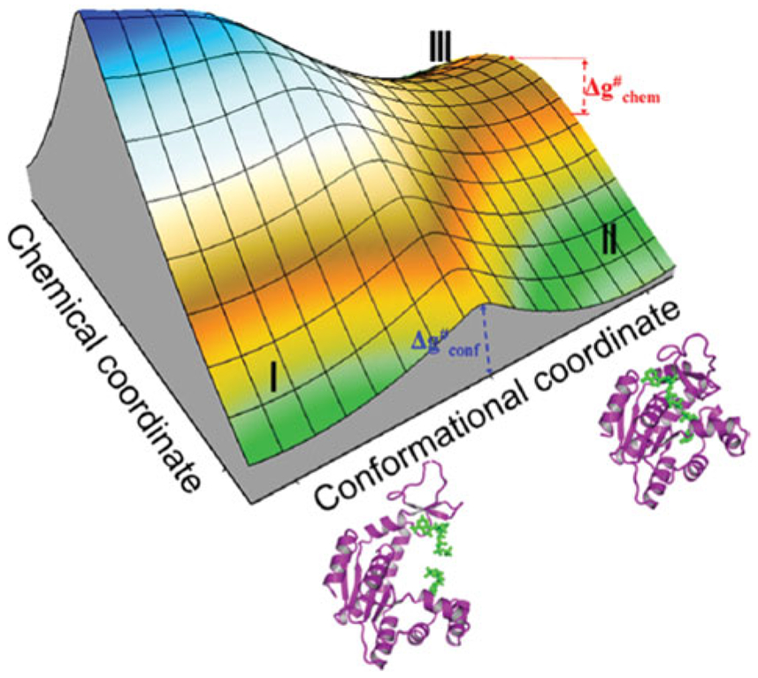
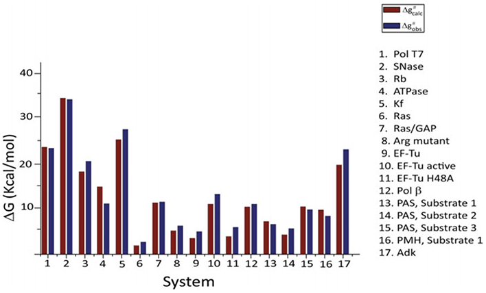
Phosphoryl transfer plays key roles in signaling, energy transduction, protein synthesis, and maintaining the integrity of the genetic material. On the surface, it would appear to be a simple nucleophile displacement reaction. However, this simplicity is deceptive, as, even in aqueous solution, the low-lying d-orbitals on the phosphorus atom allow for eight distinct mechanistic possibilities, before even introducing the complexities of the enzyme catalyzed reactions. To further complicate matters, while powerful, traditional experimental techniques such as the use of linear free-energy relationships (LFER) or measuring isotope effects cannot make unique distinctions between different potential mechanisms. A quarter of a century has passed since Westheimer wrote his seminal review, 'Why Nature Chose Phosphate' (Science 235 (1987), 1173), and a lot has changed in the field since then. The present review revisits this biologically crucial issue, exploring both relevant enzymatic systems as well as the corresponding chemistry in aqueous solution, and demonstrating that the only way key questions in this field are likely to be resolved is through careful theoretical studies (which of course should be able to reproduce all relevant experimental data). Finally, we demonstrate that the reason that nature really chose phosphate is due to interplay between two counteracting effects: on the one hand, phosphates are negatively charged and the resulting charge-charge repulsion with the attacking nucleophile contributes to the very high barrier for hydrolysis, making phosphate esters among the most inert compounds known. However, biology is not only about reducing the barrier to unfavorable chemical reactions. That is, the same charge-charge repulsion that makes phosphate ester hydrolysis so unfavorable also makes it possible to regulate, by exploiting the electrostatics. This means that phosphate ester hydrolysis can not only be turned on, but also be turned off, by fine tuning the electrostatic environment and the present review demonstrates numerous examples where this is the case. Without this capacity for regulation, it would be impossible to have for instance a signaling or metabolic cascade, where the action of each participant is determined by the fine-tuned activity of the previous piece in the production line. This makes phosphate esters the ideal compounds to facilitate life as we know it.


Figures

Fig. 1.
Fig. 1.
Representative bond distances in phosphoric acid, based on (Smith et al. 1955).
Fig. 2.
Fig. 2.
A comparison of (a) phosphoric acid, (b) a phosphate monoester, (c) a phosphate diester, and (d) a phosphate triester.
Fig. 3.
Fig. 3.
Fundamental mechanisms for phosphate ester hydrolysis (see also Gani & Wilkie, 1995).
Fig. 4.
Fig. 4.
Generalized pathways for phosphate monoester hydrolysis, using the illustrative example of hydroxide attack on a phosphate dianion. Shown here are stepwise (a) dissociative and (b) associative mechanisms.
Fig. 5.
Fig. 5.
The pseudo-rotation mechanism for the hydrolysis of phosphate diesters.
Fig. 6.
Fig. 6.
A sample MFJ diagram (Jencks, 1972; More O’Ferrall, 1970) for examining phosphate ester hydrolysis. The particular example in this figure illustrates a hypothetic surface for water attack on phosphate monoester dianions. The surface is defined as a function of the distance between the phosphorus atom and the leaving (x-axis) and entering ( y-axis) oxygen atoms, respectively. This figure was originally presented in (Klähn et al. 2006).
Fig. 7.
Fig. 7.
Phosphate ester hydrolysis in the presence of an (a) acid or (b) base catalyst.
Fig. 8.
Fig. 8.
A cubic reaction coordinate diagram. The three edge coordinates are represented by the x, y and z-axes. (0,0,0) and (1,1,1) are the starting point and product, respectively. All other points represent the corner intermediates (Guthrie, 1996).
Fig. 9.
Fig. 9.
Free-energy surfaces for hydroxide attack on dineopentyl phosphate, as functions of (a) bond order and (b) bond distance. This figure is adapted from (Kamerlin et al. 2008b).
Fig. 10.
Fig. 10.
Reaction coordinate diagram, defined in terms of bond order, for a reaction involving a symmetrical concerted TS.
Fig. 11.
Fig. 11.
A VB description of the energy surfaces for phosphate hydrolysis in three limiting cases for associative (a), dissociative (b), and concerted (c) mechanisms. The surfaces for the hydrolysis reaction were generated by mixing four states using an EVB formulation (the effect of the off-diagonal term is in included for simplicity). RS, IS, and PS represent, respectively, reactant, intermediate, and product states and the indices as and ds stand for associative and dissociative, respectively. The dots on the surfaces designate the minima of the corresponding diabatic surfaces (Klähn et al. 2006). The change of the highest TS with ΔGRS→PS defines the corresponding LFER
Fig. 12.
Fig. 12.
Sample free-energy surfaces for the hydrolysis of phosphate monoester dianions, adapted from (Klähn et al. 2006). Systems with leaving groups with high pKa, prefer a concerted associative pathway (A, left), whereas those with more acidic leaving groups prefer a dissociative pathway (B, right).
Fig. 13.
Fig. 13.
Schematic 3D representation for stepwise versus concerted PT. Here, ΔgCON and ΔgSW represent the Δg for concerted and stepwise pathways respectively, and ΔGPT denotes the free energy required for PT to the phosphate through a pre-equilibrium step. The concerted pathway is depicted in purple, and the stepwise pathway in green. Finally, the positions of the TSs on this figure are only provided for illustration, and the actual TS can occur at any point along the pathway. This figure was originally presented in (Kamerlin et al. 2008b).
Fig. 14.
Fig. 14.
Dineopentyl phosphate (Np2P).
Fig. 15.
Fig. 15.
Alternative kinetically equivalent mechanisms for the pH-independent hydrolysis of the Np2P anion (Schroeder et al. 2006).
Fig. 16.
Fig. 16.
Calculated free-energy surfaces for (a) the acid-catalyzed, (b) the pH-independent, and (c) the base catalyzed hydrolysis of dineopentyl phosphate. RS, IS, and PS denote reactant, intermediate and product states, respectively. ≠1 and ≠2 denote TSs for P–Onuc formation and P–Olg cleavage in an AN + DN pathway, respectively, and ≠denotes a single concerted ANDN TS. This figure is adapted from (Kamerlin et al. 2008b).
Fig. 17.
Fig. 17.
Free-energy diagrams depicting different diabatic states along (a, b) a stepwise associative pathway involving pre-equilibrium PT to the phosphate group, (c, d) a stepwise dissociative pathway involving unimolecular elimination of metaphosphate from the dianion, and (e,f) a concerted pathway also involving PT. Reactions ad are rate limiting by leaving group bond fission. The dashed curves show the shift in free energy upon reducing the pKa of the nucleophile (a, c, e) or leaving group (b, d, f). Taken with minor corrections from (Aqvist et al. 1999).
Fig. 18.
Fig. 18.
A comparison of experimental (circles) and calculated (squares) LFER for the hydrolysis of a series of phosphate and pyrophosphate monoesters in aqueous solution. This figure was originally presented in (Rosta et al. 2008).
Fig. 19.
Fig. 19.
The calculated free energy surfaces along the associative (A) and concerted/Late PT (B) pathways for the hydrolysis of monomethyl pyrophosphate trianion in solution utilizing both the 1 W and 2 W mechanisms. As seen from the figure the rate determining TSs for the 2 W and 1 W have similar energies. The surfaces are drawn as functions of R1, and R2 for several values of proton transfer coordinate X (R1, R2, and X are defined in Fig. 1A). TSassoc describes the transition state along the associative pathway, TS1conc and TS2conc describes the transition states along the concerted pathway. The values of the TS energies evaluated by different approaches are given near the corresponding TSs figures.
Fig. 20.
Fig. 20.
Focusing on the calculated free energy surfaces for the reaction paths that starts with an associative or dissociative paths that follows by a late PT to the phosphate oxygen. As seen from the figure the rate determining TSs for the 2 W and 1 W paths have similar energies. Note that the activation entropy of the 2 W path (which is not included) should be larger than that of the 1 W barrier. The surfaces are drawn as functions of R1, and R2 for several values of the proton transfer coordinate X, and focuses on comparing the PT barriers that starts from the intermediate where R2 is around 2 Å and R1 is partially broken.
Fig. 21.
Fig. 21.
The effect of the change in pKa, of the attacking water on the PT energetics in the 1 W mechanism. The figure displays the R2 dependence of the energy of the “reactant” (where the proton is bound to the nucleophilic water and the water oxygen is in a short distance of 2.2 Å), the “product” (where the proton is attached to the acceptor phosphate oxygen) and the corresponding TSs for the PT process. As seen from the figure, the attacking water becomes much more acidic upon formation of the O-P and the PT occur. However, the PT does not become spontaneous because of the extra energy required for bending the O-P-O angel for an optimal PT.
Fig. 22.
Fig. 22.
The effect of the change in pKa) of the attacking water on the PT energetics in the 2 W mechanism. The figure displays the R2 dependence of the energies of the “reactant” (where the proton is attached to nucleophilic water) and “product” (where the proton is attached to the second water) and the corresponding TSs for the PT process. As seen from the figure, the attacking water becomes much more acidic upon formation of the O-P and the PT becomes basically spontaneous.
Fig. 23.
Fig. 23.
Possible mechanistic pathways for the GTPase reaction mechanism. The reaction is considered as a two-step mechanism where the GTP serves as a general base. In the first step, a, the water molecule attacks the GTP and a penta-coordinated intermediate is formed. In the second step, b, the PβO bond is broken and the GDP and a phosphate group are formed. Note that the attack of the water molecule (step a) may be either concerted or stepwise mechanism. In principle, the steps b and d could also proceed with the assistance of another water or a base.
Fig. 24.
Fig. 24.
(a) The calculated energy surface for the hydrolysis of GTP in RasGAP in the space defined by the P–O distance to the leaving group and nucleophile (R1 and R2, respectively), (b) The calculated TS structure for the hydrolysis of the GTP substrate in RasGAP. This figure was originally presented in (Klähn et al. 2006).
Fig. 25.
Fig. 25.
A schematic diagram of the free-energy surface and the representative geometries for the hydrolysis of monomethyl pyrophosphate trianion in solution following the catalytic mechanism explored in (Grogorenko et al. JPCB, 2006, 110, 4407). This mechanism corresponds to the associative (2 W) pathway depicted in Fig. 20.
Fig. 26.
Fig. 26.
(a) The GTPase reaction profile. The free-energy profiles for the GTPase reaction in water (circles), Ras (squares) and in the RasGAP complex (stars) obtained from an average over five different profiles, calculated using the EVB FEP/Umbrella sampling procedure.(b) Mutation effects on the GTPase reaction profile. The free-energy profiles of the GTPase reaction in the native RasGAP complex (stars) and its different mutations, R789RNP (squares), Q61QNP (triangles) and Q61L (circles). The profiles were obtained from an average over five different profiles, calculated using the EVB FEP/Umbrella sampling procedure. NP stands for mutation to the non-polar form of the indicated residue. This figure was originally presented in (Shurki & Warshel, 2004).
Fig. 27.
Fig. 27.
An illustration of the ‘Arginine Finger’.
Fig. 28.
Fig. 28.
Electrostatic effects due to the Q61L mutation. The main changes in the electrostatic contributions from the Ras residues due to the Q61L mutation. The changes in the RS are compared with those in the TS in (a) and (b), respectively. Negative and positive values correspond, respectively, to stabilization and destabilization due to the mutation. The changes in the key areas are highlighted as in Fig. 29, P-loop (yellow), Switch I (light green), parts of β3 and L4 of the switch II (orange) and the Mg2+ (dark green). The calculated changes were obtained by the LRA procedure and then scaled by a dielectric constant of 20 and 4 for the ionized and polar residues, respectively. This figure was originally presented in (Shurki & Warshel, 2004).
Fig. 29.
Fig. 29.
An overview of the key regions in the RasGAP complex. Shown here are the P-loop (yellow), Switch I (light green), parts of Switch II (orange) and the Mg2+ ion (dark green). This figure was originally presented in (Shurki & Warshel, 2004).
Fig. 30.
Fig. 30.
An illustration of the change in electrostatic interactions between the protein residues and the γ- and β-phosphates of GTP, for the reaction catalyzed by (a) Ras, (b) Ras–GAP, and (c) Ras’. Blue indicates a stabilizing shift and red indicates a destabilizing shift in the interactions, with the degree of stabilization or destabilization being proportional to the intensity of the color. Note that what is presented here is not the change in the electrostatic potential, but rather, the actual change in electrostatic energy. This figure was originally presented in (Glennon et al. 2000).
Fig. 31.
Fig. 31.
Calculated and observed activation barriers (in kcal mol−1) for different reacting systems. The notation NP (nonpolar) indicates that all the residual charges of the given residue or set of residues are set to zero. The notation ‘rib’ indicates the presence of the ribosome in the simulations. This figure was originally presented in (Adamczyk & Warshel, 2011).
Fig. 32.
Fig. 32.
An approximated catalytic free-energy landscape (kcal mol−1) for the coupling between the chemical coordinate (i.e., the movement from the RS to the PS) and the conformational space that connects the EF-Tu and EF-Tu’ conformations in the (a) native and (b) H84A mutant systems. This figure was originally presented in (Adamczyk & Warshel, 2011).
Fig. 33.
Fig. 33.
The structures of the active site in both the EF-Tu (gray, from PDB code: 1EFT) and EF–Tu’/ribosome (yellow, from PDB code: 2XQD) from our simulations are compared, respectively. The changes in the backbone of this region do not occur in the EF-Tu protein alone. Critical regions (P-Loop, Switch I, and Switch II) are labeled. GTP and water are included in the RS configuration of EF–Tu’/ribosome. Mg2+, aa-tRNA, and ribosome are not shown for the sake of clarity in this diagram. This figure was originally presented in (Adamczyk & Warshel, 2011). Note that even though His 84 is not presented as explicitly protonated here, the actual simulations were carried out considering His 84 as protonated.
Fig. 34.
Fig. 34.
The general features of a proposed energy surface for phosphate hydrolysis, adapted from (Admiraal & Herschlag, 1995), where it was suggested (without quantitative considerations) that an associative pathway will have an essentially insurmountably large energy barrier compared with a dissociative pathway (which theoretical studies have subsequently proven to be incorrect, see discussion in main text).
Fig. 35.
Fig. 35.
Schematic representation of the ATP hydrolysis cycle of F1-ATPase, during the 120° rotation of the γ-subunit. Scheme I and II describes two of the most likely mechanisms. An atomistic picture of the system is shown on top. The stage between C and B” may involve three nucleotides supporting a tri-site mechanism (not shown explicitly). This figure was originally presented in (Mukherjee & Warshel, 2011).
Fig. 36.
Fig. 36.
Overall ΔGcalc for each of the steps of the reaction shown in Eq. (26), averaged over two different metal cations (Mg2+ and Ca2+), as well as all three solvation models (QM/MM-FEP, COSMO and PCM), in the presence of explicit water molecules. All energies are given in kcal mol−1, and the error bars show the standard deviation over three solvation models. This figure was originally presented in (Kamerlin & Warshel, 2009).
Fig. 37.
Fig. 37.
Energetics of F1-ATPase in kcal mol−1. Shown here are: (a) the calculated electrostatic free-energy surface in the space defined by the rotation of the central γ subunit (vertical coordinate) and the subunit alterations (horizontal coordinate). (b) Semiqualitative energetics (in the standard state without concentration effect and with the p concentration effect in magenta) along the least energy path based on the considerations in the Supporting Information of (Mukherjee & Warshel, 2011). This figure was originally presented in (Mukherjee & Warshel, 2011).
Fig. 38.
Fig. 38.
The conformational/catalytic landscape of F1-ATPase. The figure describes the energetics of the system along the chemical coordinate of one nucleotide (bound to subunit 2 or 3) as well as the conformational coordinate of the combined system. The functional path is shown using a dashed white line. This figure was originally presented in (Mukherjee & Warshel, 2011).
Fig. 39.
Fig. 39.
An example of a typical polymerase active site. Shown here are key residues in the exo site of the Klenow fragment, outlining the position of the substrate, nucleophile, catalytic metal ions and key active site residues. This figure was originally presented in (Fothergill et al. 1995).
Fig. 40.
Fig. 40.
A schematic description of the chemical steps of the nucleotide incorporation reaction catalyzed by Pol β. The initial deprotonation of the 3′-hydroxyl group of the primer is followed by nucleophilic attack on the Pα of the incoming dNTP. The formation of the O3′–Pα bond then results in the release of the pyrophosphate leaving group. As shown in this figure, after the initial PT step, the reaction may proceed via either a stepwise pathway involving either a trivalent (metaphosphate) or pentavalent (phosphorane) intermediate, or a concerted pathway which can in turn also be associative or dissociative in nature depending on the extent of bond formation to the incoming nucleophile and bond cleavage to the departing leaving group. However, it is worth noting that previous studies have suggested that the hydrolysis of a phosphate diester is unlikely to proceed via a stepwise dissociative mechanism. (Kamerlin et al. 2008b).
Fig. 41.
Fig. 41.
A comparison of different mechanistic possibilities for the nucleotide transfer catalyzed by T7 polymerase. Shown here are the energetics of the ‘asp-as-base’ mechanism as well as a mechanism in which nucleophile activation occurs by PT from the 3′-OH group of the primer to bulk water. For a discussion of the mechanistic details, see the main text and (Florián et al. 2003a). This figure was originally presented in (Florián et al. 2003a).
Fig. 42.
Fig. 42.
Schematic free-energy surfaces, illustrating potential coupling between the conformational and chemical coordinates, for the insertion of (a) correct and (b) incorrect dNTP by a DNA polymerase. rO, and rc, designate the open and closed configurations respectively, and TSC designates the TS associated with the conformational motion. Finally, RS, TS, and PS designate the reactant (ES), transition and product states, respectively, and (R) and (W) designate correct and incorrect nucleotide insertion. This figure was originally presented in (Prasad & Warshel, 2011).
Fig. 43.
Fig. 43.
Illustrating the origin of the allosteric effect. This figure uses the calculations from Fig. 10(C) of (Prasad & Warshel, 2011) and illustrates the relevant changes in the R and W systems at different protein configurations. Specifically, shown here are the protein rearrangement upon binding of R and W at r(ES(R)) and r(ES(W)). In the R case, the protein provides optimal sites for both the chemical and base-pairing parts of the process. On the other hand, in the W case, the protein has to relax in the base-pairing site in order to provide good binding, and this relaxation destroys the preorganization in the chemical part. The diagrams that represent the different configurations illustrate this allosteric effect, with the arrows indicating the protein dipoles. In the R state, these dipoles are preorganized in an optimal way, and thus provide maximum TS stabilization. In contrast, in the W state, the dipoles are forced to assume less effective preorganization, and thus yield correspondingly less TS stabilization. This figure was originally presented in (Prasad & Warshel, 2011).
Fig. 44.
Fig. 44.
A schematic illustration of free energies for correct (dCTP) and incorrect (dGTP) nucleotide incorporation reactions for the T7 DNA polymerase, Here, EDnN represents the enzyme in complex with a DNA primer strand of n residues in length, N represents the incoming dNTP, and FDnN represents the closed state of the enzyme with the DNA and nucleotide bound to it. This figure was originally presented in (Prasad & Warshel, 2011), and is adapted from (Johnson, 2008).
Fig. 45.
Fig. 45.
An overview of (a) the dNTP analogs presented in (Kamerlin et al. 2009c; Sucato et al. 2007, 2008)], where X=O, CH2, CHF, CHC1, CHBr (‘monohalogenated’ compounds, where for simplicity X=O and CH2 are also under this label) or CF2, CCl2, CBr2, CFCl (‘dihalogenated’ compounds), and the corresponding LFER for (b) correct, and (c) nucleotide insertion in DNA polymerase β, as well as (d) the corresponding reference reaction in aqueous solution.
Fig. 46.
Fig. 46.
An overview of cellular signal transduction pathways, highlighting the central role of Ras. Adapted from the Wikimedia Commons file ‘Image: Signal_transduction_pathways.png’ http://en.wikipedia.org/wiki/File:Signal_transduction_pathways.png
Fig. 47.
Fig. 47.
A detailed description of the Ras activation and deactivation.
Fig. 48.
Fig. 48.
Showing the ionized residues involved in the Rap/Raf interaction (Taken from (Nassar et al. 1995).
Fig. 49.
Fig. 49.
Outlining a way for exploiting structural information from the Ras /Raf interface, for designing drugs that deactivate defected Ras. In this strategy we (a) identify the regions in Raf with strong contribution to binding, (b, c) examine if the corresponding short peptides also bind strongly, (d) and then examine if cyclic analogues of those peptides also retain strong binding. The resulting constructs would be good drug candidates.
Fig. 50.
Fig. 50.
Electrostatic contributions of individual residues to the binding of GTP to p21ras. x indicates residues whose mutation leads to significant change in nucleotide-binding activity of p21ras. Highly conserved residues are designated by filled diamonds, whereas those that belong to structural motifs involved in nucleotide binding, and are not that highly conserved, are designated by unfilled diamonds (below the x symbols) (Muegge et al. 1996).
Fig. 51.
Fig. 51.
Explaining the nature of the transfer of the signal from the transformation of GTP to GDP to the Ras/Raf interaction. The change from GTP to GDP involves a loss of a negative charge. This reduction of negative charge is transmitted to the Ras surface as a reduction of the Ras/Raf electrostatic interaction, thus leading to the dissociation of Raf.
Fig. 52.
Fig. 52.
(a) The correlation between theoretical and calculated activation barriers and (b) overlay of the electrostatic group contributions for the hydrolysis of (1) p-nitrophenyl sulfate, (b) ethyl-p-nitrophenyl phosphate, (c) bis-p-nitrophenyl phosphate and (d) p-nitrophenyl phosphate by the arylsulfatase from Pseudomonas aeruginosa. This figure was originally presented in (Luo et al. 2012b).
Fig. 53.
Fig. 53.
Calculated free-energy profiles for the reaction catalyzed by SNase in aqueous solution (squares) and in the enzyme active site (triangles). Shown here are also the corresponding valence bond structures for the different reacting states. This figure was originally presented in (Åqvist & Warshel, 1989).
Fig. 54.
Fig. 54.
Different mechanistic possibilities for the reaction catalyzed by cyclin-dependent kinase 2. This figure is adapted from (Smith et al. 2011).
Fig. 55.
Fig. 55.
Calculated free-energy profiles for the reaction catalyzed by adenylate kinase in aqueous solution (blue) and in the enzyme active site (red). Shown here are also the corresponding valence bond structures for the different reacting states. This figure was originally presented in (Pisliakov et al. 2009).
Fig. 56.
Fig. 56.
Effective 2D catalytic landscape for adenylate kinase, evaluated in the space defined the conformational and chemical coordinates. Here, I, II, and III correspond to the open reactant state, the closed reactant state and the closed product state respectively. This figure was originally presented in (Pisliakov et al. 2009).
Fig. 57.
Fig. 57.
The catalytic effect (in kcal mol−1) on phosphate hydrolysis in different enzymes. The type of the system considered and the source of the analysis are: (1) T7 DNA polymerase (Florián et al. 2003a), (2) SNase (Åqvist & Warshel, 1989), (3) Ribonuclease (dianionic intermediate mechanism) (Glennon & Warshel, 1998), (4) ATPase (Štrajbl et al. 2003), (5) The exonuclease activity of the Klenow fragment or DNA polymerase I (Fothergill et al. 1995), (6–8) Ras GTPase alone, in complex with GAP and the arginine mutant respectively (Shurki & Warshel, 2004), (9–11) EF-Tu before activation, after activation and the H48A mutant respectively (Adamczyk & Warshel, 2011), (12) DNA polymerase β (Prasad & Warshel, 2011), (13–15). P. aeruginosa arylsulfatase (Luo et al. 2012a), Substrate 1 =p-nitrophenyl phosphate monoester, Substrate 2=ethyl-p-nitrophenyl phosphate, Substrate 3=bis-p-nitrophenyl phosphate (Luo et al. in press). Note that all are promiscuous activities, (16) Phosphonate monoester hydrolase from R. leguminosarum (Substrate 1, Unpublished results), (17) Adenylate kinase (Pisliakov et al. 2009).

Similar articles

Cited by

References

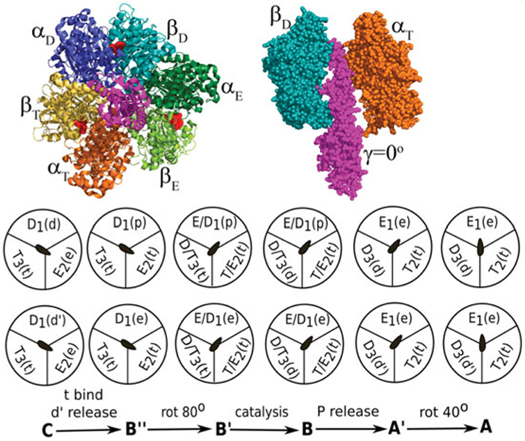
    1. Abrahams JP, Leslie AGW, Lutter R & Walker JE (1994). Structure at 2·8 Å resolution of F1-ATPase from bovine heart mitochondria. Nature 370, 621–628. - PubMed
    1. Adachi K, Oiwa K, Nishizaka T, Furuike S, Noji H, Itoh H, Yoshida M & Kinosita K (2007). Coupling of rotation and catalysis in F1-ATPase revealed by single-molecule imaging and manipulation. Cell 130, 309–321. - PubMed
    1. Adamczyk AJ, Cao J, Kamerlin SCL & Warshel A(2011). Catalysis by dihydrofolate reductase and other enzymes arises from electrostatic preorganization, not conformational motions. Proceedings of the National Academy of Sciences of the United States of America 108, 14115–14120. - PMC - PubMed
    1. Adamczyk AJ & Warshel A (2011). Converting structural information into an allosteric-energy-based picture for elongation factor Tu activation by the ribosome. Proceedings of the National Academy of Sciences of the United States of America 108, 9827–9832. - PMC - PubMed
    1. Adams JA (2001). Kinetic and catalytic mechanisms of protein kinases. Chemical Reviews 101, 2271–2290. - PubMed

Publication types

LinkOut - more resources