. 2013 Jan 31;3(1):92-102.
doi: 10.1016/j.celrep.2012.12.009. Epub 2013 Jan 10.

Chd5 requires PHD-mediated histone 3 binding for tumor suppression

Affiliations

Chd5 requires PHD-mediated histone 3 binding for tumor suppression

Shilpi Paul et al. Cell Rep. .

Abstract

Chromodomain Helicase DNA binding protein 5 (CHD5) is a tumor suppressor mapping to 1p36, a genomic region that is frequently deleted in human cancer. Although CHD5 belongs to the CHD family of chromatin-remodeling proteins, whether its tumor-suppressive role involves an interaction with chromatin is unknown. Here we report that Chd5 binds the unmodified N terminus of H3 through its tandem plant homeodomains (PHDs). Genome-wide chromatin immunoprecipitation studies reveal preferential binding of Chd5 to loci lacking the active mark H3K4me3 and also identify Chd5 targets implicated in cancer. Chd5 mutations that abrogate H3 binding are unable to inhibit proliferation or transcriptionally modulate target genes, which leads to tumorigenesis in vivo. Unlike wild-type Chd5, Chd5-PHD mutants are unable to induce differentiation or efficiently suppress the growth of human neuroblastoma in vivo. Our work defines Chd5 as an N-terminally unmodified H3-binding protein and provides functional evidence that this interaction orchestrates chromatin-mediated transcriptional programs critical for tumor suppression.


Figures

Figure 1
Figure 1. PHDs Mediate Binding of Chd5 with the N-terminus of Unmodified H3
(A) Schematic diagram of mouse Chd5: HMG-box (161–201), PHD1 (345–403), PHD2 (408–465), Chromodomains (468–646), DNA/RNA helicase C (700–1140), Homeodomain-like (1425–1481), CHD-C2 (1728–1901). Numbers in parentheses depict amino acid number. (B) Histone peptide array probed with GST-Chd5-PHD1–2, red spots (left) and corresponding red text (right) indicate GST-specific signals. H, histone; me, methylation; ac, acetylation; ph, phosphorylation; s, symmetric; a, asymmetric. (C) Peptide pull-down assays with biotinylated histone peptides and recombinant tandem PHDs of Chd5. Chd5 PHDs bind the unmodified H3 (1–21) peptide; tri-methylation of H3K4 but not H3K9 disrupts binding. (D) Peptide pull-down assays with biotinylated H3 peptides and nuclear extracts prepared from wild-type adult mouse brain, followed by immunoblotting with an anti-Chd5 antibody shows preferential binding with H3K4me0. (E) Co-immunofluorescence indicates that Chd5 (red) and H3K4me3 (green) do not overlap (Pearson coefficient of correlation ranges from −0.01 to 0.12). Cherry-tagged Chd5 expressed in mefs (upper panels) and endogenous Chd5 immunostained with anti-Chd5 antibody (lower panels). Scale bar, 15μm. (F) The majority of Chd5-peaks identified by ChIP-seq lie within 2 kb of a TSS. (G) The majority (67.2%) of nucleotides mapping to H3K4me3 and Chd5 peaks do not overlap. (H) Only 38% (represented by the gray box) of Chd5 and H3K4me3 overlapping peaks are within 100 bp of each other. Negative and positive distance indicates that the H3K4me3 peak is upstream and downstream, respectively of the Chd5-peak, relative to the transcriptional orientation of the gene. The distribution is skewed towards Chd5-bound peaks being upstream of the H3K4me3-bound peaks (positive distance) i.e. away from the gene body.
Figure 2
Figure 2. Characterization of the PHD-histone Interaction
(A) Multiple sequence alignment of PHDs of mouse Chd5, Chd4 and Chd3. The zinc binding residues (yellow) and the Chd5-PHDs residues that were mutated (green and purple) are depicted. (B) Mutant residues (green and purple) are mapped on the CHD4-PHD1 structure (2L5U) with a modeled peptide (left) and CHD4-PHD2 structure (2L75) with H3K9me3 peptide (right). (C) Peptide pull-down assays with biotinylated histone peptides and recombinant tandem PHD fingers of Chd5 containing single amino acid mutations D346A, D361A, or D434A. Whereas Chd5-PHD mutant D346A binds the H3K4me0 peptide, mutants D361A and D434A have compromised binding. (D) FP measurements for GST-cleaved wild-type and mutant PHDs binding to unmodified H3 (1–21)-Fluorescein peptides, represented as plots of average anisotropies of two or more experiments vs. protein concentration. D361A and D434A disrupt the interaction with the H3K4me0 peptide, whereas D346A retains H3K4me0 binding. Error bars denote S.E.M. (E) H3 peptide pull-down assays with nuclear extracts prepared from mefs expressing Flag-tagged wild-type Chd5 or the Chd5-PHD mutants, followed by immunoblot with an anti-Flag antibody indicates that whereas wild-type Chd5 and Chd5-D346A mutant are pulled down by H3 peptide, the Chd5-D361A and D434A mutant proteins are not.
Figure 3
Figure 3. Abrogation of PHD-mediated Chd5: H3 Binding Causes an Inability to Inhibit Proliferation, Transcriptional De-repression, and Tumorigenesis in vivo.
(A) Proliferation of mefs expressing Chd5 or PHD-mutants. Induction of wild-type Chd5 and the Chd5-PHD1-D346A mutant inhibits cellular proliferation, whereas Chd5-PHD1-D361A, Chd5-PHD2-D434A, and Chd5-PHD1-D346A/PHD2-D434A fail to do so. Mefs expressing dox-inducible wild-type Chd5 or PHD-mutants dox-treated (+dox, blue squares) or untreated (−dox, red circles) are quantitated. Data represented as mean ± S.D. Student two-tailed t-test (p-value are * 2.4E-07 and ** 4.02E-06). (B) Chd5-bound loci encode proteins involved in cancer pathways. KEGGS and Ingenuity Pathway Analysis for potential Chd5-target genes. (C) Chd5 transcriptionally represses target gene expression, whereas H3-binding mutants fail to do so. qRT-PCR analyses of RNA derived from mefs with either enhanced (wild-type+) or compromised (Chd5-KD) Chd5, vector control, or expressing Chd5-PHD mutants indicates that whereas both wild-type Chd5 and Chd5 PHD-mutant D346A repress target gene expression, Chd5 PHD-mutants unable to bind H3 (D361A, D434A, or D346A/D434A) fail to do so. Data normalized with actin, presented as mean ± S.D. Fold change is calculated by comparing mefs with (+) vs. without (−) dox. Asterisk shows student two-tailed t-test (p-value <0.001). (D) Chd5 PHD-mutants that cannot bind H3 form robust tumors in vivo. Mefs expressing RasG12D and either dox-inducible wild-type Chd5 or Chd5 PHD-mutants were injected subcutaneously into athymic nude mice and tumor formation was monitored. Y-axes denotes tumor volume, X-axes denotes the days after providing dox in the diet. Kinetics of tumor growth of untreated (red circles) and treated (blue squares) mice. Data represented as mean ± S.D. [NB. Although some injection sites developed lesions, these were extremely small (*). The large tumor made it necessary to sacrifice this mouse at day 26 (**)]. See Table S5 for detailed tumor data. (E) Tumors developing from cells expressing Chd5 PHD-mutants (D361A and D434A) have de-repression of Chd5-target genes. qRT-PCR analysis of tumor RNA. Data was normalized with actin, and fold change was derived by comparing tumors with the small lesion that developed in mefs expressing wild-type Chd5. Data presented as mean ± S.E.M.
Figure 4
Figure 4. The Chd5: H3 Interaction is Essential for Chd5 to Inhibit Proliferation, to Induce Differentiation, and to Suppress Tumor Growth of Human Neuroblastoma Cells in vivo
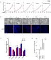
(A) Proliferation of SK-N-AS cells expressing rtTA together with either wild-type Chd5 or the PHD-mutants. Induction of wild-type Chd5 and Chd5-PHD1-D346A mutant inhibits cellular proliferation, whereas no such effects are observed with an additional mutation in D434A or with Chd5-PHD1-D361A mutants. Mefs expressing dox-inducible wild-type Chd5 or PHD-mutants dox-treated (+dox, blue squares) or untreated (−dox, red circles) are quantitated. Data represented as mean ± S.D. Y-axes denote cell numbers and X-axes denote the days after dox treatment. Student two-tailed t-test (p-values are * 0.004 and ** 0.008). (B) Morphology (upper panel) and immunofluorescence staining showing expression of the neuron-specific differentiation marker MAP2 (lower panel) of human neuroblastoma cells (SK-N-AS) expressing vector control, Chd5 or PHD-mutants. SK-N-AS cells expressing wild-type Chd5 acquire a differentiated cellular morphology and a corresponding increase in MAP2 expression compared to vector or Chd5-PHD mutant expressing SK-N-AS cells, which grow as polygonal cells in focal clusters. Cells expressing the D346A mutant have an intermediate differentiation phenotype. Scale bar 100μm. (C) Expression of wild-type Chd5 in SK-N-AS cells reduces tumorigenesis in vivo much more efficiently than does expression of vector or the Chd5 PHD-mutants that are defective in H3 binding. SK-N-AS cells expressing either dox-induced control vector, wild-type Chd5, or Chd5-PHD mutants were injected subcutaneously into athymic nude mice and tumor formation monitored. Y-axes denotes fold increase of tumor volume over a 17-day period. Mice that were untreated (red) or treated with dox (blue) are shown. Data represented as mean ± S.E.M. (D) Percentage change of tumor growth in dox-treated mice compared with untreated mice. Expression of wild-type Chd5 reduces tumorigenesis by ~31%, whereas expression of Chd5-PHD mutants D361A and D434A result in a ~30–50% increase in tumorigenesis.

Similar articles

Cited by

References

    1. Agrawal N, Frederick MJ, Pickering CR, Bettegowda C, Chang K, Li RJ, Fakhry C, Xie TX, Zhang J, Wang J, et al. Exome sequencing of head and neck squamous cell carcinoma reveals inactivating mutations in NOTCH1. Science. 2011;333:1154–1157. - PMC - PubMed
    1. Bagchi A, Mills AA. The quest for the 1p36 tumor suppressor. Cancer Res. 2008;68:2551–2556. - PMC - PubMed
    1. Bagchi A, Papazoglu C, Wu Y, Capurso D, Brodt M, Francis D, Bredel M, Vogel H, Mills AA. CHD5 is a tumor suppressor at human 1p36. Cell. 2007;128:459–475. - PubMed
    1. Baker LA, Allis CD, Wang GG. PHD fingers in human diseases: disorders arising from misinterpreting epigenetic marks. Mutat Res. 2008;647:3–12. - PMC - PubMed
    1. Barski A, Cuddapah S, Cui K, Roh TY, Schones DE, Wang Z, Wei G, Chepelev I, Zhao K. High-resolution profiling of histone methylations in the human genome. Cell. 2007;129:823–837. - PubMed

Publication types

MeSH terms