

Caffeic acid phenethyl ester induces adrenoleukodystrophy (Abcd2) gene in human X-ALD fibroblasts and inhibits the proinflammatory response in Abcd1/2 silenced mouse primary astrocytes
- PMID: 23318275
- PMCID: PMC3607212
- DOI: 10.1016/j.bbalip.2013.01.004
Caffeic acid phenethyl ester induces adrenoleukodystrophy (Abcd2) gene in human X-ALD fibroblasts and inhibits the proinflammatory response in Abcd1/2 silenced mouse primary astrocytes
Abstract
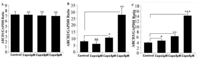
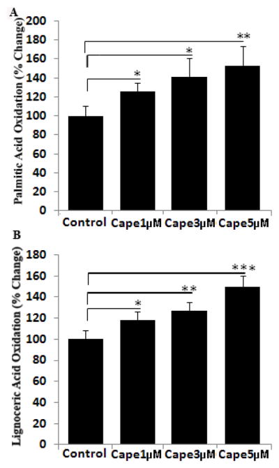
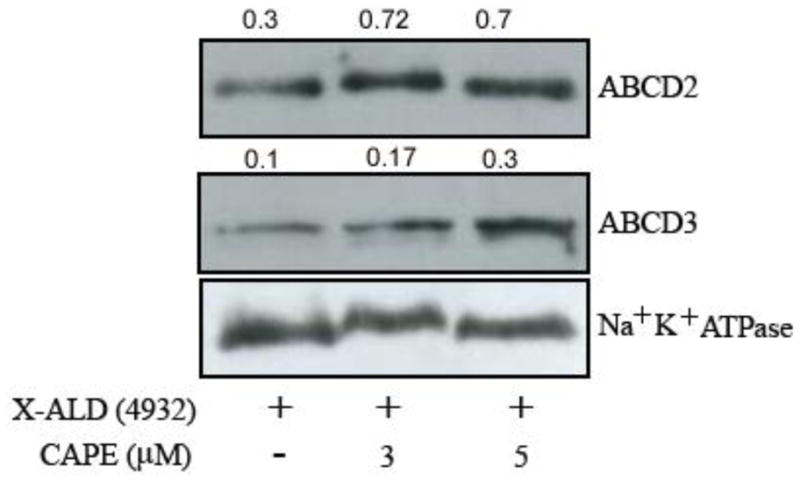
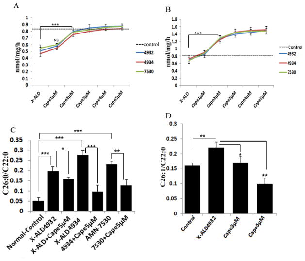
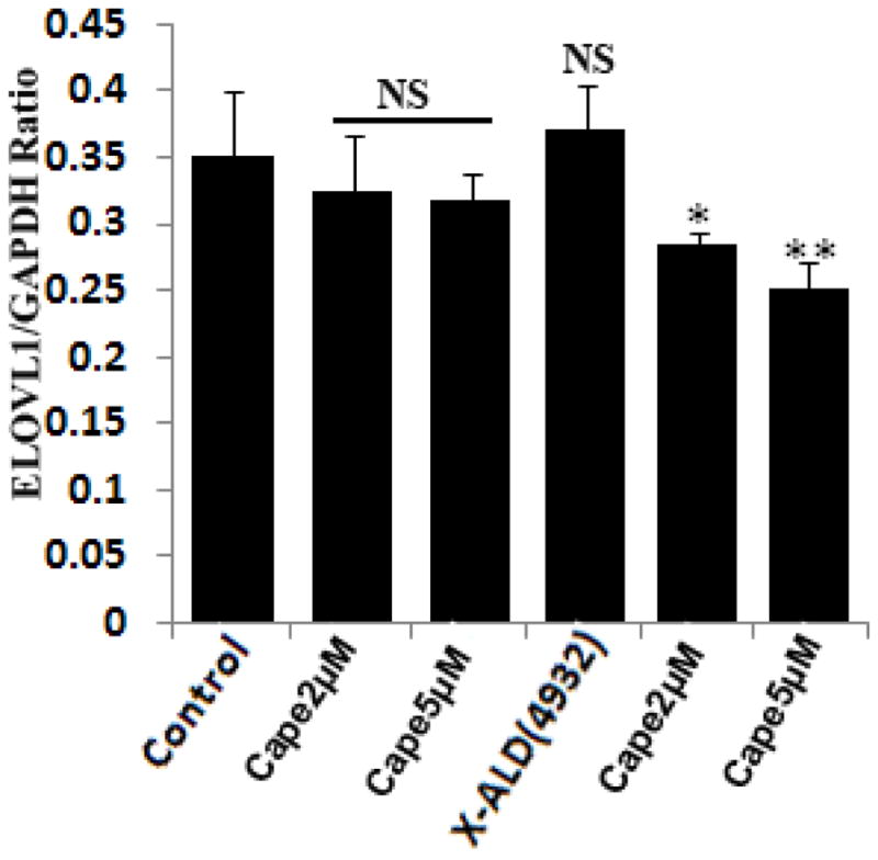
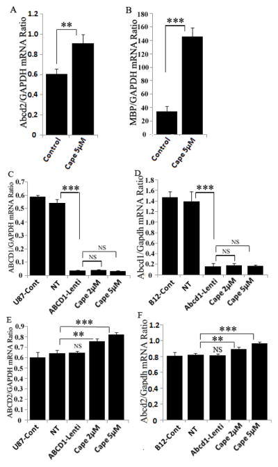
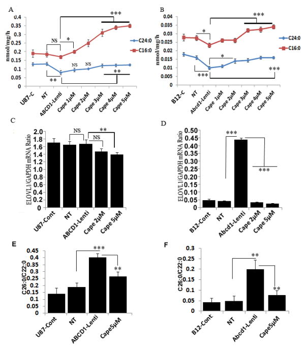
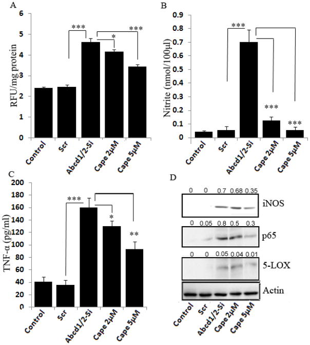
X-linked adrenoleukodystrophy (X-ALD) is a peroxisomal disorder caused by mutations in the ABCD1 gene. Accumulation of very long chain fatty acids (VLCFA) that have been attributed to reduced peroxisomal VLCFA β-oxidation activity are the hallmark of the disease. Overexpression of ABCD2 gene, the closest homolog of ABCD1, has been shown to compensate for ABCD1, thus correcting the VLCFA derangement. The accumulation of VLCFA leads to a neuroinflammatory disease process associated with demyelination of the cerebral white matter. The present study underlines the importance of caffeic acid phenethyl ester (CAPE) in inducing the expression of ABCD2 (ALDRP), and normalizing the peroxisomal β-oxidation as well as the levels of saturated and monounsaturated VLCFAs in cultured human skin fibroblasts of X-ALD patients. The expression of ELOVL1, the single elongase catalyzing the synthesis of both saturated VLCFA (C26:0) and mono-unsaturated VLCFA (C26:1), was also reduced by CAPE treatment. Importantly, CAPE upregulated Abcd2 expression and peroxisomal β-oxidation and lowered the VLCFA levels in Abcd1-deficient U87 astrocytes and B12 oligodendrocytes. In addition, using Abcd1/Abcd2-silenced mouse primary astrocytes we examined the effects of CAPE in VLCFA-induced inflammatory response. CAPE treatment decreased the inflammatory response as the expression of inducible nitric oxide synthase, inflammatory cytokine, and activation of NF-κB in Abcd1/Abcd2-silenced mouse primary astrocytes was reduced. The observations indicate that CAPE corrects both the metabolic disease of VLCFA as well as secondary inflammatory disease; therefore, it may be a potential drug candidate to be tested for X-ALD therapy in humans.
Copyright © 2013 Elsevier B.V. All rights reserved.
Figures


Similar articles
-
Abcd2 is a strong modifier of the metabolic impairments in peritoneal macrophages of ABCD1-deficient mice.PLoS One. 2014 Sep 25;9(9):e108655. doi: 10.1371/journal.pone.0108655. eCollection 2014. PLoS One. 2014. PMID: 25255441 Free PMC article.
-
HDAC inhibitor SAHA normalizes the levels of VLCFAs in human skin fibroblasts from X-ALD patients and downregulates the expression of proinflammatory cytokines in Abcd1/2-silenced mouse astrocytes.J Lipid Res. 2011 Nov;52(11):2056-69. doi: 10.1194/jlr.M017491. Epub 2011 Sep 4. J Lipid Res. 2011. PMID: 21891797 Free PMC article.
-
Silencing of Abcd1 and Abcd2 genes sensitizes astrocytes for inflammation: implication for X-adrenoleukodystrophy.J Lipid Res. 2009 Jan;50(1):135-47. doi: 10.1194/jlr.M800321-JLR200. Epub 2008 Aug 21. J Lipid Res. 2009. PMID: 18723473 Free PMC article.
-
Biochemical aspects of X-linked adrenoleukodystrophy.Brain Pathol. 2010 Jul;20(4):831-7. doi: 10.1111/j.1750-3639.2010.00391.x. Brain Pathol. 2010. PMID: 20626744 Free PMC article. Review.
-
ABC subfamily D proteins and very long chain fatty acid metabolism as novel targets in adrenoleukodystrophy.Curr Drug Targets. 2011 May;12(5):694-706. doi: 10.2174/138945011795378577. Curr Drug Targets. 2011. PMID: 21039332 Review.
Cited by
-
Drug discovery for X-linked adrenoleukodystrophy: An unbiased screen for compounds that lower very long-chain fatty acids.J Cell Biochem. 2021 Oct;122(10):1337-1349. doi: 10.1002/jcb.30014. Epub 2021 May 30. J Cell Biochem. 2021. PMID: 34056752 Free PMC article.
-
Abcd2 is a strong modifier of the metabolic impairments in peritoneal macrophages of ABCD1-deficient mice.PLoS One. 2014 Sep 25;9(9):e108655. doi: 10.1371/journal.pone.0108655. eCollection 2014. PLoS One. 2014. PMID: 25255441 Free PMC article.
-
ABCD1 deletion-induced mitochondrial dysfunction is corrected by SAHA: implication for adrenoleukodystrophy.J Neurochem. 2015 May;133(3):380-96. doi: 10.1111/jnc.12992. Epub 2015 Jan 13. J Neurochem. 2015. PMID: 25393703 Free PMC article.
-
Protective Effects of α-Tocopherol, γ-Tocopherol and Oleic Acid, Three Compounds of Olive Oils, and No Effect of Trolox, on 7-Ketocholesterol-Induced Mitochondrial and Peroxisomal Dysfunction in Microglial BV-2 Cells.Int J Mol Sci. 2016 Nov 25;17(12):1973. doi: 10.3390/ijms17121973. Int J Mol Sci. 2016. PMID: 27897980 Free PMC article.
-
Caffeic acid phenethyl ester restores mitochondrial homeostasis against peritoneal fibrosis induced by peritoneal dialysis through the AMPK/SIRT1 pathway.Ren Fail. 2024 Dec;46(1):2350235. doi: 10.1080/0886022X.2024.2350235. Epub 2024 May 9. Ren Fail. 2024. PMID: 38721924 Free PMC article.
References
-
- Moser HW. Lorenzo oil therapy for adrenoleukodystrophy: a prematurely amplified hope. Ann Neurol. 1993;34:121–122. - PubMed
-
- Contreras M, Mosser J, Mandel JL, Aubourg P, Singh I. The protein coded by the X-adrenoleukodystrophy gene is a peroxisomal integral membrane protein. FEBS Lett. 1994;344:211–215. - PubMed
-
- Contreras M, Sengupta TK, Sheikh F, Aubourg P, Singh I. Topology of ATP-binding domain of adrenoleukodystrophy gene product in peroxisomes. Arch Biochem Biophys. 1996;334:369–379. - PubMed
-
- Di Biase A, Di Benedetto R, Fiorentini C, Travaglione S, Salvati S, Attorri L, Pietraforte D. Free radical release in C6 glial cells enriched in hexacosanoic acid: implication for X-linked adrenoleukodystrophy pathogenesis. Neurochem Int. 2004;44:215–221. - PubMed
Publication types
MeSH terms
Substances
Grants and funding
LinkOut - more resources
Full Text Sources
Other Literature Sources
Research Materials
