. 2014 Jan 16;33(3):300-7.
doi: 10.1038/onc.2012.601. Epub 2013 Jan 14.

The ARF tumor-suppressor controls Drosha translation to prevent Ras-driven transformation

Affiliations

The ARF tumor-suppressor controls Drosha translation to prevent Ras-driven transformation

M J Kuchenreuther et al. Oncogene. .

Abstract

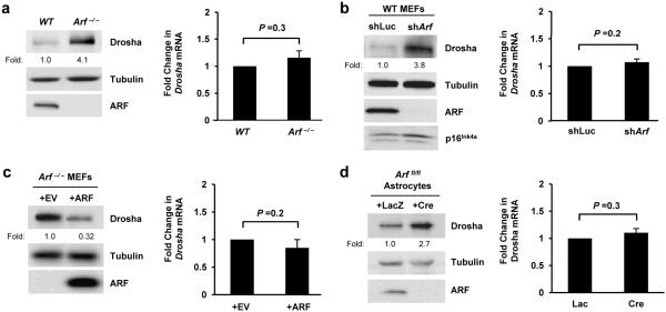
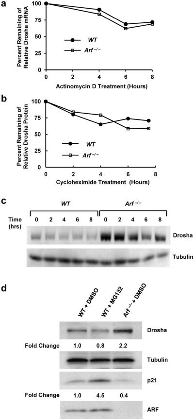
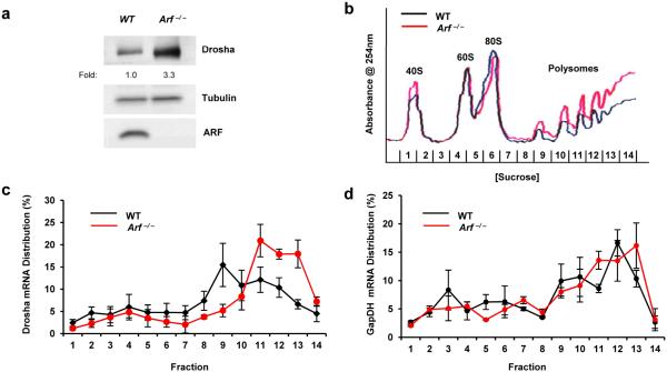
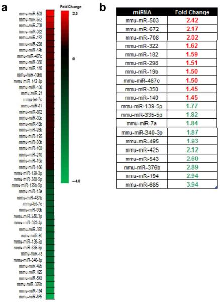
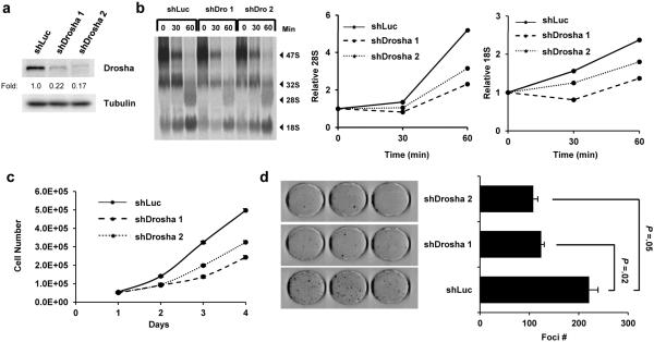
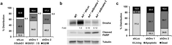
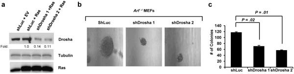
ARF is a multifunctional tumor suppressor that acts as both a sensor of oncogenic stimuli and as a key regulator of ribosome biogenesis. Recently, our group established the DEAD-box RNA helicase and microRNA (miRNA) microprocessor accessory subunit, DDX5, as a critical target of basal ARF function. To identify other molecular targets of ARF, we focused on known interacting proteins of DDX5 in the microprocessor complex. Drosha, the catalytic core of the microprocessor complex, has a critical role in the maturation of specific non-coding RNAs, including miRNAs and ribosomal RNAs (rRNAs). Here, we report that chronic or acute loss of Arf enhanced Drosha protein expression. This induction did not involve Drosha mRNA transcription or protein stability but rather relied on the increased translation of existing Drosha mRNAs. Enhanced Drosha expression did not alter global miRNA production but rather modified expression of a subset of miRNAs in the absence of Arf. Elevated Drosha protein levels were required to maintain the increased rRNA synthesis and cellular proliferation observed in the absence of Arf. Arf-deficient cells transformed by oncogenic Ras(V12) were dependent on increased Drosha expression as Drosha knockdown was sufficient to inhibit Ras-dependent cellular transformation. Thus, we propose that ARF regulates Drosha mRNA translation to prevent aberrant cell proliferation and Ras-dependent transformation.


Figures

Figure 1
Figure 1. Arf negatively regulates Drosha protein expression in a transcriptionally independent manner
(a-d, left column) Cells of the indicated genotype were lysed, and separated proteins were immunoblotted for the indicated proteins. Arf flox/flox astrocytes were infected with adenoviruses encoding β-galactosidase (LacZ) or Cre recombinase and were harvested at 5 days post-infection for gene expression analysis. Drosha expression fold change relative to WT or control infected cells is indicated. (a-d, right column) Quantitative RT-PCR analysis was performed. Drosha mRNA levels were normalized to Gadph mRNA levels. Fold change was calculated using the ΔΔCT method. Data are the mean ± SEM (N=3).
Figure 2
Figure 2. Loss of Arf has no effect on Drosha mRNA or protein stability
(a) WT and Arf −/− MEFs were treated with 4 μg/ml actinomycin D for the indicated times. Quantitative RT-PCR analysis was performed. Data are represented as the percent of Drosha mRNA remaining after normalization to Gadph levels at t=0. (b-c) Cells were treated with 25 μg/ml cycloheximide (CHX) and were harvested at the indicated times for immunoblot analysis. Densitometry quantification is depicted in panel B and data are represented as percent remaining of Drosha protein levels normalized to γ-tubulin. (d) WT MEFs were treated with 40 μM MG-132 or DMSO for 8 hours and changes in Drosha and p21 (positive control) protein levels were measured.
Figure 3
Figure 3. Translation of Drosha mRNAs is augmented upon loss of Arf
(a) Endogenous Drosha protein levels are elevated in MEFs that lack Arf compared to WT MEFs. (b) Cytosolic extracts were prepared from equal number WT and Arf −/− MEFs that had been treated for 5 min with CHX (10 μg/ml). Extracts were then subjected to differential density centrifugation and analyzed via constant UV monitoring (254 nm). (c-d) Monosome-, disome-, and polysome-associated Drosha mRNA levels were measured with qRT-PCR and were calculated as a percentage of total Drosha mRNA present in all fractions. Data are the mean ± SEM (N=3).
Figure 4
Figure 4. The expression of only a subset of miRNAs is altered upon ARF knockdown
(a) Global miRNA expression profiles of WT MEFs infected with shLuc or shArf-encoded lentivirus were determined by TaqMan MicroRNA qRT-PCR in three separate experiments. Only miRNAs (N = 147), that were present at appreciable quantities in at least one condition (CT value is less than 31) were used for analysis. miRNA expression fold changes were calculated for each replicate and then averaged. The heat map shows the fold-changes in expression for a subset of miRNAs in WT shArf MEFs relative to WT shLuc MEFs. Each colored block represents the expression of 1 miRNA (labeled on the left). Expression signals are converted into color (red, high signal; green, low signal). Color intensities are proportional to the variation of expression as indicated in the scale bar. (b) Table depicting the 10 most up- and down-regulated miRNAs in WT shArf cells relative to WT shLuc cells.
Figure 5
Figure 5. Drosha knockdown reduces proliferation and impairs ribosomal RNA maturation
(a) Infected Arf −/− MEFs were lysed, and separated proteins were immunoblotted to confirm Drosha knockdown. (b) shLuc and shDrosha Arf −/− MEFs were labeled with [methyl-3H]-methionine and chased for the indicated times. Radiolabeled RNA was separated on an agarose gel, transferred to a membrane and visualized by autoradiography (left panel). Relative band intensities were determined for rRNA in the processing assay and plotted over time (right panels). The band intensities for all conditions were first individually normalized to their respective 47S levels at T=0 and then fold change was calculated. (c-d) Following Drosha knockdown, cells were plated in triplicate at a density of 5×10⁴ per well in a 6-well plate for a proliferation assay and counted over a 4-day time period (c). These cells were also seeded in triplicate at 5×10³ cells per dish in parallel and grown for 12 days. Foci were fixed in methanol, stained with Giemsa and counted (d).
Figure 6
Figure 6. shRNA-mediated knockdown of Drosha in MEFs promotes cell death via apoptosis
(a) Quantification of the cell cycle distribution of shLuc and shDrosha Arf −/− MEFs as determined by flow cytometry. (b) Immunoblot analysis examining PARP cleavage in response to Drosha knockdown. (c) Percentage of living, apoptotic (annexin V-positive), and dead (PI-positive and double-positive) shLuc and shDrosha cells determined by flow cytometry. Data are expressed as the mean ± SD of 10 000 events performed in triplicate.
Figure 7
Figure 7. Drosha knockdown significantly inhibits Ras-induced colony formation of Arf −/− MEFs
(a) Immunoblot analysis to confirm Ras overexpression and Drosha knockdown in Arf −/− MEFs. (b-c) A total of 5×10⁴ infected cells per condition were seeded in triplicate onto soft agar plates and were grown for 3 weeks. Colonies were examined under a microscope and counted. Colony number is expressed as the mean ± SEM.

Similar articles

Cited by

References

    1. Quelle DE, Zindy F, Ashmun RA, Sherr CJ. Alternative reading frames of the INK4a tumor suppressor gene encode two unrelated proteins capable of inducing cell cycle arrest. Cell. 1995;83:993–1000. - PubMed
    1. Kamijo T, Weber JD, Zambetti G, Zindy F, Roussel MF, Sherr CJ. Functional and physical interactions of the ARF tumor suppressor with p53 and Mdm2. Proc Natl Acad Sci U S A. 1998;95:8292–8297. - PMC - PubMed
    1. Weber JD, Taylor LJ, Roussel MF, Sherr CJ, Bar-Sagi D. Nucleolar Arf sequesters Mdm2 and activates p53. Nat Cell Biol. 1999;1:20–26. - PubMed
    1. Pomerantz J, Schreiber-Agus N, Liegeois NJ, Silverman A, Alland L, Chin L, et al. The Ink4a tumor suppressor gene product, p19Arf, interacts with MDM2 and neutralizes MDM2's inhibition of p53. Cell. 1998;92:713–723. - PubMed
    1. Zhang Y, Xiong Y, Yarbrough WG. ARF promotes MDM2 degradation and stabilizes p53: ARF-INK4a locus deletion impairs both the Rb and p53 tumor suppression pathways. Cell. 1998;92:725–734. - PubMed

Publication types

MeSH terms