

Reduced FANCD2 influences spontaneous SCE and RAD51 foci formation in uveal melanoma and Fanconi anaemia
- PMID: 23318456
- PMCID: PMC3898318
- DOI: 10.1038/onc.2012.627
Reduced FANCD2 influences spontaneous SCE and RAD51 foci formation in uveal melanoma and Fanconi anaemia
Abstract
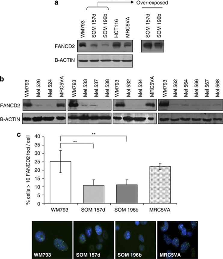
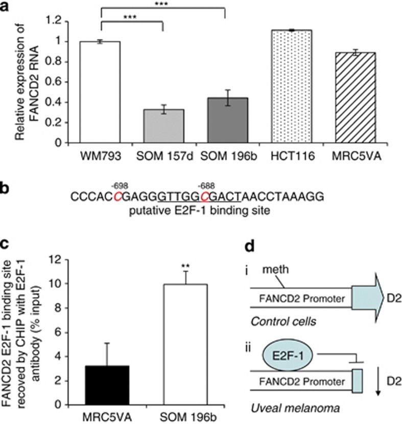
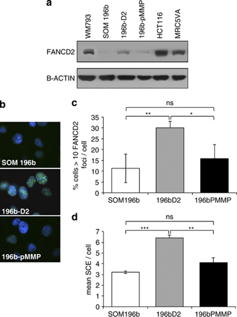
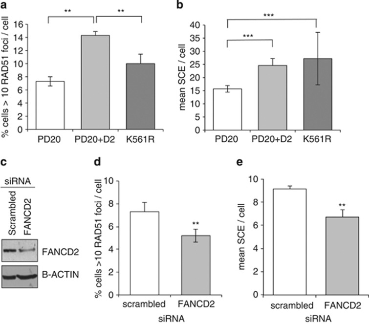
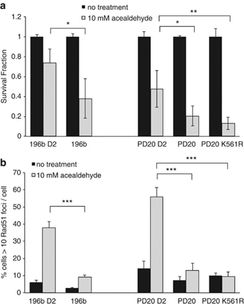
Uveal melanoma (UM) is unique among cancers in displaying reduced endogenous levels of sister chromatid exchange (SCE). Here we demonstrate that FANCD2 expression is reduced in UM and that ectopic expression of FANCD2 increased SCE. Similarly, FANCD2-deficient fibroblasts (PD20) derived from Fanconi anaemia patients displayed reduced spontaneous SCE formation relative to their FANCD2-complemented counterparts, suggesting that this observation is not specific to UM. In addition, spontaneous RAD51 foci were reduced in UM and PD20 cells compared with FANCD2-proficient cells. This is consistent with a model where spontaneous SCEs are the end product of endogenous recombination events and implicates FANCD2 in the promotion of recombination-mediated repair of endogenous DNA damage and in SCE formation during normal DNA replication. In both UM and PD20 cells, low SCE was reversed by inhibiting DNA-PKcs (DNA-dependent protein kinase, catalytic subunit). Finally, we demonstrate that both PD20 and UM are sensitive to acetaldehyde, supporting a role for FANCD2 in repair of lesions induced by such endogenous metabolites. Together, these data suggest FANCD2 may promote spontaneous SCE by influencing which double-strand break repair pathway predominates during normal S-phase progression.
Figures

Similar articles
-
Fanconi anemia protein FANCD2 promotes immunoglobulin gene conversion and DNA repair through a mechanism related to homologous recombination.Mol Cell Biol. 2005 Jan;25(1):34-43. doi: 10.1128/MCB.25.1.34-43.2005. Mol Cell Biol. 2005. PMID: 15601828 Free PMC article.
-
FANCD2 influences replication fork processes and genome stability in response to clustered DSBs.Cell Cycle. 2015;14(12):1809-22. doi: 10.1080/15384101.2015.1036210. Cell Cycle. 2015. PMID: 26083937 Free PMC article.
-
Functional cross talk between the Fanconi anemia and ATRX/DAXX histone chaperone pathways promotes replication fork recovery.Hum Mol Genet. 2020 May 8;29(7):1083-1095. doi: 10.1093/hmg/ddz250. Hum Mol Genet. 2020. PMID: 31628488 Free PMC article.
-
Beyond interstrand crosslinks repair: contribution of FANCD2 and other Fanconi Anemia proteins to the replication of DNA.Mutat Res. 2018 Mar;808:83-92. doi: 10.1016/j.mrfmmm.2017.09.004. Epub 2017 Sep 14. Mutat Res. 2018. PMID: 29031493 Review.
-
FANCD2 and DNA Damage.Int J Mol Sci. 2017 Aug 19;18(8):1804. doi: 10.3390/ijms18081804. Int J Mol Sci. 2017. PMID: 28825622 Free PMC article. Review.
Cited by
-
Increased Non-Homologous End Joining Makes DNA-PK a Promising Target for Therapeutic Intervention in Uveal Melanoma.Cancers (Basel). 2019 Aug 30;11(9):1278. doi: 10.3390/cancers11091278. Cancers (Basel). 2019. PMID: 31480356 Free PMC article.
-
FANCD2 limits acetaldehyde-induced genomic instability during DNA replication in esophageal keratinocytes.Mol Oncol. 2021 Nov;15(11):3109-3124. doi: 10.1002/1878-0261.13072. Epub 2021 Aug 8. Mol Oncol. 2021. PMID: 34328261 Free PMC article.
-
Bloom syndrome radials are predominantly non-homologous and are suppressed by phosphorylated BLM.Cytogenet Genome Res. 2014;144(4):255-263. doi: 10.1159/000375247. Epub 2015 Feb 28. Cytogenet Genome Res. 2014. PMID: 25766002 Free PMC article.
-
Chromosomal Integrity after UV Irradiation Requires FANCD2-Mediated Repair of Double Strand Breaks.PLoS Genet. 2016 Jan 14;12(1):e1005792. doi: 10.1371/journal.pgen.1005792. eCollection 2016 Jan. PLoS Genet. 2016. PMID: 26765540 Free PMC article.
-
Sister Chromatid Exchange and Genomic Instability in Soft Tissue Sarcomas: Potential Implications for Response to DNA-Damaging Treatments.Sarcoma. 2018 May 7;2018:3082526. doi: 10.1155/2018/3082526. eCollection 2018. Sarcoma. 2018. PMID: 29853780 Free PMC article.
References
-
- Miranda M, Ligas C, Amicarelli F, D'Alessandro E, Brisdelli F, Zarivi O, et al. Sister chromatid exchange (SCE) rates in human melanoma cells as an index of mutagenesis. Mutagenesis. 1997;12:233–236. - PubMed
-
- Goyanes V, Fernandez JL, Pereira S, Campos A, Rodriguez E, Gonzalez M, et al. Frequency of sister chromatid exchanges in humans at 14–16 week foetal stage and at birth. Cytobios. 1994;77:67–72. - PubMed
-
- Kato H, Stich HF. Sister chromatid exchanges in ageing and repair-deficient human fibroblasts. Nature. 1976;260:447–448. - PubMed
-
- Nagasawa H, Fornace D, Little JB. Induction of sister-chromatid exchanges by DNA-damaging agents and 12-O-tetradecanoyl-phorbol-13-acetate (TPA) in synchronous Chinese hamster ovary (CHO) cells. Mutat Res. 1983;107:315–327. - PubMed
-
- Wojcik A, Bruckmann E, Obe G. Insights into the mechanisms of sister chromatid exchange formation. Cytogenet Genome Res. 2004;104:304–309. - PubMed
Publication types
MeSH terms
Substances
LinkOut - more resources
Full Text Sources
Other Literature Sources
Medical
Research Materials
Miscellaneous
