

Dehydroepiandrosterone sulfate (DHEAS) as an endocrine marker of aging in calorie restriction studies
- PMID: 23318475
- PMCID: PMC3641169
- DOI: 10.1016/j.exger.2013.01.001
Dehydroepiandrosterone sulfate (DHEAS) as an endocrine marker of aging in calorie restriction studies
Abstract
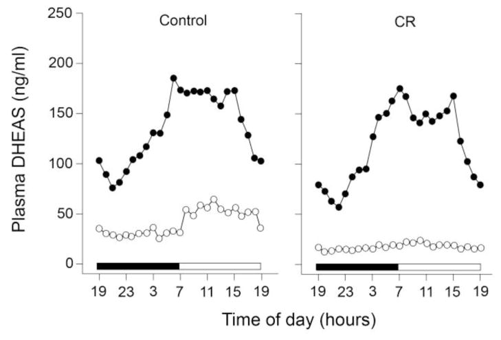
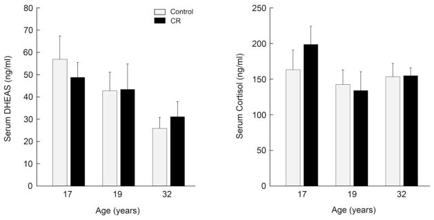
The adrenal steroid, dehydroepiandrosterone sulfate (DHEAS), is generally regarded as being a reliable endocrine marker of aging, because in humans and nonhuman primates its circulating concentrations are very high during young adulthood, and the concentrations then decline markedly during aging. Despite promising results from early studies, we were recently surprised to find that caloric restriction (CR) did little to prevent or delay the decline of DHEAS concentrations in old rhesus macaques. Here we summarize the use of circulating DHEAS concentrations as a biomarker of aging in CR studies and suggest reasons for its limited value. Although DHEAS can reliably predict aging in animals maintained on a standard diet, dietary manipulations may affect liver enzymes involved in the metabolism of steroid hormones. Consequently, in CR studies the reliability of using DHEAS as a biomarker of aging may be compromised.
Keywords: Adrenal gland; Biomarker; Cortisol; DHEAS; Rhesus macaque.
Copyright © 2013 Elsevier Inc. All rights reserved.
Figures
Similar articles
-
Effect of caloric restriction on the 24-hour plasma DHEAS and cortisol profiles of young and old male rhesus macaques.Ann N Y Acad Sci. 2004 Jun;1019:443-7. doi: 10.1196/annals.1297.081. Ann N Y Acad Sci. 2004. PMID: 15247063
-
Effect of age and caloric restriction on circadian adrenal steroid rhythms in rhesus macaques.Neurobiol Aging. 2008 Sep;29(9):1412-22. doi: 10.1016/j.neurobiolaging.2007.03.011. Epub 2007 Apr 8. Neurobiol Aging. 2008. PMID: 17420071 Free PMC article.
-
Dehydroepiandrosterone sulfate: a biomarker of primate aging slowed by calorie restriction.J Clin Endocrinol Metab. 1997 Jul;82(7):2093-6. doi: 10.1210/jcem.82.7.4038. J Clin Endocrinol Metab. 1997. PMID: 9215277
-
Adrenal aging and its effects on the stress response and immunosenescence.Maturitas. 2023 Feb;168:13-19. doi: 10.1016/j.maturitas.2022.10.006. Epub 2022 Nov 4. Maturitas. 2023. PMID: 36370489 Free PMC article. Review.
-
Dehydroepiandrosterone: a springboard hormone for female sexuality.Fertil Steril. 2002 Apr;77 Suppl 4:S19-25. doi: 10.1016/s0015-0282(02)02987-4. Fertil Steril. 2002. PMID: 12007898 Review.
Cited by
-
Fasting or caloric restriction for healthy aging.Exp Gerontol. 2013 Oct;48(10):1003-5. doi: 10.1016/j.exger.2013.04.011. Epub 2013 Apr 29. Exp Gerontol. 2013. PMID: 23639403 Free PMC article.
-
Can longer lifespan be associated with gut microbiota involvement in lipid metabolism?FEMS Microbiol Ecol. 2024 Oct 25;100(11):fiae135. doi: 10.1093/femsec/fiae135. FEMS Microbiol Ecol. 2024. PMID: 39354675 Free PMC article.
-
Cognition in aged rhesus monkeys: effect of DHEA and correlation with steroidogenic gene expression.Genes Brain Behav. 2017 Mar;16(3):361-368. doi: 10.1111/gbb.12351. Epub 2016 Nov 24. Genes Brain Behav. 2017. PMID: 27736018 Free PMC article.
-
Chronic Obstructive Pulmonary Disease as a Main Factor of Premature Aging.Int J Environ Res Public Health. 2019 Feb 13;16(4):540. doi: 10.3390/ijerph16040540. Int J Environ Res Public Health. 2019. PMID: 30781849 Free PMC article.
-
Nutrigenomics at the Interface of Aging, Lifespan, and Cancer Prevention.J Nutr. 2016 Oct;146(10):1931-1939. doi: 10.3945/jn.116.235119. Epub 2016 Aug 24. J Nutr. 2016. PMID: 27558581 Free PMC article. Review.
References
-
- Barrows CH, Kokkonen GC. Dietary restriction and life extension, biological mechanisms. In: Moment GB, editor. Nutritional approaches to aging research. Boca Raton, FL: CRC Press Inc; 1982. pp. 219–43.
-
- Bartke A, Wright JC, Mattison JA, Ingram DK, Miller RA, Roth GS. Extending the lifespan of long-lived mice. Nature. 2001;414:412. - PubMed
-
- Bliwise DL. Normal aging. In: Kryger MH, Roth T, Dement WC, editors. Principles and Practice of Sleep Medicine. 3. WB Saunders; Philadelphia: 2000. pp. 26–42.
-
- Bodkin NL, Alexander TM, Ortmeyer HK, Johnson E, Hansen BC. Mortality and morbidity in laboratory-maintained rhesus monkeys and effects of long-term dietary restriction. J Gerontol. 2003;58A:212–219. - PubMed
Publication types
MeSH terms
Substances
Grants and funding
LinkOut - more resources
Full Text Sources
Other Literature Sources
Medical
