. 2013 Jan;70(1):34-43.
doi: 10.1001/jamaneurol.2013.573.

Relationships between retinal axonal and neuronal measures and global central nervous system pathology in multiple sclerosis

Affiliations

Relationships between retinal axonal and neuronal measures and global central nervous system pathology in multiple sclerosis

Shiv Saidha et al. JAMA Neurol. 2013 Jan.

Abstract

Objective: To determine the relationships between conventional and segmentation-derived optical coherence tomography (OCT) retinal layer thickness measures with intracranial volume (a surrogate of head size) and brain substructure volumes in multiple sclerosis (MS).

Design: Cross-sectional study.

Setting: Johns Hopkins University, Baltimore, Maryland.

Participants: A total of 84 patients with MS and 24 healthy control subjects.

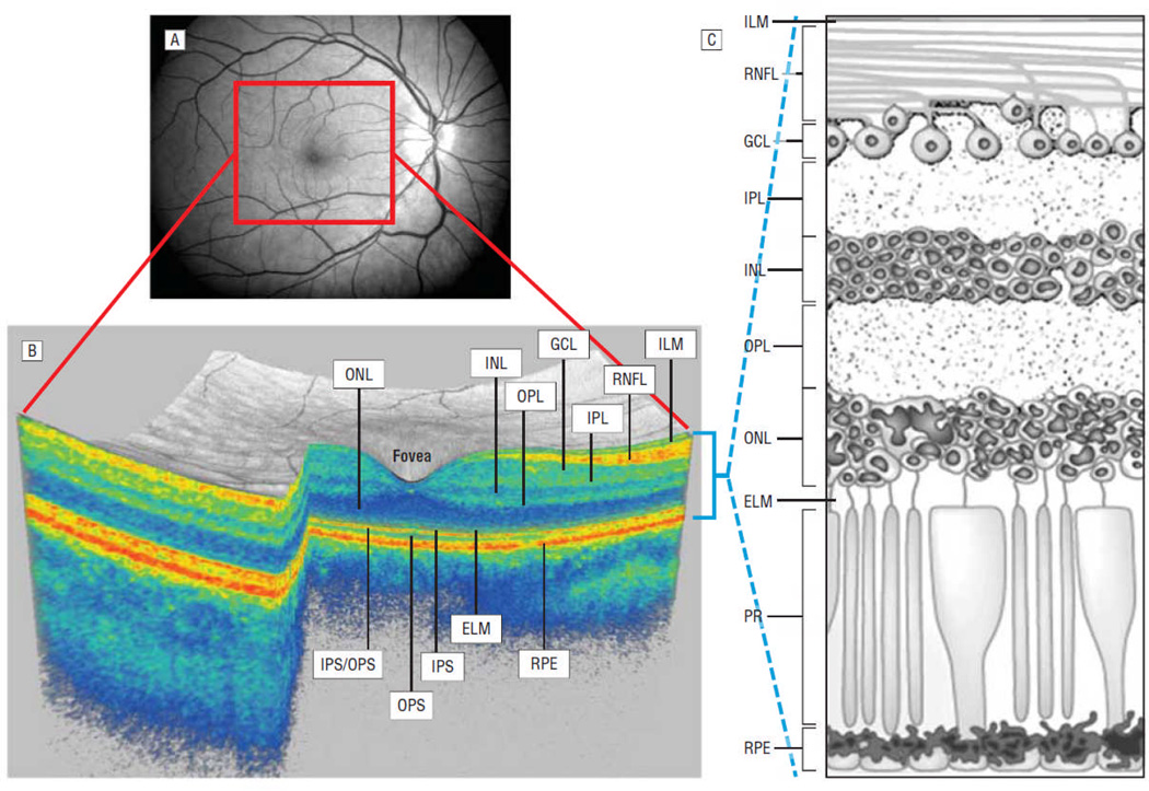
Main outcome measures: High-definition spectral-domain OCT conventional and automated segmentation-derived discrete retinal layer thicknesses and 3-T magnetic resonance imaging brain substructure volumes.

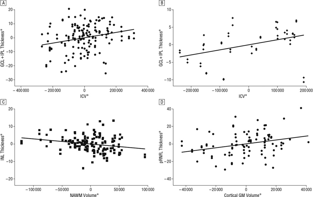
Results: Peripapillary retinal nerve fiber layer as well as composite ganglion cell layer+inner plexiform layer thicknesses in the eyes of patients with MS without a history of optic neuritis were associated with cortical gray matter (P=.01 and P=.04, respectively) and caudate (P=.04 and P=.03, respectively) volumes. Inner nuclear layer thickness, also in eyes without a history of optic neuritis, was associated with fluid-attenuated inversion recovery lesion volume (P=.007) and inversely associated with normal-appearing white matter volume (P=.005) in relapsing-remitting MS. As intracranial volume was found to be related with several of the OCT measures in patients with MS and healthy control subjects and is already known to be associated with brain substructure volumes, all OCT-brain substructure relationships were adjusted for intracranial volume. CONCLUSIONS Retinal measures reflect global central nervous system pathology in multiple sclerosis, with thicknesses of discrete retinal layers each appearing to be associated with distinct central nervous system processes. Moreover, OCT measures appear to correlate with intracranial volume in patients with MS and healthy control subjects, an important unexpected factor unaccounted for in prior studies examining the relationships between peripapillary retinal nerve fiber layer thickness and brain substructure volumes.


Conflict of interest statement

Competing interests

The authors report no conflicts of interest

Figures

Figure 1
Figure 1
Panel A represents a fundus photograph from a healthy control. Panel B is a 3-D macular volume cube generated by Cirrus HD-OCT from the macular region denoted by the red box in Panel A from the same healthy control. Note the individual layers of the retina are readily discernible, except for the ganglion cell layer (GCL) and inner plexform layer (IPL) which are difficult to distinguish. During the segmentation process (performed in 3-D), the segmentation software identifies the outer boundaries of the macular retinal nerve fiber layer (RNFL), inner plexiform layer (IPL) and outer plexiform layer (OPL), as well as the inner boundary of the retinal pigment epithelium (RPE) which is identified by the conventional Cirrus HD-OCT algorithm. The identification of these boundaries facilitates OCT-segmentation, enabling determination of the thicknesses of the macular-RNFL, GCL+IPL (GCIP), the inner nuclear layer (INL)+OPL, and the outer nuclear layer (ONL) including the inner and outer photoreceptor segments. Panel C illustrates the cellular composition of the retinal layers depicted in panel B. Abbreviations; IS: inner photoreceptor segments, OS: outer photoreceptor segments, IS/OS: IS/OS junction, PR: photoreceptors, ILM: inner limiting membrane, ELM: external limiting membrane
Figure 2
Figure 2
*residual values from multivariate regression models Panel A represents an adjusted variables plot of ganglion cell layer+inner plexiform layer (GCIP) thickness and intracranial volume (ICV) in multiple sclerosis (MS), adjusted for age, sex and disease duration. The solid red line graphically illustrates the independent relationship between GCIP thickness and ICV in MS. Note that as ICV increases, GCIP thickness similarly increases, consistent with the detection of significant associations between GCIP thickness and ICV in MS (p=0.008). Panel B represents an adjusted variables plot of ganglion cell layer+inner plexiform layer (GCIP) thickness and intracranial volume (ICV) in healthy controls (HCs), adjusted for age and sex. The solid red line graphically illustrates the independent relationship between GCIP thickness and ICV in HCs. Note that as ICV increases, GCIP thickness similarly increases, consistent with the detection of significant associations between GCIP thickness and ICV in HCs (p=0.04). Panel C represents an adjusted variables plot of inner nuclear layer (INL) thickness and normal appearing white matter (NAWM) volume in MS, adjusted for age, sex, disease duration and ICV. The solid red line graphically illustrates the independent relationship between INL thickness and NAWM volume in MS. Note that as INL thickness increases, NAWM volume decreases, consistent with the detection of significant inverse associations between INL thickness and NAWM volume in MS (p=0.01). Moreover, although not depicted in this figure, higher INL thickness was also associated with higher FLAIR-lesion volume in relapsing-remitting MS (RRMS) (p=0.02). Since INL pathology in MS is thought to result from primary retinal mechanisms of pathology, rather than being related to optic neuropathy, these findings raise the possibility that the potential mechanism underlying the proposed occurrence of primary retinal pathology affecting the INL in MS may be inflammatory, such as related to retinal periphlebitis. Panel D represents an adjusted variables plot of peri-papillary retinal nerve fiber layer (RNFL) thickness and cortical-gray matter (GM) volume in RRMS, adjusted for age, sex, disease duration and ICV. The solid red line graphically illustrates the independent relationship between RNFL thickness and cortical-GM volume in RRMS. Note that as RNFL thickness decreases, cortical-GM volume similarly decreases, consistent with the detection of significant associations between RNFL thickness and cortical-GM volume in RRMS (p=0.01).

Comment in

Similar articles

Cited by

References

    1. Saidha S, Syc SB, Ibrahim MA, et al. Primary retinal pathology in multiple sclerosis as detected by optical coherence tomography. Brain. 2011;134(Pt 2):518–533. - PubMed
    1. Saidha S, Syc SB, Durbin MK, et al. Visual dysfunction in multiple sclerosis correlates better with optical coherence tomography derived estimates of macular ganglion cell layer thickness than peripapillary retinal nerve fiber layer thickness. Mult Scler. 2011;17(12):1449–1463. - PubMed
    1. Syc SB, Saidha S, Newsome SD, et al. Optical coherence tomography segmentation reveals ganglion cell layer pathology after optic neuritis. Brain. 2012;135(Pt 2):521–533. - PMC - PubMed
    1. Seigo MA, Sotirchos ES, Newsome S, et al. In vivo assessment of retinal neuronal layers in multiple sclerosis with manual and automated optical coherence tomography segmentation techniques. J Neurol. 2012 [Epub ahead of print] - PubMed
    1. Frohman EM, Fujimoto JG, Frohman TC, Calabresi PA, Cutter G, Balcer LJ. Optical coherence tomography: A window into the mechanisms of multiple sclerosis. Nat Clin Pract Neurol. 2008;4(12):664–675. - PMC - PubMed

Publication types