. 2013 Apr;23(4):473-90.
doi: 10.1038/cr.2013.7. Epub 2013 Jan 15.

Reelin induces EphB activation

Affiliations

Reelin induces EphB activation

Elisabeth Bouché et al. Cell Res. 2013 Apr.

Abstract

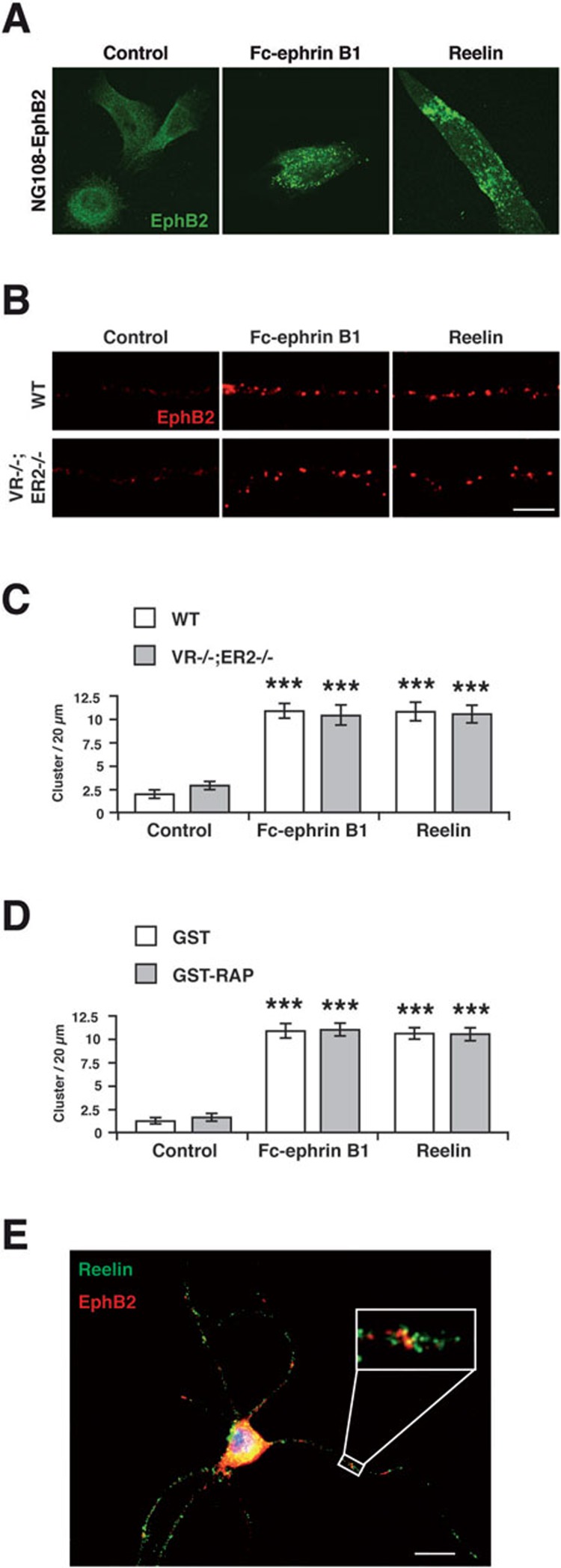
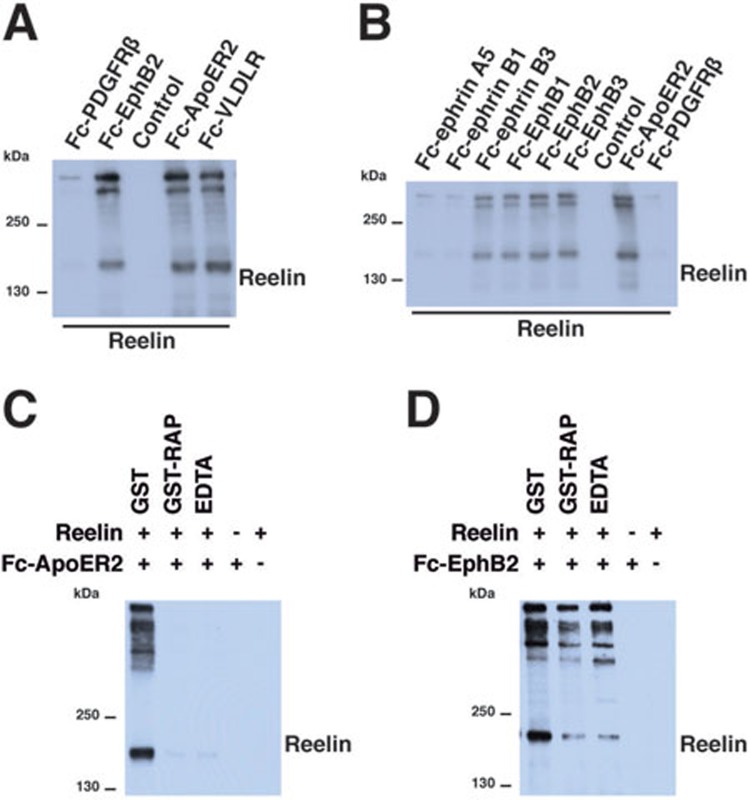
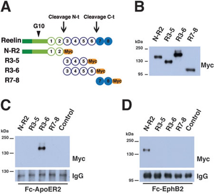
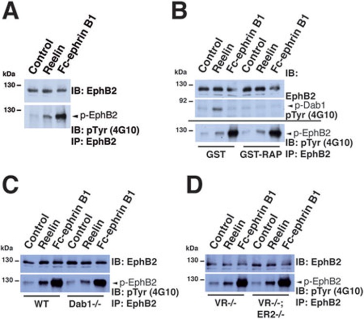
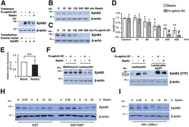
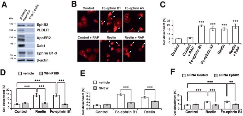
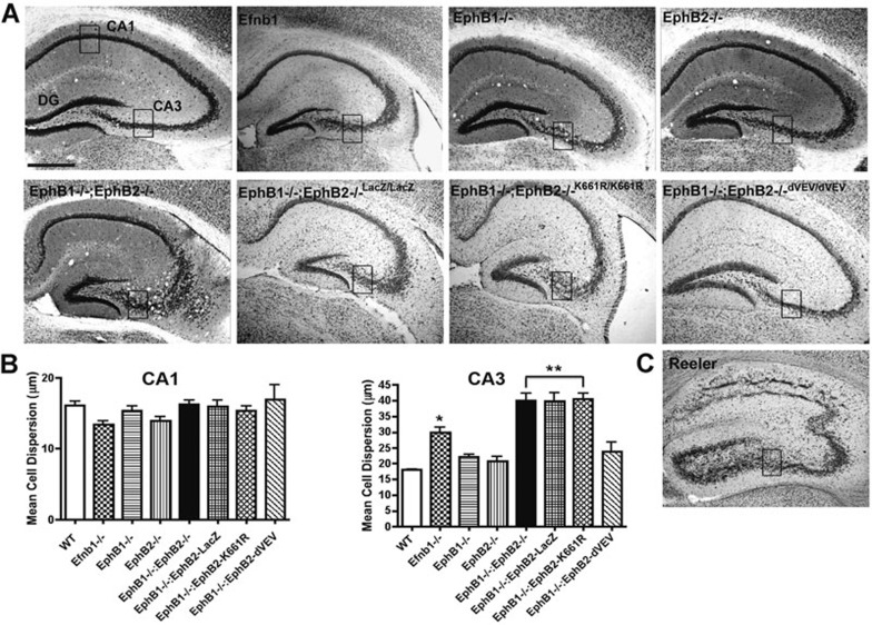
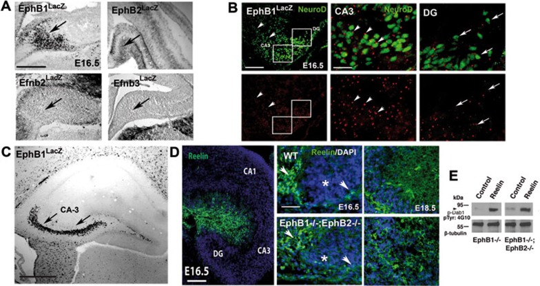
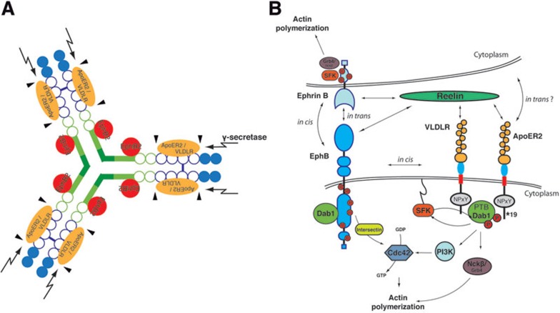
The integration of newborn neurons into functional neuronal networks requires migration of cells to their final position in the developing brain, the growth and arborization of neuronal processes and the formation of synaptic contacts with other neurons. A central player among the signals that coordinate this complex sequence of differentiation events is the secreted glycoprotein Reelin, which also modulates synaptic plasticity, learning and memory formation in the adult brain. Binding of Reelin to ApoER2 and VLDL receptor, two members of the LDL receptor family, initiates a signaling cascade involving tyrosine phosphorylation of the intracellular cytoplasmic adaptor protein Disabled-1, which targets the neuronal cytoskeleton and ultimately controls the positioning of neurons throughout the developing brain. However, it is possible that Reelin signals interact with other receptor-mediated signaling cascades to regulate different aspects of brain development and plasticity. EphB tyrosine kinases regulate cell adhesion and repulsion-dependent processes via bidirectional signaling through ephrin B transmembrane proteins. Here, we demonstrate that Reelin binds to the extracellular domains of EphB transmembrane proteins, inducing receptor clustering and activation of EphB forward signaling in neurons, independently of the 'classical' Reelin receptors, ApoER2 and VLDLR. Accordingly, mice lacking EphB1 and EphB2 display a positioning defect of CA3 hippocampal pyramidal neurons, similar to that in Reelin-deficient mice, and this cell migration defect depends on the kinase activity of EphB proteins. Together, our data provide biochemical and functional evidence for signal integration between Reelin and EphB forward signaling.


Figures

Figure 1
Figure 1
Reelin induces clustering of EphB2 proteins. (A) Stimulation with preclustered ephrin B1 extracellular domain or Reelin induces clustering of EphB2 (green) in stably transfected NG-108 cells. (B) Clustering of endogenous EphB2 (red) in primary cortical neurons after stimulation with preclustered ephrin B1 or Reelin. Clustering is observed in wild-type (WT) neurons as well as in neurons lacking both ApoER2 and VLDLR (VR−/−;ER2−/−). Scale bar, 5 μm. (C) Quantification of EphB2 cluster density in ephrin B1- or Reelin-treated primary neurons prepared from WT or ApoER2/VLDLR double knockout embryos (VR−/−;ER2−/−) (S.E.M., ***P < 0.001 compared with control, n = 15 per condition, 3 knockout embryous). (D) Quantification of EphB2 cluster density in ephrin B1- or Reelin-treated primary neurons preincubated with GST or GST-RAP, a lipoprotein receptor antagonist (S.E.M., ***P < 0.001 compared with control, n = 15 per condition). (E) Co-localization of EphB2 and Reelin in cortical neurons. Cultured cortical neurons were treated with recombinant Reelin, washed twice, fixed and immunostained at DIV 6 for EphB2 and Reelin. Scale bar, 10 μm. A higher magnification of a dendrite is shown (boxed area). Co-localization of EphB2 (red) and Reelin (green) appears as yellow puncta.
Figure 2
Figure 2
Reelin binds to the extracellular domain of EphB proteins. (A) Supernatant of Reelin-expressing HEK-293 cells was incubated with the ectodomains of different transmembrane receptors. The Fc-fused ectodomains were precipitated with protein A/G-agarose beads. Reelin binds to the ectodomains of the lipoprotein receptors ApoER2 and VLDLR (positive controls), and to the ectodomain of EphB2. The ectodomain of platelet-derived growth factor receptor-β (Fc-PDGFRβ) and protein A/G-agarose beads without Fc-coupled ectodomain (control) served as negative controls. (B) Biochemical interaction of Reelin with members of the EphB and ephrin B gene families. Recombinant Reelin was incubated with the ectodomains of ephrin A5, ephrin B1 and ephrin B3, EphB1, EphB2 and EphB3, and of PDGFRβ as a negative control. The Fc-fused ectodomains were precipitated with protein A/G-beads, and bound Reelin was detected by immunoblotting. Fc-ApoER2 served as a positive control. Reelin was bound by the ectodomains of EphB1-3. Moderate binding was also observed for Fc-ephrin B3. (C) GST-RAP (30 μg/ml) and the calcium chelator EDTA (30 mmol/l) blocked binding of Reelin to its lipoprotein receptors, shown here for ApoER2. (D) The EphB2-Reelin interaction was not blocked by GST-RAP or EDTA.
Figure 3
Figure 3
EphB2 interacts with the amino-terminal domain of Reelin. (A) Schematic diagram of partial Reelin constructs. The dark- and bright-green rectangles indicate the signal peptide, the F-spondin homology region and the unique region that is recognized by the G10 anti-Reelin monoclonal antibody (arrowhead). The circles with numbers represent the eight Reelin repeats. The carboxy-terminal myc-tags of the partial Reelin constructs are highlighted in orange. Arrows indicate sites of proteolytic Reelin cleavage. (B) The Reelin constructs shown in A were transfected into HEK-293 cells. Supernatants were separated by SDS-PAGE and proteins were detected with an anti-myc antibody. Comparable amounts of the constructs were used for the coprecipitation experiments shown in C and D. (C) Fc-tagged ApoER2 or (D) EphB2 ectodomains were incubated with supernatants containing similar amounts of the different myc-tagged partial Reelin polypeptides and precipitated with protein A/G-sepharose. Coprecipitated Reelin constructs were visualized by immunoblotting with an anti-myc antibody. The central fragment containing Reelin repeats R3-6 binds to the lipoprotein receptor ectodomains (C), whereas EphB2 binds to the amino-terminal Reelin fragment N-R2 (D).
Figure 4
Figure 4
Reelin induces EphB2 tyrosine phosphorylation in cortical neurons. (A) Primary neurons were treated with recombinant Reelin or preclustered Fc-ephrin B1. Before immunoprecipitation with an anti-EphB2 antibody, the cell lysates were analyzed by western blotting with the same antibody (IB: EphB2; input). The immunoprecipitated samples (IP: EphB2) were analyzed by western blotting with a phosphotyrosine-specific monoclonal antibody (IB: pTyr) to determine tyrosine phosphorylation levels of the EphB2 receptor in Reelin- or Fc-ephrin B1-treated neurons compared to control treatment. (B) Primary neurons were preincubated with GST or GST-RAP before stimulation with Reelin or preclustered soluble ephrin B1. The lipoprotein receptor antagonist RAP prevented the Reelin-induced tyrosine phosphorylation of Dab1 as detected by immunoblotting with the phosphotyrosine-specific antibody (middle blot, lane 5), but did not block the induction of EphB2 tyrosine phosphorylation (bottom). Shown are representative blots of at least three independent experiments (A-B). (C) Primary neurons lacking the adapter protein Dab1 (Dab1−/−) were treated with Reelin or preclustered Fc-ephrin B1 (n = 2 knockout embryos). Deficiency in the Src family kinase switch protein Dab1 did not prevent Reelin- or ephrin B1-induced EphB2 phosphorylation. (D) Primary neurons deficient in VLDLR or both Reelin receptors ApoER2 and VLDLR were treated with Reelin or preclustered soluble ephrin B1. Lack of VLDLR (VR−/−) or VLDLR and ApoER2 (VR−/−;ER2−/−) did not block the induction of EphB2 tyrosine phosphorylation by Reelin or ephrin B1. Neurons were prepared from three different double knockout embryos.
Figure 5
Figure 5
Reelin induces proteolytic processing of EphB2. (A) Treatment with Reelin induces decrease of full-length EphB2 in transfected HEK-293 cells. HEK-293 cells were transiently transfected with a plasmid encoding full-length EphB2 and treated with preclustered soluble ephrin B1 or Reelin for 4 h. Cell lysates were analyzed by western blotting. Treatment with both preclustered ephrin B1 or Reelin led to a significant decrease of full-length EphB2 protein levels. Actin served as a loading control. (B, C) Decrease of EphB2 levels in cortical neurons after stimulation with Reelin (B) or Fc-ephrin B1 (C). Cortical neurons (E15.5, DIV5) were treated for the indicated times, lysed in RIPA buffer and analyzed by western blotting for EphB2 protein levels. Treatment with Reelin induced a moderate decrease in the levels of full-length EphB2 after 2-8 h. Actin served as a loading control. (D) Quantification of the EphB2 signal intensity after treatment with Reelin (white bars) or Fc-ephrin B1 (grey bars) is shown (S.D., *P < 0.05, **P < 0.01 for Reelin and #P < 0.005 for Fc-ephrin B1 as compared to controls, n = 3). (E) Quantification of relative Ephb2 mRNA levels in primary neurons treated for 6 h with recombinant Reelin (n = 3) as determined by qRT-PCR. n.s., non-significant. (F) Cortical neurons were preincubated with bafilomycin A1 or lactacystin for 30 min and then treated with Reelin or preclustered Fc-ephrin B1 for 7.5 h. The endosomal inhibitor bafilomycin prevented degradation of the full-length form of EphB2, whereas the proteasomal inhibitor lactacystin had no effect. (G) EphB2-transfected HEK-293 cells were preincubated with lactacytin or lactacystin + DAPT, a γ-secretase inhibitor, and then treated with Reelin or preclustered Fc-ephrin B1 for 8 h. Inhibition of the proteasome led to an increase of a carboxy-terminal fragment of EphB2 (CTF2), whereas pretreatment with lactacystin and DAPT leads to the accumulation of CTF1, a slightly larger carboxy-terminal fragment of EphB2 that serves as a substrate for the γ-secretase complex. (H) Cortical neurons were pretreated with GST or GST-RAP (30 μg/ml), then stimulated with Reelin for the indicated times, lysed in RIPA buffer and analyzed by western blotting for EphB2 protein levels. GST-RAP did not prevent the Reelin-induced EphB2 degradation. Actin served as a loading control. One experiment representative of 3 is shown. (I) Cortical neurons were prepared from embryos lacking both VLDLR and ApoER2 (VR−/−;ER2−/−), stimulated with Reelin for the indicated times and analyzed as described above. Shown is one out of two independent experiments.
Figure 6
Figure 6
Reelin-dependent activation of EphB2 induces deadhesive cytoskeletal changes in Cos cells. (A) Expression of components of the Reelin and EphB2 signaling cascades in primary neurons and Cos-1 cells was analyzed by western blotting. Cos-1 cells express EphB2 and ephrin B proteins but do not express detectable levels of VLDLR, ApoER2 or Dab-1. Actin was used as a loading control. (B) Treatment with preclustered ephrin B1 (4 μg/ml), ephrin A5 (20 μg/ml) or Reelin but not with control medium induces deadhesive cytoskeletal changes in Cos-1 cells, resulting in rounding and loss of attachment (arrowheads) after 2.5 h of treatment. The effect of Reelin was not blocked by the lipoprotein receptor antagonist RAP, and treatment with RAP alone has no effect. (C) Quantification of the deadhesive changes in Cos-1 cells shown in B as compared with control (S.E.M., ***P < 0.001, 3 independent experiments, n = 10 fields of view with a minimum of 1 000 cells per group analyzed). (D) Pharmacological EphB2 kinase inhibition with WHI-P180 (10 μmol/l) blocks the Reelin- or Fc-ephrin B1-induced detachment of Cos-1 cells. Quantification of deadhesive changes in Cos-1 cells (S.E.M., n = 10 fields of view per group analyzed, ***P < 0.001) is shown. (E) Pretreatment with the EphB2-selective antagonistic 12-mer peptide SNEW (400 μmol/l) blocks the Reelin- or Fc-ephrin B1-induced detachment of Cos-1 cells. Quantification of the deadhesive changes in Cos-1 cells is shown (S.E.M., n = 10 fields of view per group analyzed, ***P < 0.001). (F) Knockdown of endogenous EphB2 in Cos-1 cells by exogenous siRNA against human EphB2 (siRNA EphB2) prevented the Reelin- and ephrin B1-mediated detachment of Cos-1 cells, as compared with control-treated cells (siRNA control). Quantification of the cell detachment is shown (S.E.M., n = 10 fields of view per group analyzed, ***P < 0.001, **P < 0.01).
Figure 7
Figure 7
Defective forward signaling leads to a neuronal migration defect in the hippocampus CA3 region of EphB1/2-deficient mice. (A) H&E staining of coronal hippocampal sections of WT, Ephrin B1 (Efnb1) knockout, EphB1 knockout, EphB2 knockout and compound Ephb1−/−;Ephb2−/− knockout mice, those with a lacZ reporter motif replacing the EphB2 kinase domain (EphB2-lacZ) and those with either a point mutation disrupting EphB2 kinase activity (K661R) or with inactivation of the PDZ domain-binding motif of EphB2 (dVEV994) on an EphB1-deficient background. Scale bar, 500 μm. CA1 and CA3, cornu ammonis subfields 1 and 3 of the hippocampus proper; DG, dentate gyrus. (B) Quantification of the mean cell dispersion (in μm) in the CA1 vs CA3 hippocampal subfield (boxed areas in A; mean ± standard deviation (S.D.), n = 4-8 animals per genotype; *P ≤ 0.01 for Efnb1 knockout mice vs control, **P ≤ 0.001 for compound KO mice vs other genotypes; ANOVA followed by Newman-Keuls multiple comparison test). The compound knockout mice lacking EphB2 or expressing either carboxy-terminally truncated EphB2-lacZ or EphB2 carrying point mutation K661R that inactivates the tyrosine kinase catalytic domain on an EphB1-deficient background display an increased dispersion of the CA3 pyramidal layer that is not seen in the CA1 region. (C) H&E staining of reeler hippocampus showed cellular dispersion in all CA subfields. Specifically, the cellular defects of the medial CA3 region in reeler mice (boxed area) are comparable to those observed in the Ephb1−/−;Ephb2−/− mice.
Figure 8
Figure 8
Reelin and EphB/ephrin B expression in the hippocampus. (A) Expression of EphB1, EphB2 and their ligands ephrin B2 (Efnb2) and ephrin B3 (Efnb3) in the developing hippocampus. Using animals in which most of the intracellular domains of the EphB and ephrin B transmembrane proteins is substituted by the lacZ gene, we determined their expression patterns at E16.5 by β-galactosidase staining. EphB1 protein is almost exclusively expressed by migratory CA3 pyramidal cell precursors. EphB2-expressing cells were also located throughout the CA3 field (arrow). Both Efnb2 and Efnb3 are not expressed in this region prenatally. (B) Double immunofluorescence of β-galactosidase and NeuroD, a speficic marker for CA3 and DG neuronal precursors, in the developing EphB1lacZ brain demonstrated the neural expression of EphB1 in CA3 precursor cells. Arrowheads in the left panel showed β-galactosidase-positive cells throughout the CA3 subfield. Middle, higher magnification of the CA3 region (boxed area) showed robust NeuroD and β-galactosidase co-localization, confirming the expression of EphB1 (arrowheads) by CA3 migrating neurons. In contrast, NeuroD-positive cells in the dentate gyrus area (right, higher magnification of boxed area) showed reduced EphB1 expression (arrows). (C) β-galactosidase staining in adult EphB1lacZ hippocampus further confirmed that EphB1 is expressed almost exclusively in the CA3 region (arrows). (D) Reelin (green) is expressed medially to the developing CA3 region prenatally (left). The levels of Reelin expression (green, arrowheads) in both WT (top) and Ephb1−/−;Ephb2−/− mice (bottom) are comparable at both E16.5 (left) and E18.5 (right). Counterstain with DAPI (blue). (E) Normal Dab1 phosphorylation in primary neurons from mice lacking EphB1 (left) or both EphB1 and EphB2 (right) after treatment with Reelin for 15 min. β-tubulin served as a loading control. Scale bars, 100 μm (A), 100 μm (B, left), 20 μm (B, middle and right), 500 μm (C) and 200 μm (D, left) and 50 μm (D, middle and right).
Figure 9
Figure 9
Model of Reelin-EphB signaling crosstalk. (A) Oligomerized Reelin binds to the lipoprotein receptors ApoerER2 or VLDLR (orange) with its central fragment (Reelin repeats 3-6), whereas the amino-terminal domain (green) interacts with EphB2 (red). The composition and turnover of the Reelin-receptor supramolecular complex and thereby its signaling output are dynamically regulated, depending on the developmental stage, tissue expression and activity of additional molecules interacting with Reelin and its receptors, and the proteolytic cleavage of Reelin and its receptors by metalloproteases (arrowheads) and the intramembrane γ-secretase complex (arrows). (B) Model of how Reelin-induced ApoE receptor and Eph/ephrin-dependent signaling cascades may interact to regulate cellular behavior in responsive neurons. Reelin might activate different receptor systems individually or recruit them into larger macromolecular complexes, either in cis or in trans, which would differentially modulate the signaling output of its components. Abbreviations: SFK, Src family kinase; PTB: phosphotyrosine binding domain; NPXY, Asn-Pro-X-Tyr tetra-amino-acid motif; PI3K, phosphatidylinositol-3-kinase; *19, alternatively spliced exon 19 of ApoER2.

References

    1. Ayala R, Shu T, Tsai L-H. Trekking across the brain: the journey of neuronal migration. Cell. 2007;128:29–43. - PubMed
    1. Rice DS, Curran T. Role of the reelin signaling pathway in central nervous system development. Annu Rev Neurosci. 2001;24:1005–1039. - PubMed
    1. Herz J, Chen Y. Reelin, lipoprotein receptors and synaptic plasticity. Nat Rev Neurosci. 2006;7:850–859. - PubMed
    1. Tissir F, Goffinet AM. Reelin and brain development. Nat Rev Neurosci. 2003;4:496–505. - PubMed
    1. Stolt PC, Bock HH. Modulation of lipoprotein receptor functions by intracellular adaptor proteins. Cell Signal. 2006;18:1560–1571. - PubMed

Publication types

MeSH terms