. 2013 Feb;140(4):719-29.
doi: 10.1242/dev.083741. Epub 2013 Jan 14.

pbx is required for pole and eye regeneration in planarians

Affiliations

pbx is required for pole and eye regeneration in planarians

Chun-Chieh G Chen et al. Development. 2013 Feb.

Abstract

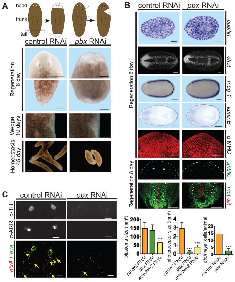
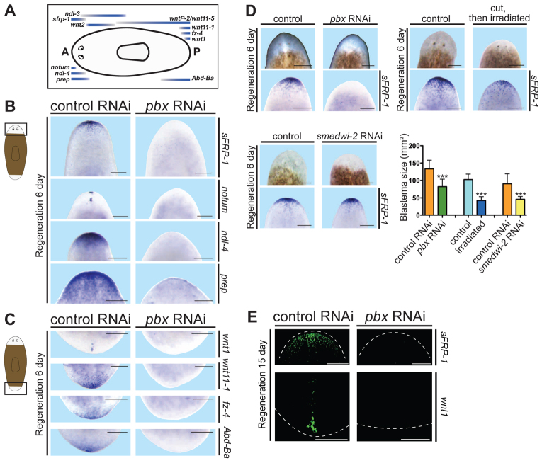
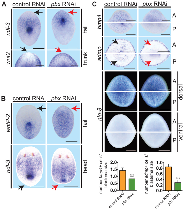
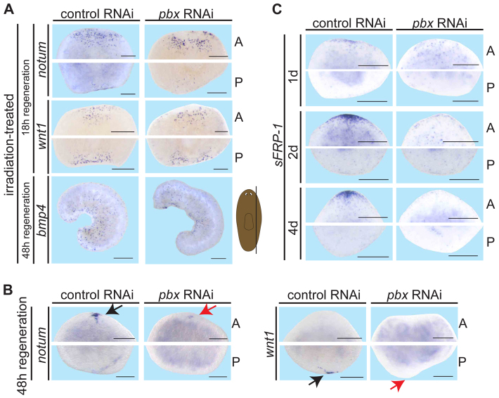
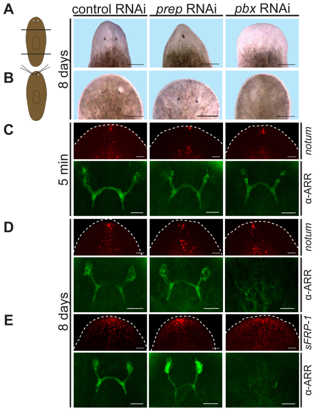
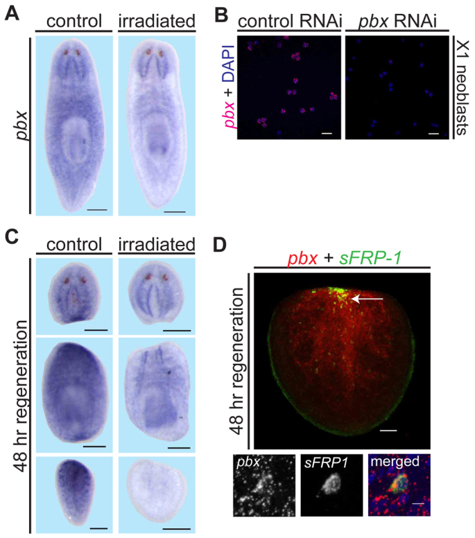
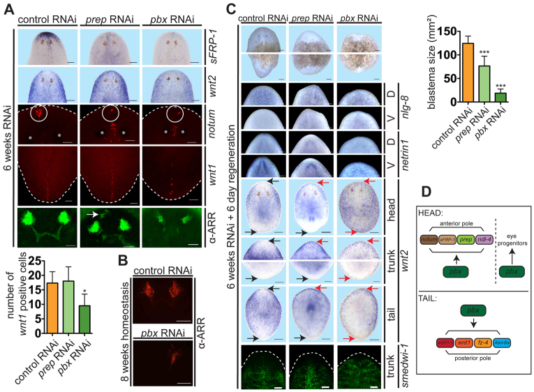
Planarian regeneration involves regionalized gene expression that specifies the body plan. After amputation, planarians are capable of regenerating new anterior and posterior poles, as well as tissues polarized along the anterior-posterior, dorsal-ventral and medial-lateral axes. Wnt and several Hox genes are expressed at the posterior pole, whereas Wnt inhibitory genes, Fgf inhibitory genes, and prep, which encodes a TALE-family homeodomain protein, are expressed at the anterior pole. We found that Smed-pbx (pbx for short), which encodes a second planarian TALE-family homeodomain transcription factor, is required for restored expression of these genes at anterior and posterior poles during regeneration. Moreover, pbx(RNAi) animals gradually lose pole gene expression during homeostasis. By contrast, pbx was not required for initial anterior-posterior polarized responses to wounds, indicating that pbx is required after wound responses for development and maintenance of poles during regeneration and homeostatic tissue turnover. Independently of the requirement for pbx in pole regeneration, pbx is required for eye precursor formation and, consequently, eye regeneration and eye replacement in homeostasis. Together, these data indicate that pbx promotes pole formation of body axes and formation of regenerative progenitors for eyes.


Figures

Fig. 1.
Fig. 1.
pbx(RNAi) planarians fail to regenerate eyes. (A) pbx(RNAi) animals regenerated blastemas without eyes 6 days after cutting (n=96) and displayed locomotion defects when exposed to long-term RNAi (n=20). pbx(RNAi) animals generated unpigmented tissue at the site of wedge amputations at 10 days after amputation (control, n=8/8; pbx(RNAi), n=8/8). Left cartoon depicts head and tail amputation sites; right cartoon depicts wedge regeneration sites. Scale bars: 200 μm for pole regeneration, 1 mm for wedge regeneration, 1.26 mm for homeostasis. (B) Whole-mount ISH 6 days after regeneration demonstrates that pbx(RNAi) animals regenerated protonephridia (cubilin, n=5/5), the nervous system with morphological defects (chat, n=13/13), subepidermal marginal adhesive gland cells (mag-1, n=6/6), lamin B-positive lateral tissue (n=12/12) and muscle fibers (α-MHC, n=16/16). At 8 days, pbx(RNAi) sensory cells (cintillo+) regenerated with normal cell number (P=0.325, t-test), but failed to distribute into two lateral domains (n=6/6). pbx(RNAi) intestinal branches were generated, but slight defects in medial branch length were present (mat, n=2/7), and midline cells failed to extend completely posteriorly (slit, n=6/7). Feeding RNAi was used for lamin B and α-MHC images; similar results were obtained with dsRNA injection. Anterior is to the left except for α-MHC, cintillo and mat/slit panels, which are oriented anterior to the top. Asterisks, photoreceptors; white dashed lines, head rim; yellow dotted lines, primary posterior intestinal branches. Scale bars: 200 μm except chat, 100 μm; and α-MHC, 50 μm. (C) pbx(RNAi) animals failed to regenerate pigment cup cells (α-TH immunostaining, n=0/10 were normal) and photoreceptor neurons (α-ARR immunostaining, n=0/10 were normal); eye progenitor numbers (double-labeled with otxA and eya RNA probes) were greatly reduced (n=13, right-hand graph, ***P<0.0001, t-test). Animals are oriented anterior to the top. Yellow arrows, double-positive cells. The eye regeneration defect in pbx(RNAi) animals is not explained only by reduced blastema size (n≥8 each). Left and middle graphs, one-way ANOVA tests followed by a Dunnet post-hoc test; ***P≤0.001 for experimental condition versus control. Graphs show mean±s.e.m. Scale bars: 50 μm.
Fig. 2.
Fig. 2.
pbx is required for expression of genes at AP poles in regeneration. (A) Cartoon shows AP expression locations of genes used in this study to characterize body axis regionalization; eyes (in the head) and the pharynx (centrally located) are depicted. A, anterior; P, posterior. (B) Whole-mount ISH showing missing or reduced gene expression in the anterior blastema of pbx(RNAi) planarians: sFRP-1 (control n=14/14; pbx n=0/15), notum (control n=6/6; pbx n=4/6 greatly reduced and mislocalized, n=2/6 not present), ndl-4 (control n=5/6; pbx n=0/6) and prep (control n=12/12; pbx n=0/12). Cartoon depicts area shown in images. (C) Expression of posterior markers, wnt1 (control n=6/6; pbx n=1/6), wnt11-1 (control n=4/6; pbx n=0/6), fz-4 (control n=6/6; pbx n=1/6) and Abd-Ba (control n=6/6; pbx n=0/5) was absent in pbx(RNAi) animals. Cartoon depicts area shown in images. (D) Animals with reduced anterior blastema size displayed more sFRP-1 expression than did pbx(RNAi) animals. Upper left panel: pbx RNAi involved dsRNA injection with 3-5 mg/ml dsRNA (n=10 for both conditions); upper right panel: small blastemas were generated by exposing cut worms to 6000 rads of γ-radiation 24 hours after cutting (control n=10; irradiated n=8); lower left panel: small blastemas were generated by weak RNAi of smedwi-2 (one feeding with amputation 3 days later) (n=10). Quantification of blastema size is shown in the bottom graph (mean±s.e.m.; ***P=0.0001, left pair; ***P<0.0001, middle pair; ***P=0.0002, right pair; t-test for each pair of conditions). (E) Pole markers sFRP-1 (control, n=6/6, pbx RNAi, n=0/6) and wnt1 (control, n=6/6, pbx RNAi, n=1/6) were absent in pbx(RNAi) animals after 15 days of regeneration. All animals are at regeneration day 6 except for panel E and are oriented anterior at the top. Scale bars: in E, 100 μm; in B,C,D, 200 μm.
Fig. 3.
Fig. 3.
pbx(RNAi) animals display abnormal pole gene expression and fail to re-scale expression gradients during regeneration. (A-C) Whole-mount ISH of regenerating pieces of planarians at day 6. Anterior is to the top. Black arrows, normal gene expression location (control); red arrows, aberrant expression in pbx(RNAi) animals. (A) pbx(RNAi) animals exhibited aberrant anterior blastema gene expression: ndl-3 (control, n=15/15; pbx RNAi, n=1/16 display restriction from the head tip of tail fragments) and wnt2 (control, n=10/11; pbx RNAi, n=0/13 display restriction from the head tip; head blastema of trunk fragment shown). (B) pbx(RNAi) animals failed to re-scale the wntP-2 expression gradient (control, n=17/18; pbx RNAi, n=4/20 tail fragments show re-scaling) and ndl-3 expression (control, n=22/25; pbx RNAi, n=1/22 head fragments show re-scaling). (C) Dorsal bmp4 expression (control, n=18; pbx RNAi, n=20) and ventral admp expression (control, n=13; pbx RNAi, n=10) were reduced in pbx(RNAi) animals, but dorsal expression of nlg-8 was normal in both anterior (A) and posterior (P) blastemas (control, n=13/13; pbx RNAi, n=12/12). ‘Head’, ‘trunk’ and ‘tail’ refer to a head fragment regenerating a tail, a fragment with head and tail amputated transversely, and a tail fragment regenerating a new head after transverse amputation, respectively. Graphs show quantification of bmp4+ and admp+ cells normalized to blastema size (mean±s.e.m.; ***P=0.0,001, t-test). Scale bars: 200 μm.
Fig. 4.
Fig. 4.
Wound-induced patterning gene expression is not affected in pbx(RNAi) planarians. (A) Anterior (A) and posterior (P) blastemas of irradiated pbx(RNAi) trunk pieces exhibited normal irradiation-insensitive, wound-induced expression of notum (control, n=5/7; pbx RNAi, n=6/6; transversely amputated), wnt1 (control, n=7/7; pbx RNAi, n=8/8; transversely amputated) and bmp4 (control, n=8/8; pbx RNAi, n=13/13; parasagitally amputated). Cartoon depicts amputation site; thin fragments were used. (B) notum expression (control, n=6/6; pbx RNAi, n=2/6 no expression, n=3/6 reduced expression) in the anterior blastema and wnt1 expression (control, n=6/6; pbx(RNAi), n=0/7 trunk pieces) in the posterior blastema of transversely cut animals 48 hours post-cutting. Black and red arrows indicate normal and reduced/absent expression, respectively. (C) sFRP-1 expression in transversely cut trunk pieces during regeneration; 1, 2 and 4 days (d) shown (pbx RNAi, n=14 trunk pieces for each condition). Scale bars: 200 μm.
Fig. 5.
Fig. 5.
The requirement of pbx for eye regeneration is separable from its requirement for pole regeneration. (A,B) Cartoons depict head or eye removal. (A) pbx (n=0/20 no eyes) and prep (n=18/19 one eye; n=1/19 no eyes) RNAi planarians exhibited different eye regeneration phenotypes after transverse amputation. (B) Both control (n=25/25) and prep RNAi (n=24/24) animals regenerated eyes but pbx(RNAi) animals (n=0/25) did not 8 days after eye removal with glass needles. (C-E) Double-fluorescent labeling of notum or sFRP-1 FISH with α-ARRESTIN immunostaining were used to assess anterior pole gene expression and eye regeneration in each animal. (C) Photoreceptor cell bodies were mostly absent 5 minutes after eye removal, with axons (green fluorescence) remaining (control, n=9/9; prep RNAi, n=10/10; pbx RNAi, n=10/10). notum expression was normal. (D,E) Eye regeneration defects were observed in pbx(RNAi) animals but not in prep(RNAi) or control animals. The size of the cluster of photoreceptor cells increased in prep(RNAi) (n=24/24) and control (n=25/25) animals with time, but did not in pbx(RNAi) (n=25/25) animals with only sparse pre-existing axon fragments remaining. notum and sFRP-1 expression were normal. Dashed line depicts animal boundary. Anterior is to the top. Scale bars: in A,B, 200 μm; in C-E, 50 μm.
Fig. 6.
Fig. 6.
pbx displays broad expression, including in neoblasts. (A) Whole-mount ISH showing pbx expression in non-irradiated (control) and irradiated planarians (n=6 for each). Scale bars: 200 μm. (B) pbx expression in neoblasts (referred to as X1 cells) sorted using FACS (Hayashi et al., 2006) was detected using FISH. Expression was absent following pbx RNAi. Scale bars: 20 μm. (C) pbx mRNA levels were strong and irradiation sensitive near wounds, 48 hours after amputation (n=6 for each). Scale bars: 200 μm. (D) Double FISH of pbx (red) and sFRP-1 (green) with DAPI (blue) in a regenerating tail (n=10) fragment at 48 hours. Some cells displayed both pbx and sFRP-1 signal, indicated by an arrow. Scale bars: upper panel, 50 μm; lower panels, 5 μm.
Fig. 7.
Fig. 7.
pbx is required for maintenance of pole gene expression and eyes during homeostatic tissue turnover. (A) Six weeks of prep and pbx RNAi caused reduced sFRP-1 expression (control, n=0/13; prep RNAi, n=13/13; pbx RNAi, n=10/10) and aberrant wnt-2 (control, n=0/4; prep RNAi, n=3/3; pbx RNAi, n=3/3) and notum (control, n=0/6; prep RNAi, n=6/6; pbx RNAi, n=3/3; white circle) expression. pbx RNAi, but not prep RNAi, caused wnt1+ cell reduction at the posterior pole (control, n=7; prep RNAi, n=7; pbx RNAi, n=6; shown are mean±s.e.m.; one-way ANOVA test followed by a Dunnet post-hoc test; *P≤0.05 between experimental condition and control). pbx(RNAi) animals, labeled with α-ARRESTIN antibody gradually lost photoreceptors and prep(RNAi) animals exhibited extra photoreceptors (white arrow) (control, n=6; prep RNAi, n=6; pbx RNAi, n=3). Asterisks in images indicate eyes. Scale bars: 50 μm. (B) Unamputated pbx(RNAi) animals failed to homeostatically maintain photoreceptor neurons after 8 weeks of RNAi (labeled with α-ARRESTIN antibody; pbx RNAi, n=8/10; control, n=0/10). Scale bars: 50 μm. (C) Control, prep or pbx RNAi animals at 6 days of regeneration. Blastema size quantification is shown at right (one-way ANOVA test followed by a Dunnet post-hoc test; ***P≤0.001 between experimental condition and control; n=10 for each condition). Dorsal expression of nlg-8 and ventral expression of netrin in trunk anterior blastemas (n=14/14 for all conditions). Both prep and pbx RNAi animals exhibited aberrant wnt-2 expression at the anterior tip of all regenerating pieces (control, n=0/16; prep RNAi, n=16/16; pbx RNAi, n=11/12). However, only pbx(RNAi) animals showed aberrant wnt-2 expression at posterior blastemas (control, n=1/16; prep RNAi, n=1/16; pbx RNAi, n=11/12). Black arrows, normal expression; red arrows, aberrant expression. Neoblasts (smedwi-1+) were similar in distribution among all RNAi treatments (control RNAi n=15/15; prep RNAi n=16/16; pbx RNAi, n=14/14 regenerating pieces; shown are mean±s.e.m.). Scale bars: 100 μm. (D) Data summary. pbx is required for expression of anterior and posterior pole genes in head and tail blastemas, respectively. pbx is also required for eye progenitor formation. Dashed lines indicate animal boundaries. All animals are oriented with anterior to the top.

Similar articles

Cited by

References

    1. Aboobaker A. A. (2011). Planarian stem cells: a simple paradigm for regeneration. Trends Cell Biol. 21, 304-311 - PubMed
    1. Adell T., Salò E., Boutros M., Bartscherer K. (2009). Smed-Evi/Wntless is required for beta-catenin-dependent and -independent processes during planarian regeneration. Development 136, 905-910 - PubMed
    1. Adell T., Cebrià F., Saló E. (2010). Gradients in planarian regeneration and homeostasis. Cold Spring Harb. Perspect. Biol. 2, a000505 - PMC - PubMed
    1. Almuedo-Castillo M., Sureda-Gomez M., Adell T. (2012). Wnt signaling in planarians: new answers to old questions. Dev. Biol. 56, 53-65 - PubMed
    1. Arata Y., Kouike H., Zhang Y., Herman M. A., Okano H., Sawa H. (2006). Wnt signaling and a Hox protein cooperatively regulate psa-3/Meis to determine daughter cell fate after asymmetric cell division in C. elegans. Dev. Cell 11, 105-115 - PubMed

Publication types

MeSH terms