. 2013 Mar;62(4):510-20.
doi: 10.1016/j.neuint.2013.01.003. Epub 2013 Jan 11.

Rearing environment, sex and developmental lead exposure modify gene expression in the hippocampus of behaviorally naïve animals

Affiliations

Rearing environment, sex and developmental lead exposure modify gene expression in the hippocampus of behaviorally naïve animals

D W Anderson et al. Neurochem Int. 2013 Mar.

Abstract

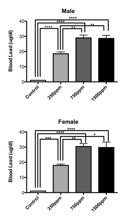
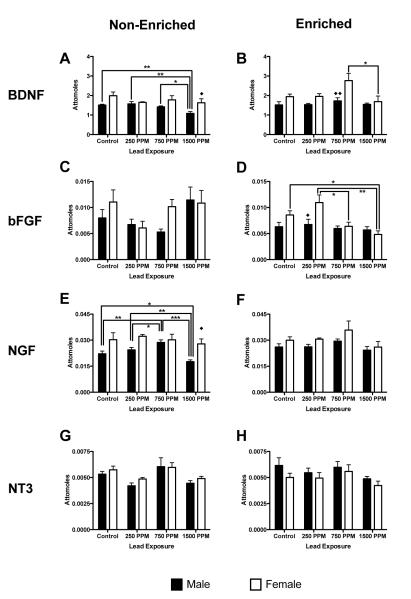
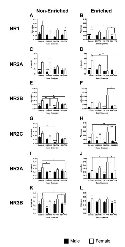
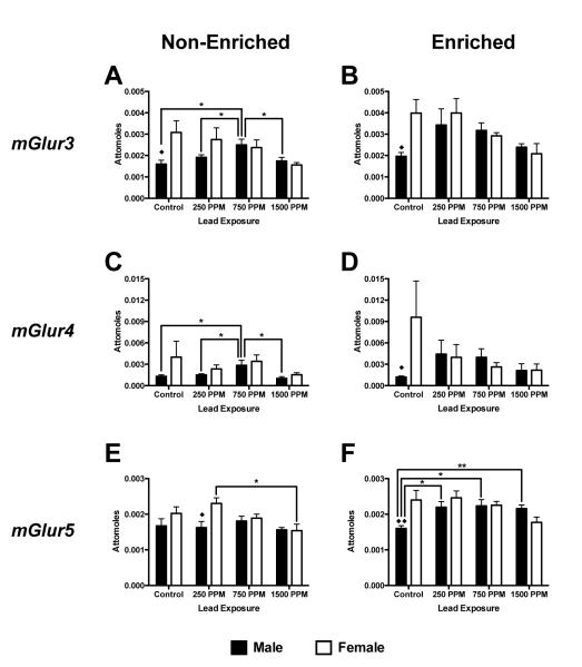
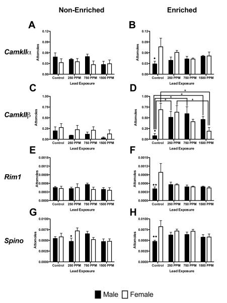
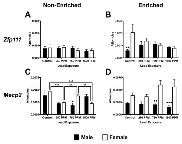
Developmental lead (Pb) exposure impairs various cognitive processes and behaviors in both humans and animals. In particular, specific deficits in spatial learning and memory have been described in Pb-exposed rats. It is also known that rearing environment (i.e., non-enriched vs. enriched) can have significant influences on cognitive performance and that rearing environment and sex may modify the influence of Pb exposure on learning and memory processes. It is also known that behavioral testing can alter hippocampal gene expression and interactive effects of environment. Little is known however about the molecular correlates of developmental Pb-exposure on expression of key sets of cognition-relevant genes in the hippocampus and how sex and environmental rearing condition may modify these effects. The present study examined expression profiles of neurobiologically-relevant genes (i.e., neurotrophic factors, NMDA receptors, metabotropic glutamate receptors, synaptic function/plasticity, and transcription/gene regulation) in behaviorally naïve rats with perinatal exposure (i.e., gestation through weaning) to different levels of Pb (250, 750 and 1,500 ppm Pb acetate) in males and females raised in a non-enriched environment (standard housing without toys) or an enriched environment (large cage containing toys changed twice weekly). Unlike previous studies identifying gene changes following behavioral testing, which alters expression analysis, we identified both sex and environmental related changes in hippocampal genes following Pb exposure alone. The gene expression changes described may be associated with learning and memory and may pre-determine how cognitive profiles develop following Pb exposure.


Figures

Figure 1
Figure 1
Blood lead levels (BLL) in animals on the last day of lead exposure. Both male and female postnatal day 21 animals showed significantly elevated BLL when compared to age matched controls not exposed to lead. There were no significant differences between males and females in BLL at any level of lead exposure. Interestingly, BLL’s did not increase in 1500ppm exposed animals when compared to 750ppm exposed animals. Data are mean blood lead level ± S.E.M. All statistically significant differences between exposures are designated on the graphs using connecting lines. * = p<0.05, ** = p<0.01, *** = p<0.001, **** = p<0.0001
Figure 2
Figure 2
Effects of perinatal lead-exposure on neurotrophic factor gene expression profiles in the hippocampus of male and female rats housed in either a non-enriched or enriched environment. Quantitative PCR analysis of mRNA expression levels of brain derived neurotrophic factor (Bdnf; A, B), basic fibroblast growth factor (Fgf2; C, D), nerve growth factor (Ngf; E, F) and neurotrophic factor-3 (Nt-3; G, H) in the hippocampus of control and lead-exposed animals. Sex, environment and dose specific patterns of response to lead were observed for Bdnf, Ffg2 and Ngf gene expression. Data are mean number of attomoles of mRNA ± S.E.M. * p < 0.05, ** p < 0.01, and *** p < 0.001 within sex analysis. ◆ p<0.05, and ◆◆ p < 0.01 across sex analysis
Figure 3
Figure 3
Effects of perinatal lead-exposure on NMDA receptor subtype gene expression profiles in the hippocampus of male and female rats housed in either a non-enriched or enriched environment. Quantitative PCR analysis of mRNA expression levels for five NMDA receptor subtypes: Nr1 (A, B), Nr2a (C, D), Nr2b (E, F), Nr3a (G, H) and Nr3b (I, J) in the hippocampus of control and lead-exposed animals. Significant sex, environment and dose specific effects were observed. Data are mean number of attomoles of mRNA ± S.E.M. * p<0.05, ** p < 0.01, and *** p<0.001 within sex analysis. ◆ p<0.05, ◆◆ p < 0.01, ◆◆◆ p<0.001 and ◆◆◆◆ p<0.0001 across sex analysis.
Figure 4
Figure 4
Effects of perinatal lead-exposure on metabotropic glutamate receptor subtype gene expression profiles in the hippocampus of male and female rats housed in either a non-enriched or enriched environment. Quantitative PCR analysis of mRNA expression levels for metabotropic glutamate receptors mGluR3 (A, B), mGluR4 (C, D) and mGluR5 (E, F) in the hippocampus of control and lead-exposed animals. Significant sex, environment and dose effects were observed. Data are mean number of attomoles of mRNA ± S.E.M. * p<0.05 and ** p<0.01 within sex analysis. ◆ p<0.05 and ◆◆ p < 0.01 across sex analysis.
Figure 5
Figure 5
Effects of perinatal lead-exposure on genes related to synaptic function and plasticity in the hippocampus of male and female rats housed in either a non-enriched or enriched environment. Quantitative PCR analysis of mRNA expression levels for Ca2+/calmodulin-dependent protein kinase II alpha (CamkIIα; A, B), Ca2+/calmodulin-dependent protein kinase II beta (CamkIIβ; C, D), regulating synaptic membrane exocytosis 1 (Rim1; G ,H) and spinophilin (Spino; I, J) in the hippocampus of control and lead-exposed animals. Significant effects were only seen in CamkIIβ expression. Data are mean number of attomoles of mRNA ± S.E.M. * p<0.05 within sex analysis. ◆ p<0.05 and ◆◆ p < 0.01 across sex analysis.
Figure 6
Figure 6
Effects of perinatal lead-exposure on mRNA expression of genes related to transcription and regulation in the hippocampus of male and female rats housed in either a non-enriched or enriched environment. Quantitative PCR analysis of mRNA expression levels for zinc finger protein 111 (Zfp111; A, B) and Methyl CpG binding protein 2 (Mecp2; C, D) and in the hippocampus of lead exposed animals. A number of structure and dose specific effects were observed. Data are mean number of attomoles of mRNA ± S.E.M. * p<0.05, ** p < 0.01, and *** p<0.001 within sex analysis. ◆ p<0.05, ◆◆ p < 0.01 and ◆◆◆ p<0.001 across sex analysis.

References

    1. Alkondon M, Costa AC, Radhakrishnan V, Aronstam RS, Albuquerque EX. Selective blockade of NMDA-activated channel currents may be implicated in learning deficits caused by lead. FEBS Letters. 1990;261:124–130. - PubMed
    1. Anderson DW, Pothakos K, Schneider JS. Sex and rearing condition modify the effects of perinatal lead exposure on learning and memory. Neurotoxicology. 2012 - PMC - PubMed
    1. Baghurst PA, Tong S, Sawyer MG, Burns J, McMichael AJ. Sociodemographic and behavioural determinants of blood lead concentrations in children aged 11-13 years. The Port Pirie Cohort Study. The Medical journal of Australia. 1999;170:63–67. - PubMed
    1. Bellinger DC. Lead neurotoxicity and socioeconomic status: conceptual and analytical issues. Neurotoxicology. 2008;29:828–832. - PMC - PubMed
    1. Brody DJ, Pirkle JL, Kramer RA, Flegal KM, Matte TD, Gunter EW, Paschal DC. Blood lead levels in the US population. Phase 1 of the Third National Health and Nutrition Examination Survey (NHANES III, 1988 to 1991) JAMA. 1994;272:277–283. - PubMed

Publication types