. 2013;89(1):59-65.
doi: 10.2183/pjab.89.59.

Circadian clock-controlled diurnal oscillation of Ras/ERK signaling in mouse liver

Affiliations

Circadian clock-controlled diurnal oscillation of Ras/ERK signaling in mouse liver

Yoshiki Tsuchiya et al. Proc Jpn Acad Ser B Phys Biol Sci. 2013.

Abstract

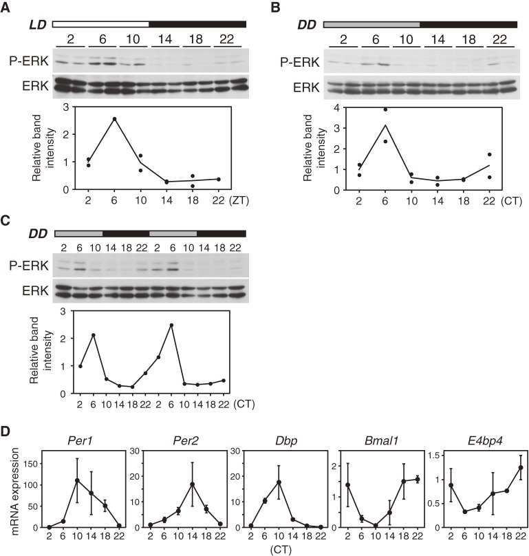
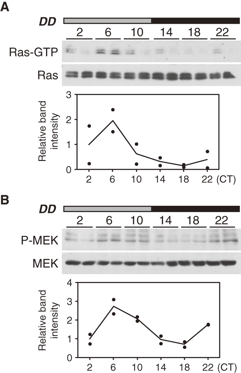
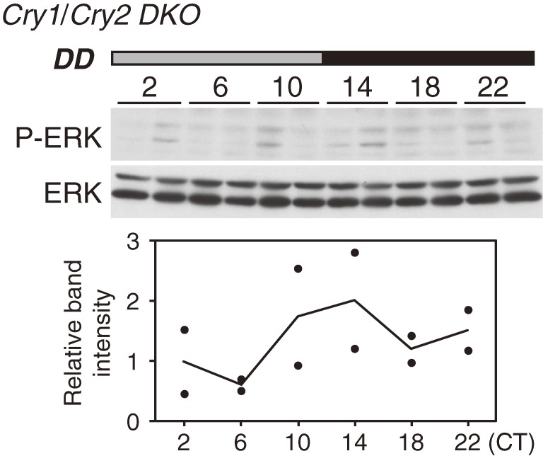
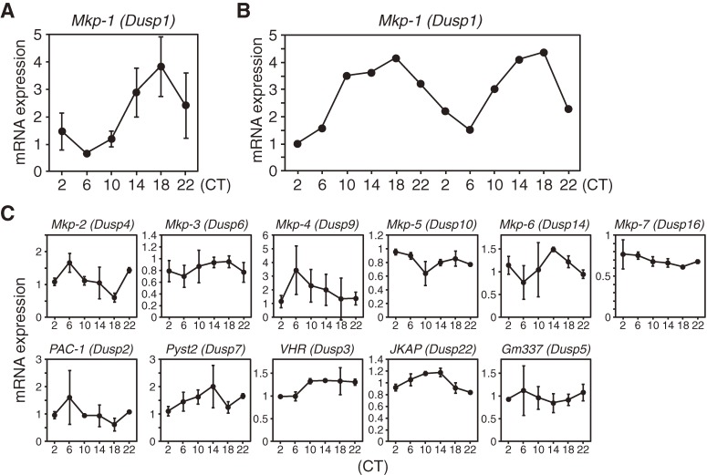
Accumulating evidence indicates that ERK MAP kinase signaling plays an important role in the regulation of the circadian clock, especially in the clock-resetting mechanism in the suprachiasmatic nucleus (SCN) in mammals. Previous studies have also shown that ERK phosphorylation exhibits diurnal variation in the SCN. However, little is known about circadian regulation of ERK signaling in peripheral tissues. Here we show that the activity of Ras/ERK signaling exhibits circadian rhythms in mouse liver. We demonstrate that Ras activation, MEK phosphorylation, and ERK phosphorylation oscillate in a circadian manner. As the oscillation of ERK phosphorylation is lost in Cry1/Cry2 double-knockout mice, Ras/ERK signaling should be under the control of the circadian clock. Furthermore, expression of MAP kinase phosphatase-1 (Mkp-1) shows diurnal changes in liver. These results indicate that Ras/ERK signaling is strictly regulated by the circadian clock in liver, and suggest that the circadian oscillation of the activities of Ras, MEK, and ERK may regulate diurnal variation of liver function and/or homeostasis.(Communicated by Shigekazu NAGATA, M.J.A.).


Figures

Figure 1.
Figure 1.
Circadian oscillation of ERK phosphorylation in mouse liver. (A) Circadian oscillation of ERK phosphorylation in LD conditions. Mice were sacrificed at the indicated time points and liver extracts were analyzed by immunoblotting with the indicated antibodies. Liver samples from two mice were analyzed at each time point. The graph shows relative band intensities of phospho-ERK that were normalized with band intensities of total ERK. The black dot indicates the value of each band and the solid line indicates the means. The mean value at ZT2 was set to 1. (B) Circadian oscillation of ERK phosphorylation in DD conditions. ERK phosphorylation was analyzed as in (A) on the first day in DD conditions. (C) Circadian oscillation of ERK phosphorylation lasts two circadian cycles in DD conditions. ERK phosphorylation was analyzed as in (A) on the first two days in DD conditions. Data from one mouse at each time point were shown. (D) Circadian rhythms of clock gene expression in liver in DD conditions. Mice were sacrificed and mRNA was collected from livers at each time point. The mRNA expression levels of the indicated clock genes were evaluated by real-time quantitative PCR. Each value was normalized to GAPDH and is presented as the means ± SD (n = 3).
Figure 2.
Figure 2.
Circadian oscillation of Ras activation and MEK phosphorylation in mouse liver. (A) Circadian oscillation of Ras activation in DD conditions. The GTP-bound active form of Ras was detected by immunoblotting. The graph shows relative band intensities of Ras-GTP that were normalized with band intensities of total Ras. The graph is presented as in Fig. 1A. (B) Circadian oscillation of MEK phosphorylation in DD conditions. The graph shows relative band intensities of phospho-MEK that were normalized with band intensities of total MEK. The graph is presented as in Fig. 1A.
Figure 3.
Figure 3.
ERK phosphorylation rhythms are lost in liver of Cry1/Cry2 double-knockout mice. Cry1/Cry2 double-knockout mice were sacrificed at the indicated time points in DD conditions and liver extracts were analyzed by immunoblotting with the indicated antibodies. ERK phosphorylation was analyzed as in Fig. 1A.
Figure 4.
Figure 4.
Expression of Mkp-1 shows circadian oscillation in mouse liver. (A) Circadian rhythms of mRNA expression of Mkp-1 in DD conditions. Mice were sacrificed and mRNA was collected from livers at each time point on the first day in DD conditions. The mRNA expression levels of Mkp-1 were evaluated by real-time quantitative PCR. Each value was normalized to GAPDH and is presented as the means ± SD (n = 3). (B) Circadian oscillation of Mkp-1 expression lasts two circadian cycles in DD conditions. The mRNA expression levels of Mkp-1 was analyzed as in (A) on the first two days in DD conditions. Data from one mouse at each time point were shown. (C) Time profiles of the mRNA expression levels of several dual-specificity phosphatases in DD conditions. The mRNA expression levels of the indicated genes were evaluated by real-time quantitative PCR. Each value was normalized to GAPDH and is presented as the means ± SD (n = 3).

Similar articles

Cited by

References

    1. Bell-Pedersen D., Cassone V.M., Earnest D.J., Golden S.S., Hardin P.E., Thomas T.L., Zoran M.J. (2005) Circadian rhythms from multiple oscillators: lessons from diverse organisms. Nat. Rev. Genet. 6, 544–556 - PMC - PubMed
    1. Reppert S.M., Weaver D.R. (2002) Coordination of circadian timing in mammals. Nature 418, 935–941 - PubMed
    1. Ko C.H., Takahashi J.S. (2006) Molecular components of the mammalian circadian clock. Hum. Mol. Genet. 15, R271–R277 - PubMed
    1. Hastings M.H., Reddy A.B., Maywood E.S. (2003) A clockwork web: circadian timing in brain and periphery, in health and disease. Nat. Rev. Neurosci. 4, 649–661 - PubMed
    1. Gachon F., Olela F.F., Schaad O., Descombes P., Schibler U. (2006) The circadian PAR-domain basic leucine zipper transcription factors DBP, TEF, and HLF modulate basal and inducible xenobiotic detoxification. Cell Metab. 4, 25–36 - PubMed

Publication types

Substances