. 2013 Apr 1;591(7):1951-66.
doi: 10.1113/jphysiol.2012.246983. Epub 2013 Jan 14.

Repeated in vivo exposure of cocaine induces long-lasting synaptic plasticity in hypocretin/orexin-producing neurons in the lateral hypothalamus in mice

Affiliations

Repeated in vivo exposure of cocaine induces long-lasting synaptic plasticity in hypocretin/orexin-producing neurons in the lateral hypothalamus in mice

Yan Rao et al. J Physiol. .

Abstract

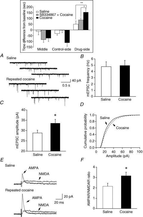
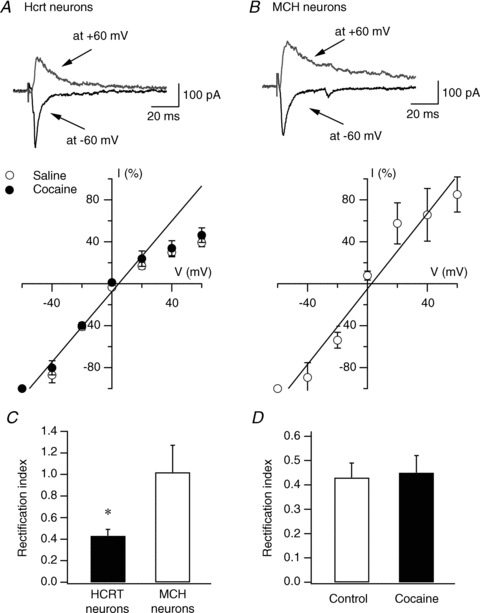
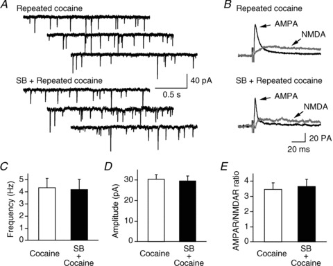
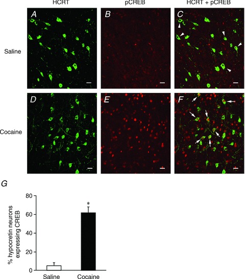
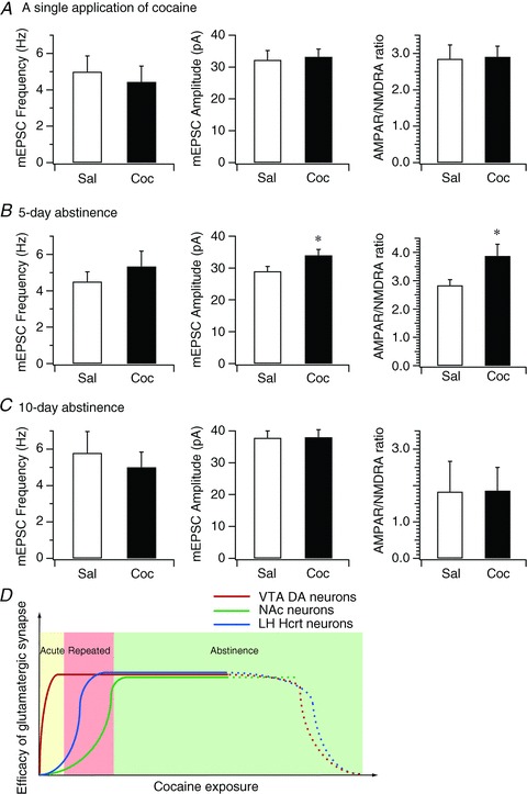
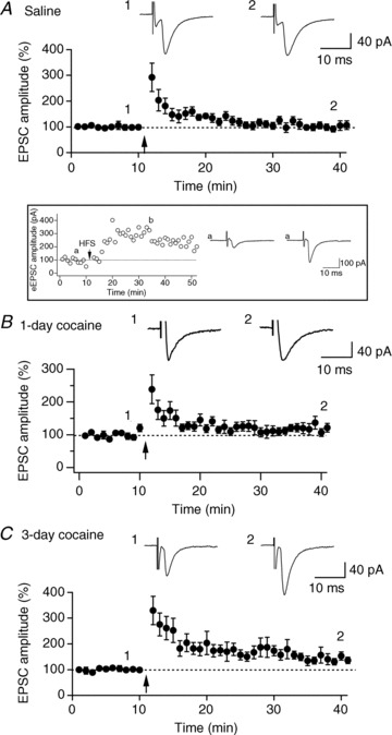
Hypocretin (orexin), a neuropeptide synthesized exclusively in the perifornical/lateral hypothalamus, is critical for drug seeking and relapse, but it is not clear how the circuitry centred on hypocretin-producing neurons (hypocretin neurons) is modified by drugs of abuse and how changes in this circuit might alter behaviours related to drug addiction. In this study, we show that repeated, but not single, in vivo cocaine administration leads to a long-lasting, experience-dependent potentiation of glutamatergic synapses on hypocretin neurons in mice following a cocaine-conditioned place preference (CPP) protocol. The synaptic potentiation occurs postsynaptically and probably involves up-regulation of AMPA-type glutamate receptors on hypocretin neurons. Phosphorylation of cAMP response element-binding protein (CREB) is also significantly increased in hypocretin neurons in cocaine-treated animals, suggesting that CREB-mediated pathways may contribute to synaptic potentiation in these cells. Furthermore, the potentiation of synaptic efficacy in hypocretin neurons persists during cocaine withdrawal, but reverses to baseline levels after prolonged abstinence. Finally, the induction of long-term potentiation (LTP) triggered by a high-frequency stimulation is facilitated in hypocretin neurons in cocaine-treated mice, suggesting that long-lasting changes in synapses onto hypocretin neurons would probably be further potentiated by other stimuli (such as concurrent environmental cues) paired with the drug. In summary, we show here that hypocretin neurons undergo experience-dependent synaptic potentiation that is distinct from that reported in other reward systems, such as the ventral tegmental area, following exposure to cocaine. These findings support the idea that the hypocretin system is important for behavioural changes associated with cocaine administration in animals and humans.


Figures

Figure 1
Figure 1. Potentiation of glutamatergic synapses on hypocretin neurons following the establishment of cocaine-conditioned place preference (CPP)
A, sample traces of mEPSCs recorded from control and cocaine-treated mice are presented. B, mean frequency of mEPSCs detected in saline- and cocaine-treated mice showed no significant difference between these two groups. C, a significant increase in the mean amplitude of mEPSC events detected in cocaine-treated mice was indicated as compared to the control group (*P < 0.05, t test). D, a significant right-shift in the cumulative probability of mEPSC amplitude was detected in cocaine-treated mice as compared to control mice (P < 0.001, Kolmogorov–Smirnov test). E, sample traces of evoked EPSCs carried by AMPA receptors (AMPAR) and NMDA receptors (NMDAR) in control and cocaine-treated mice. F, the ratio of evoked EPSCs carried by AMPAR and NMDAR was enhanced in mice treated with cocaine as compared to controls (*P < 0.05, t test). Inset, bar graph indicates that mice spent more time in the cocaine-paired side of the testing apparatus after a 3-day training session. Blockade of the hypocretin-1 receptor pathway blocks cocaine CPP in mice.
Figure 2
Figure 2. Repeated cocaine exposure does not induce changes in the current–voltage (I–V) relationship of AMPAR-mediated EPSC in hypocretin neurons
A, I–V relationship of evoked AMPAR-EPSCs recorded in hypocretin (Hcrt) neurons in naive (open circles) and cocaine-treated (filled circles) mice. Representative traces from naive mice are shown on the upper part of the panel. The straight line represents the linear regression of mean EPSC amplitude at negative potentials. B, representative traces (upper) and I–V relationship (lower) of evoked AMPAR-EPSCs recorded in melanin-concentrating hormone (MCH)-containing neurons in MCH-GFP mice. The straight line represents the linear regression of mean EPSC amplitude at negative potentials. C, mean RI in hypocretin and MCH neurons is presented. D, mean RI in hypocretin neurons from control and cocaine-treated mice is presented.
Figure 3
Figure 3. The synaptic efficacy of glutamatergic synapses on hypocretin neurons was not significantly different in cocaine-treated mice in the absence and presence of a selective hypocretin-1 receptor antagonist, SB334867 (SB)
Sample traces of mEPSCs and evoked EPSCs are presented in A and B. The frequency, amplitude and AMPAR/NMDAR ratio in mice treated with cocaine alone and SB plus cocaine are presented in C, D and E.
Figure 4
Figure 4. Enhanced phosphorylation of CREB (pCREB) induced by cocaine treatment in hypocretin neurons
Dual immunostaining of pCREB and hypocretin (Hcrt) was performed in control (n= 3) and cocaine-treated mice (n= 3) after a 3-day treatment. AC, images show that only hypocretin-immunopositive neurons (indicated by arrowheads) are present in the perifornical/LH area in saline-treated mice. DF, images show that hypocretin and pCREB-immunopositive neurons (indicated by arrows) are present in the LH area in cocaine-treated mice. Scale bar: 15 μm. G, pooled data from all examined sections from saline- (n= 24 sections) and cocaine-treated (n= 21 sections) mice indicate a significant increase in pCREB expression in hypocretin neurons following a 3-day cocaine treatment. *P < 0.001, t test.
Figure 5
Figure 5. Duration of experience-dependent synaptic plasticity at glutamatergic synapses on hypocretin neurons following cocaine exposure
A, bar graph shows the mean frequency and amplitude of mEPSCs and mean AMPAR/NMDAR ratio in hypocretin neurons in control and cocaine-treated mice 1 day after a single exposure to cocaine. B, bar graph shows the mean frequency and amplitude of mEPSCs and mean AMPAR/NMDAR ratio in hypocretin neurons in control and cocaine-treated mice on the 5th day of withdrawal following the 3-day cocaine regimen. *P < 0.05, paired t test. C, bar graph shows the mean frequency and amplitude of mEPSCs and mean AMPAR/NMDAR ratio in hypocretin neurons in control and cocaine-treated mice on the 10th day of withdrawal after the 3-day cocaine regimen. D, the diagram summarizes the time courses of synaptic potentiation in the VTA, NAc and hypocretin neurons induced by exposure to cocaine. Our data suggest that the onset of expression of experience-dependent synaptic potentiation in hypocretin neurons (blue line) is later than synaptic potentiation induced in the VTA (red line) (Ungless et al. 2001) and earlier than in the NAc (green line) (Kourrich et al. 2007), which implicates the distinctive roles of synaptic plasticity in these areas in the development of animal behaviours relevant to cocaine addiction.
Figure 6
Figure 6. Cocaine treatment facilitates the induction of LTP by HFS in hypocretin neurons
A, time course of evoked EPSCs recorded in hypocretin neurons from control mice treated with saline for 3 days. 1 and 2 are averaged traces of evoked EPSCs recorded before and 30 min after the HFS. The arrow indicates application of 4 trains of HFS (100 Hz, 1 s duration, 10 s interval) to the MFB when the recorded neurons were under current clamp. B, time course of evoked EPSCs in hypocretin neurons before and after HFS in mice given a single application of cocaine. Arrow, application of HFS. 1 and 2 are averaged traces (from 5 consecutive traces) of evoked EPSCs recorded before and 30 min after the application of HFS. C, time course of evoked EPSCs recorded in hypocretin neurons from mice treated with cocaine for 3 days. Arrow, application of HFS. 1 and 2 are averaged traces of evoked EPSCs recorded before and 30 min after the application of HFS. Inset, the time course of evoked EPSCs recorded in a control hypocretin neuron showing the expression of LTP induced by 10 trains of HFS. The arrow indicates application of 10 trains of high frequency stimulation (100 Hz, 1 s duration, 10 s interval) to the MFB when the recorded neurons were under current clamp. Symbols ‘a’ and ‘b’ indicate raw traces of evoked EPSCs recorded before and after high frequency stimulation.

Similar articles

Cited by

References

    1. Adamantidis AR, Zhang F, Aravanis AM, Deisseroth K, de Lecea L. Neural substrates of awakening probed with optogenetic control of hypocretin neurons. Nature. 2007;450:420–424. - PMC - PubMed
    1. Adams WJ, Lorens SA, Mitchell CL. Morphine enhances lateral hypothalamic self-stimulation in the rat. Proc Soc Exp Biol Med. 1972;140:770–771. - PubMed
    1. Aston-Jones G, Smith RJ, Moorman DE, Richardson KA. Role of lateral hypothalamic orexin neurons in reward processing and addiction. Neuropharmacol. 2009;56(Suppl. 1):112–121. - PMC - PubMed
    1. Badiani A, Robinson TE. Drug-induced neurobehavioral plasticity: the role of environmental context. Behav Pharmacol. 2004;15:327–339. - PubMed
    1. Balcita-Pedicino JJ, Sesack SR. Orexin axons in the rat ventral tegmental area synapse infrequently onto dopamine and γ-aminobutyric acid neurons. J Comp Neurol. 2007;503:668–684. - PubMed

Publication types

LinkOut - more resources