. 2013 Mar 25;425(6):1050-64.
doi: 10.1016/j.jmb.2013.01.004. Epub 2013 Jan 11.

Sequence-specific, RNA-protein interactions overcome electrostatic barriers preventing assembly of satellite tobacco necrosis virus coat protein

Affiliations

Sequence-specific, RNA-protein interactions overcome electrostatic barriers preventing assembly of satellite tobacco necrosis virus coat protein

Robert J Ford et al. J Mol Biol. .

Abstract

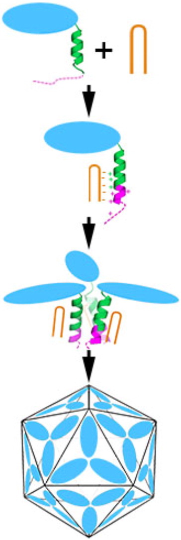
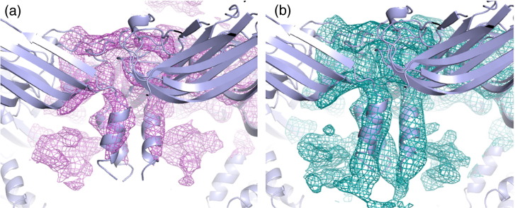
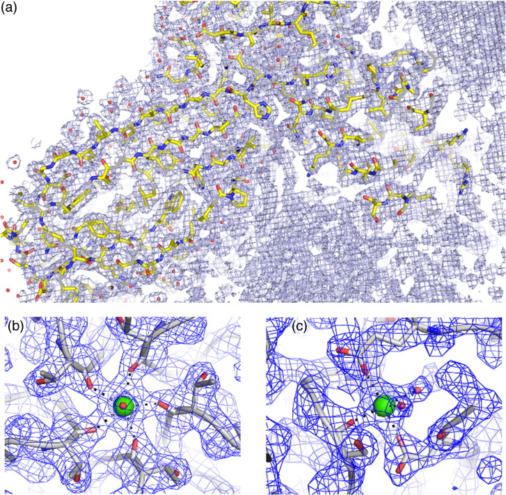
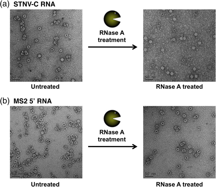
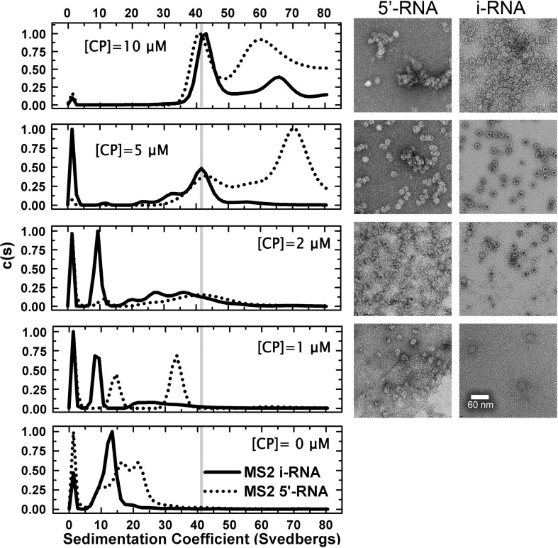
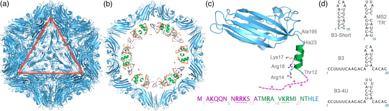
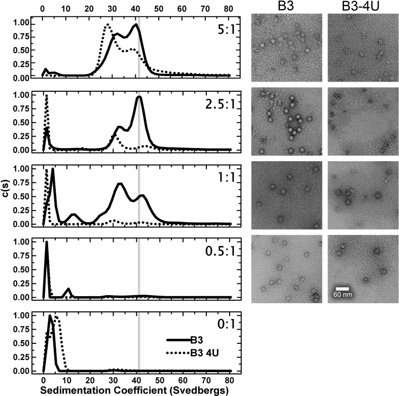
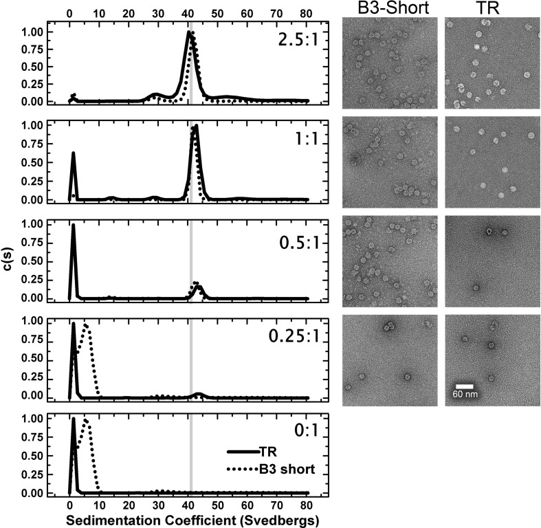
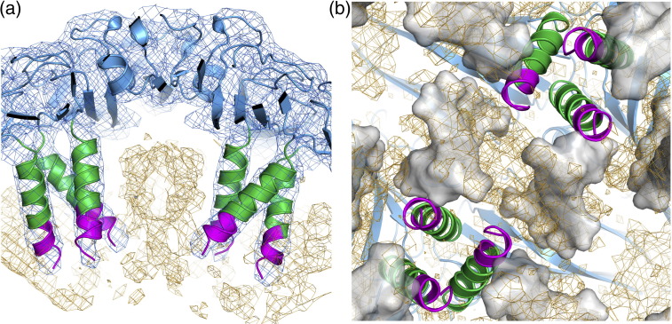
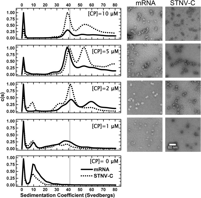
We have examined the roles of RNA-coat protein (CP) interactions in the assembly of satellite tobacco necrosis virus (STNV). The viral genomic RNA encodes only the CP, which comprises a β-barrel domain connected to a positively charged N-terminal extension. In the previous crystal structures of this system, the first 11 residues of the protein are disordered. Using variants of an RNA aptamer sequence isolated against the CP, B3, we have studied the sequence specificity of RNA-induced assembly. B3 consists of a stem-loop presenting the tetra-loop sequence ACAA. There is a clear preference for RNAs encompassing this loop sequence, as measured by the yield of T=1 capsids, which is indifferent to sequences within the stem. The B3-containing virus-like particle has been crystallised and its structure was determined to 2.3Å. A lower-resolution map encompassing density for the RNA has also been calculated. The presence of B3 results in increased ordering of the N-terminal helices located at the particle 3-fold axes, which extend by roughly one and a half turns to encompass residues 8-11, including R8 and K9. Under assembly conditions, STNV CP in the absence of RNA is monomeric and does not self-assemble. These facts suggest that a plausible model for assembly initiation is the specific RNA-induced stabilisation of a trimeric capsomere. The basic nature of the helical extension suggests that electrostatic repulsion between CPs prevents assembly in the absence of RNA and that this barrier is overcome by correct placement of appropriately orientated helical RNA stems. Such a mechanism would be consistent with the data shown here for assembly with longer RNA fragments, including an STNV genome. The results are discussed in light of a first stage of assembly involving compaction of the genomic RNA driven by multiple RNA packaging signal-CP interactions.


Figures

None
Graphical abstract
Supplementary Fig. S1
2Fo − Fc electron density for the STNV B3 complex showing the quality of the 60-fold averaged electron density, calculated using all data (217–2.3 Å) and the model fit. (a) is a 12-Å-thick slice through the capsid showing the clearly defined side chains as well as the presence of considerable non-protein internal electron density (electron density contoured at 0.038 e/Å3). (b) The calcium ion binding site at a capsid 5-fold axis and (c) at a capsid 3-fold axis. The electron densities in (b) and (c) are contoured at 5 σ.
Supplementary Fig. S2
2Fo − Fc electron density for (a) the mRNA VLP and (b) the STNV B3 complex reported here. Both maps were calculated using data to a maximum resolution of 6 Å to enable clearer visualisation of electron density that does not follow the strict icosahedral symmetry of the capsid. Both maps are contoured at the same level. Additional electron density at the N-terminus, attributed to an extra turn of the helix, is clearly visible in (b).
Supplementary Fig. S3
Reassembled STNV VLPs formed from STNV CP and either STNV-C RNA (Fig. 4) or the MS2 5′ RNA (Supplementary Fig. 4) at 5 μM [CP] appear aggregated (Fig. 4). Treatment of these particles with RNase A results in loss of the aggregates and the appearance of separated T = 1 particles. These results are consistent with the formation of fused capsids assembling on the same RNA.
Supplementary Fig. 4
An overlay of STNV reassembly using MS2 iRNA and MS2 5′ RNA analysed by analytical ultracentrifugation (left) and the corresponding images from EM (right). The RNA concentrations were as follows: iRNA, 60 nM and 5′ RNA, 24 nM. The CP concentrations are listed on the figure.
Fig. 1
Fig. 1
The components of the STNV system. (a) The STNV capsid. The X-ray structure of the recombinant STNV capsid shown as a blue cartoon (PDB entry 3S4G14). The view is along an icosahedral 3-fold axis, and the symmetry of the capsid is indicated by the red triangle, which corresponds to one face of the icosahedral particle. (b) A central (45 Å thick) slab through the STNV structure. The N-terminal helices are coloured green, and the structure of 7 nt per CP monomer of ordered RNA is also shown. (c) Close-up view of the STNV CP subunit. The subunit is a wedge-shaped jelly roll β-sandwich. The N-terminus is positively charged and visible in 3S4G up to the threonine at position 12. (Note that the amino acid numbering follows the PDB entries that begin with the genetically encoded N-terminal methionine as residue − 1). The unstructured portion (magenta) of the N-terminus contains a further four basic amino acid side chains as indicated in the sequence shown. (d) The sequences and predicted secondary structures of the oligoribonucleotides used in this study.
Fig. 2
Fig. 2
A comparison of STNV CP reassembly efficiency using B3 4U and B3 RNAs. Left panels show a titration of increasing molar ratios in reassembly reactions carried out as described in Materials and Methods analysed by svAUC. The corresponding images on the right are TEMs of the samples at the end of the reactions. The RNA concentrations were held constant at 2 μM and the molar ratios of CP:RNA are shown in each panel. The sedimentation coefficient of the recombinant VLP, here and throughout, is shown as vertical grey lines.
Fig. 3
Fig. 3
A comparison of STNV CP reassembly efficiency using B3 short and MS2 TR stem–loops. The RNA concentrations were held constant at 4 μM. All other details, here and in Fig. 5 and Supplementary Fig. 2, are as in the legend to Fig. 2.
Fig. 4
Fig. 4
The X-ray structure of B3 VLP. (a) Ordered density in the N-terminal helices. The STNV CP is shown in cartoon representation and coloured as in Fig. 1. The orange mesh is the 60-fold averaged 2Fo − Fc electron density of the B3 VLP structure with the parts of the map corresponding to the CP shell masked away and contoured at 0.03 e/Å3. The blue mesh is the unmasked electron density contoured at ~ 0.07 e/Å3. The extra density at the N-terminal end of the helices is clearly visible, and residues 8–13 are shown in magenta. Residues 12 and 13 are present in other STNV structures but they adopt a different conformation in the mRNA containing VLP. (b) The B3 RNA density. The view is from the inside of the capsid, outward along an icosahedral 2-fold axis. The protein cartoon and orange mesh are as described in (a), while the grey surface is the solvent-accessible surface of the mRNA modelled in the study by Lane et al. (PDB entry 3S4G).
Fig. 5
Fig. 5
A comparison of STNV CP reassembly efficiency using STNV-C and STNV mRNAs. The RNA concentrations were as follows: mRNA, 100 nM and STNV-C, 50 nM, corresponding to ~ 60 μM phosphodiester concentration in each case. The CP concentrations are listed on the figure.
Fig. 6
Fig. 6
Schematic model of the STNV assembly process. (a) STNV CP subunits (blue oval) have an N-terminal extension that is partly helical (green) and partly unstructured (magenta). These extensions are highly positively charged with 7 Arg/Lys residues in the 24 residues at the N-terminus. (b) Binding of RNA stem–loops, exemplified by the B3 loop, at least partly neutralises the positive charge on the N-terminus, allowing the N-terminal helix to become longer and more ordered. (c) Once the N-terminal region has had its positive charge neutralised by RNA binding, the CPs can trimerize, forming an assembly-competent trimeric capsomere. (d) These trimeric capsomeres can then assemble to form higher-order structures and ultimately the T = 1 capsid (e).

References

    1. Schneemann A. The structural and functional role of RNA in icosahedral virus assembly. Annu. Rev. Microbiol. 2006;60:51–67. - PubMed
    1. Stockley, P. G., Ranson, N. A. & Twarock, R. (2013). A new paradigm for the roles of the genome in ssRNA viruses. Future Virol. in press.
    1. Bancroft J.B., Hills G.J., Markham R. A study of the self-assembly process in a small spherical virus formation of organized structures from protein subunits in vitro. Virology. 1967;31:354–379. - PubMed
    1. Harrison S.C., Olson A.J., Schutt C.E., Winkler F.K., Bricogne G. Tomato bushy stunt virus at 2.9 Å resolution. Nature. 1978;276:368–373. - PubMed
    1. Caspar D.L.D., Klug A. Physical principles in the construction of regular viruses. Cold Spring Harbor Symp. Quant. Biol. 1962;27:1–24. - PubMed

Publication types

Substances

Associated data