. 2013 Apr 12;425(7):1172-82.
doi: 10.1016/j.jmb.2013.01.010. Epub 2013 Jan 11.

Dispersed sites of HIV Vif-dependent polyubiquitination in the DNA deaminase APOBEC3F

Affiliations

Dispersed sites of HIV Vif-dependent polyubiquitination in the DNA deaminase APOBEC3F

John S Albin et al. J Mol Biol. .

Abstract

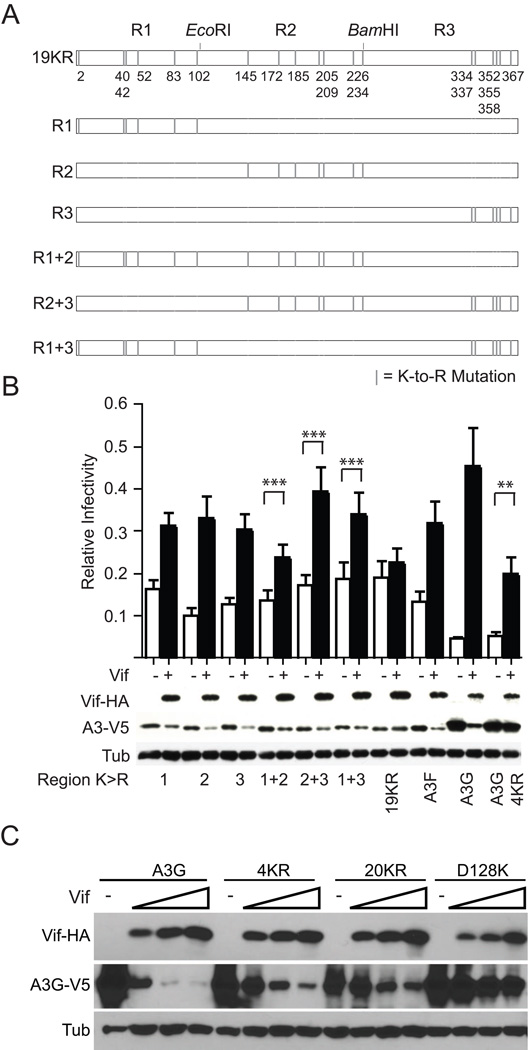
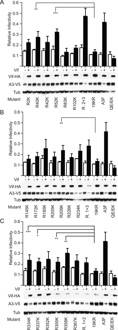
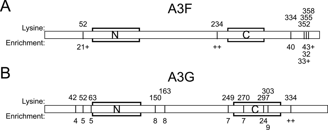
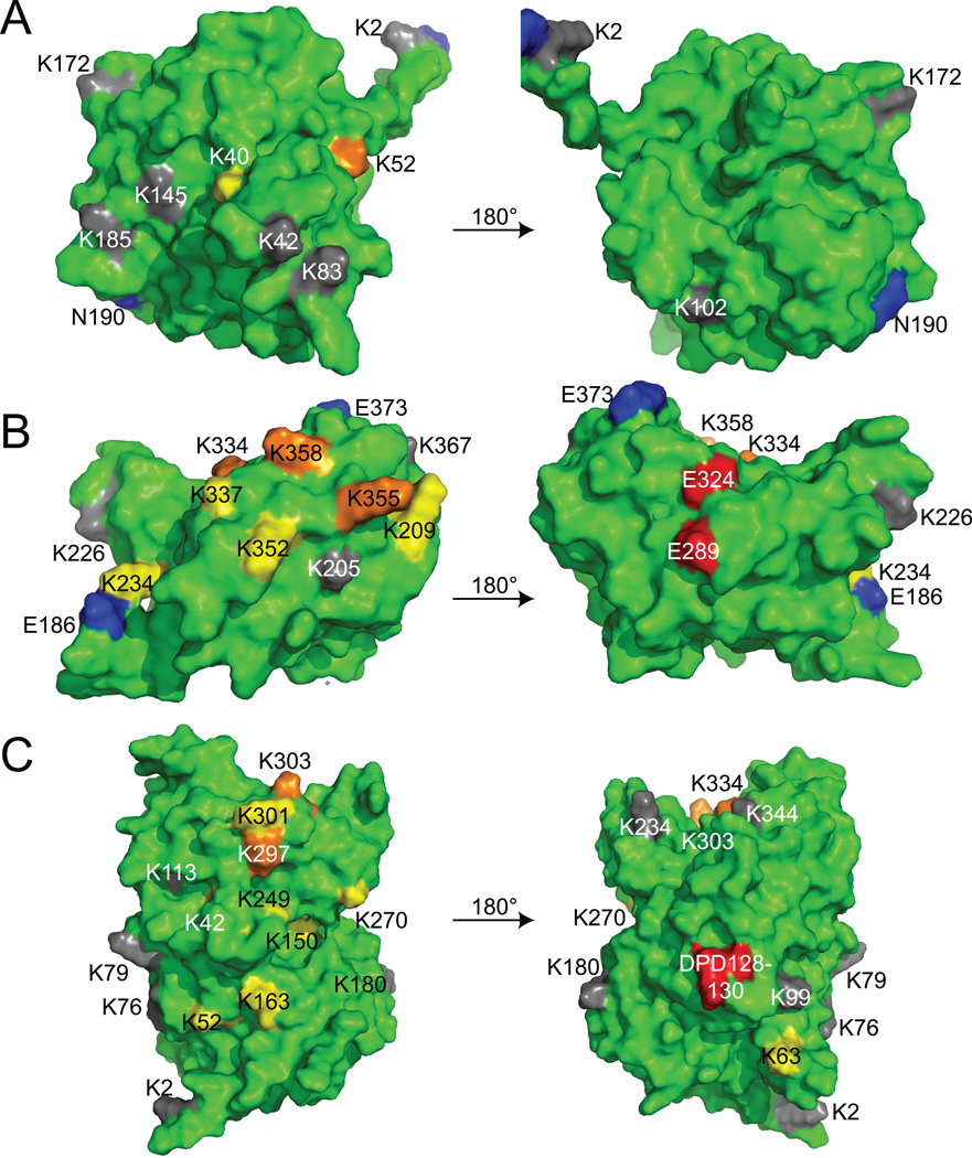
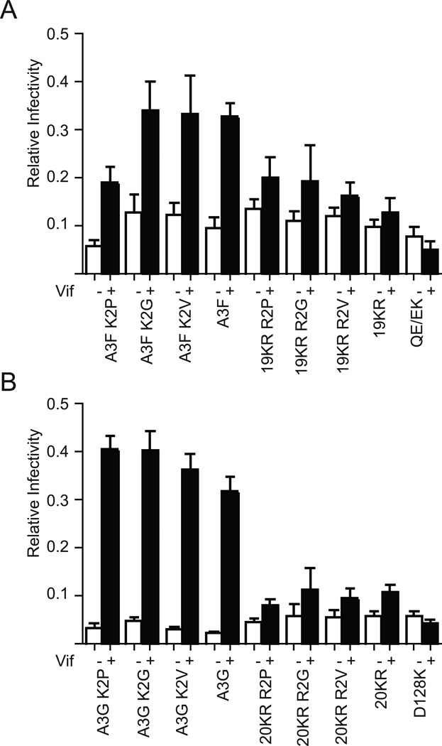
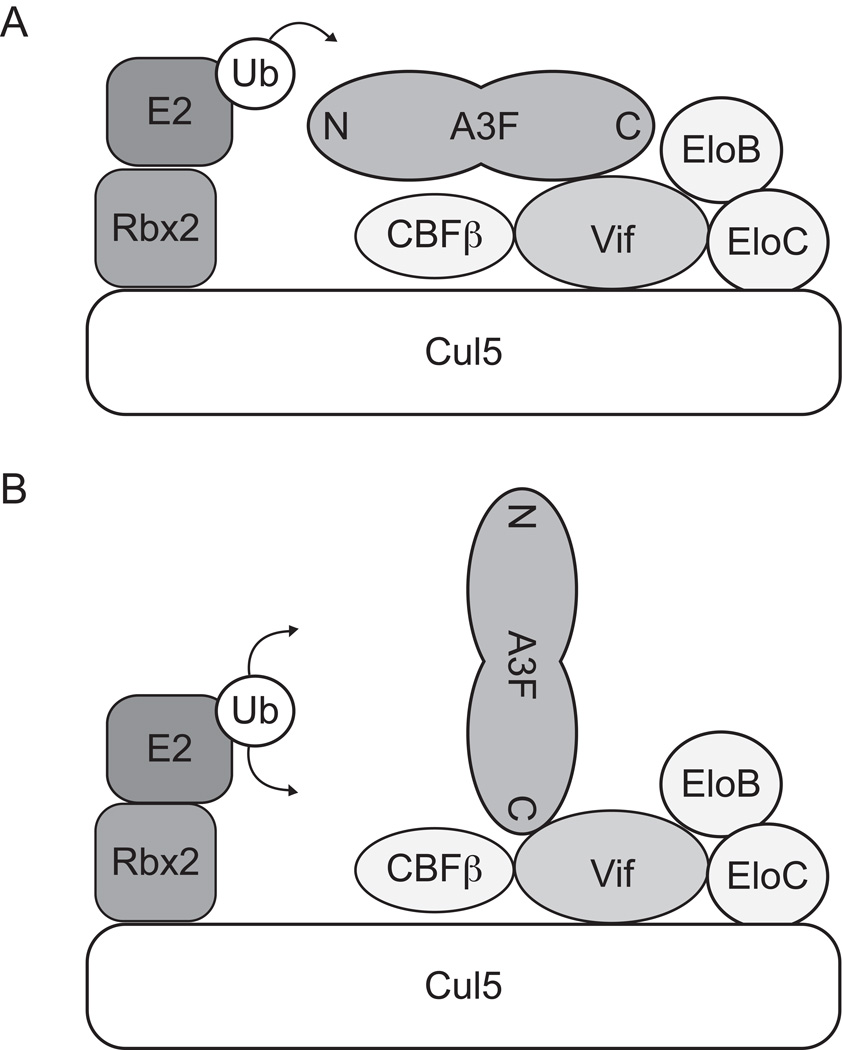
APOBEC3F (A3F) and APOBEC3G (A3G) are DNA cytosine deaminases that potently restrict human immunodeficiency virus type 1 replication when the virus is deprived of its accessory protein Vif (virion infectivity factor). Vif counteracts these restriction factors by recruiting A3F and A3G to an E3 ubiquitin (Ub) ligase complex that mediates their polyubiquitination (polyUb) and proteasomal degradation. While previous efforts have identified single amino acid residues in APOBEC3 proteins required for Vif recognition, less is known about the downstream Ub acceptor sites that are targeted. One prior report identified a cluster of polyubiquitinated residues in A3G and proposed an antiparallel model of A3G interaction with the Vif-E3 Ub ligase complex wherein Vif binding at one terminus of A3G orients the opposite terminus for polyUb [Iwatani et al. (2009). Proc. Natl. Acad. Sci. USA, 106, 19539-19544]. To test the generalizability of this model, we carried out a complete mutagenesis of the lysine residues in A3F and used a complementary, unbiased proteomic approach to identify Ub acceptor sites targeted by Vif. Our data indicate that internal lysines are the dominant Ub acceptor sites in both A3F and A3G. In contrast with the proposed antiparallel model, however, we find that the Vif-dependent polyUb of A3F and A3G can occur at multiple acceptor sites dispersed along predicted lysine-enriched surfaces of both the N- and C-terminal deaminase domains. These data suggest an alternative model for binding of APOBEC3 proteins to the Vif-E3 Ub ligase complex and diminish enthusiasm for the amenability of APOBEC3 Ub acceptor sites to therapeutic intervention.


Figures

Fig. 1
Fig. 1
The sites of functional Vif-mediated polyUb in A3F are distributed throughout the protein. (A) A schematic showing the K-to-R variants of A3F tested in Fig. 1B. (B) Single-cycle infectivity of Vif-deficient viruses produced in the presence of the indicated A3F-V5 variant −/+ transcomplementation by Vif-HA. Infectivity data represent the mean and SEM of four independent experiments; here as in all singe-cycle infectivity experiments conducted, infectivity is normalized to the infectivities of virus produced in the presence of an A3 vector control −/+ Vif. Statistics were derived by carrying out one-tailed, paired T tests comparing the infectivity of virus produced in the presence of each A3 construct −/+ Vif. ** = p < 0.05; *** = p < 0.01. Western blots showing the steady state levels of Vif-HA and A3-V5 are derived from the producer cells from one of the experiments conducted. (C) A degradation experiment in which constant amounts of A3G or mutant derivatives thereof has been cotransfected with increasing amounts of Vif. Data are representative of two independent experiments.
Fig. 2
Fig. 2
Multiple internal lysines residues in A3F are suitable substrates for functional Vif-dependent polyUb. (A–C) Single-cycle infectivity of individual single lysine revertants in an A3F-19KR background in the absence or presence of Vif where data represent the mean and SEM of three independent experiments with associated Western blots showing producer cell steady state levels of Vif-HA and A3-V5 derived from one of these experiments. Statistics represent two-way ANOVA with Dunnett’s post-test comparing the infectivity recovery of each single amino acid revertant (Vif+/Vif−) to that of the A3F-19KR control. * = p < 0.1; ** = p < 0.05; *** = p < 0.01. Controls that appear in multiple panels of this figure (e.g. A3F in A, B and C) are regraphed and reblotted for visual comparison with the mutants of each individual region; all data shown in this figure are the result of one large experiment repeated three times and presented visually as three parts.
Fig. 3
Fig. 3
Mass spectrometry indicates Ub of multiple internal lysine residues in the N- and C-termini of both A3F and A3G. (A–B). Diagrams showing Ub-modified residues in A3F (A) or A3G (B), with residues numbered at the top and the fold-enrichment of associated peptides indicated on the bottom. ++ indicates that all of the peptides detected indicating modification of the associated residues were so rare in the absence of Vif as to preclude calculation of fold-enrichment. The combination of a number with a + reflects calculation of a fold enrichment for a mixed residue in which some associated peptides permitted calculation while others were so rare in the absence of Vif as to preclude a fold-enrichment calculation. Zinc-coordination motifs of N- and C-terminal deaminase domains are indicated by boxes.
Fig. 4
Fig. 4
Lysines in the N- and C-termini of A3F and A3G cluster at distinct predicted surfaces. (A) A model of the A3F N-terminal deaminase domain is shown rotated 180° about the y-axis. (B) A model of the A3F C-terminal deaminase domain is shown rotated 180° about the y-axis. (C) A previously-described model of full-length A3G is shown rotated 180° about the y-axis . Orange indicates residues implicated by both genetic and biophysical experiments, while yellow indicates residues implicated by one or the other. A3G residues implicated by Iwatani et al. and also identified in Fig. 3B are orange in (C), while additional residues not implicated by Iwatani et al. are yellow. Lysine residues not significantly implicated by either data set are gray, while Vif interaction residues are colored red ; .
Fig. 5
Fig. 5
Changes at the A3 N-terminus do not alter Vif susceptibility. (A–B) Single-cycle infectivity experiments demonstrate the Vif sensitivity of A3F, A3G and lysine-free variants thereof to Vif when the second amino acid is altered as shown. Infectivity data represent the mean and SEM of four independent experiments.
Fig. 6
Fig. 6
An alternative model of A3 binding to the Vif-E3 ligase complex. (A) The antiparallel model previously proposed by Iwatani et al. in which binding of Vif at one deaminase domain orients an A3 protein for polyUb at its second deaminase domain . (B) An alternative, angled/perpendicular model for A3F binding to the Vif-E3 ligase complex in which binding of Vif to a lysine-poor surface orients the lysine-rich surfaces of A3F for polyUb.

References

    1. Iwatani Y, Chan DS, Liu L, Yoshii H, Shibata J, Yamamoto N, Levin JG, Gronenborn AM, Sugiura W. HIV-1 Vif-mediated ubiquitination/degradation of APOBEC3G involves four critical lysine residues in its C-terminal domain. Proc Natl Acad Sci U S A. 2009;106:19539–19544. - PMC - PubMed
    1. Chiu YL, Greene WC. The APOBEC3 cytidine deaminases: an innate defensive network opposing exogenous retroviruses and endogenous retroelements. Annu Rev Immunol. 2008;26:317–353. - PubMed
    1. Malim MH. APOBEC proteins and intrinsic resistance to HIV-1 infection. Philos Trans R Soc Lond B Biol Sci. 2009;364:675–687. - PMC - PubMed
    1. Albin JS, Harris RS. Interactions of host APOBEC3 restriction factors with HIV-1 in vivo: implications for therapeutics. Expert Reviews in Molecular Medicine. 2010;12:1–26. - PMC - PubMed
    1. Jäger S, Kim DY, Hultquist JF, Shindo K, Larue RS, Kwon E, Li M, Anderson BD, Yen L, Stanley D, Mahon C, Kane J, Franks-Skiba K, Cimermancic P, Burlingame A, Sali A, Craik CS, Harris RS, Gross JD, Krogan NJ. Vif hijacks CBFβ to degrade APOBEC3G and promote HIV-1 infection. Nature. 2011;481:371–375. - PMC - PubMed

Publication types

MeSH terms