A systematic approach to the diagnosis of suspected central nervous system lymphoma
- PMID: 23319132
- PMCID: PMC4135394
- DOI: 10.1001/jamaneurol.2013.606
A systematic approach to the diagnosis of suspected central nervous system lymphoma
Abstract
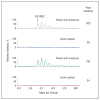
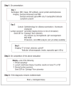
Central nervous system (CNS) lymphoma can present a diagnostic challenge. Currently, there is no consensus regarding what presurgical evaluation is warranted or how to proceed when lesions are not surgically accessible. We conducted a review of the literature on CNS lymphoma diagnosis (1966 to October 2011) to determine whether a common diagnostic algorithm can be generated. We extracted data regarding the usefulness of brain and body imaging, serum and cerebrospinal fluid (CSF) studies, ophthalmologic examination, and tissue biopsy in the diagnosis of CNS lymphoma. Contrast enhancement on imaging is highly sensitive at the time of diagnosis: 98.9% in immunocompetent lymphoma and 96.1% in human immunodeficiency virus-related CNS lymphoma. The sensitivity of CSF cytology is low (2%-32%) but increases when combined with flow cytometry. Cerebrospinal fluid lactate dehydrogenase isozyme 5, β2-microglobulin, and immunoglobulin heavy chain rearrangement studies have improved sensitivity over CSF cytology (58%-85%) but have only moderate specificity (85%). New techniques of proteomics and microRNA analysis have more than 95% specificity in the diagnosis of CNS lymphoma. Positive CSF cytology, vitreous biopsy, or brain/leptomeningeal biopsy remain the current standard for diagnosis. A combined stepwise systematic approach outlined here may facilitate an expeditious, comprehensive presurgical evaluation for cases of suspected CNS lymphoma.
Conflict of interest statement
Figures


References
-
- Bühring U, Herrlinger U, Krings T, Thiex R, Weller M, Küker W. MRI features of primary central nervous system lymphomas at presentation. Neurology. 2001;57(3):393–396. - PubMed
-
- Coulon A, Lafitte F, Hoang-Xuan K, et al. Radiographic findings in 37 cases of primary CNS lymphoma in immunocompetent patients. Eur Radiol. 2002;12 (2):329–340. - PubMed
-
- Jellinger KA, Paulus W. Primary central nervous system lymphomas—an update. J Cancer Res Clin Oncol. 1992;119(1):7–27. - PubMed
-
- Lachance DH, O’Neill BP, Macdonald DR, et al. Primary leptomeningeal lymphoma: report of 9 cases, diagnosis with immunocytochemical analysis, and review of the literature. Neurology. 1991;41(1):95–100. - PubMed
-
- Fischer L, Martus P, Weller M, et al. Meningeal dissemination in primary CNS lymphoma: prospective evaluation of 282 patients. Neurology. 2008;71(14):1102–1108. - PubMed
Publication types
MeSH terms
Substances
Grants and funding
LinkOut - more resources
Full Text Sources
Other Literature Sources
Medical

