

Characterization of proteolytic activities during intestinal regeneration of the sea cucumber, Holothuria glaberrima
- PMID: 23319344
- PMCID: PMC4068352
- DOI: 10.1387/ijdb.113473cp
Characterization of proteolytic activities during intestinal regeneration of the sea cucumber, Holothuria glaberrima
Abstract
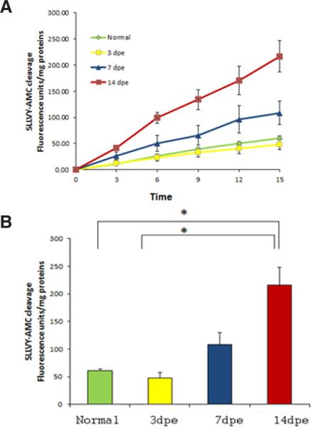
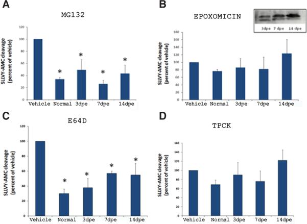
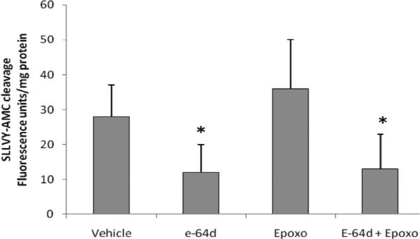
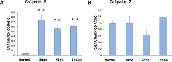
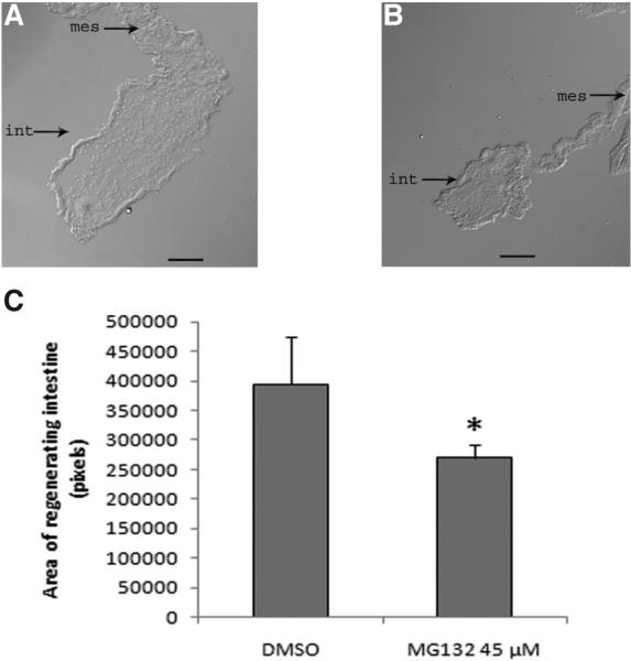
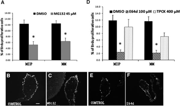
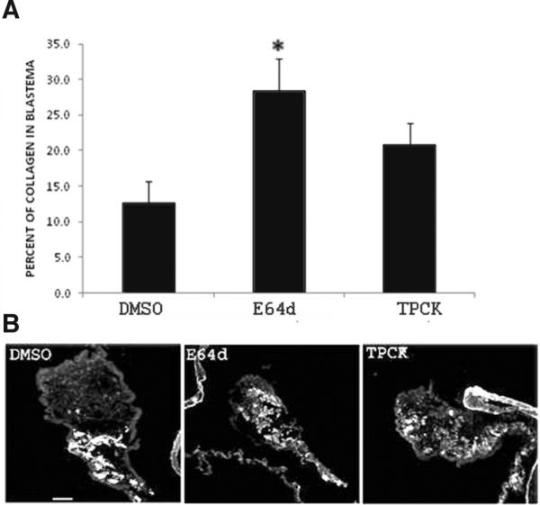
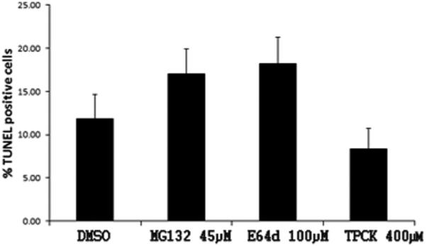
Proteolysis carried out by different proteases control cellular processes during development and regeneration. Here we investigated the function of the proteasome and other proteases in the process of intestinal regeneration using as a model the sea cucumber Holothuria glaberrima. This echinoderm possesses the ability to regenerate its viscera after a process of evisceration. Enzymatic activity assays showed that intestinal extracts at different stages of regeneration possessed chymotrypsin-like activity. This activity was inhibited by i) MG132, a reversible inhibitor of chymotrypsin and peptidylglutamyl peptidase hydrolase (PGPH) activities of the proteasome, ii) E64d, a permeable inhibitor of cysteine proteases and iii) TPCK, a serine chymotrypsin inhibitor, but not by epoxomicin, an irreversible and potent inhibitor of all enzymatic activities of the proteasome. To elucidate the role which these proteases might play during intestinal regeneration, we carried out in vivo experiments injecting MG132, E64d and TPCK into regenerating animals. The results showed effects on the size of the regenerating intestine, cell proliferation and collagen degradation. These findings suggest that proteolysis by several proteases is important in the regulation of intestinal regeneration in H. glaberrima.
Figures
Similar articles
-
Insights into intestinal regeneration signaling mechanisms.Dev Biol. 2020 Feb 1;458(1):12-31. doi: 10.1016/j.ydbio.2019.10.005. Epub 2019 Oct 9. Dev Biol. 2020. PMID: 31605680 Free PMC article.
-
Spherulocytes in the echinoderm Holothuria glaberrima and their involvement in intestinal regeneration.Dev Dyn. 2006 Dec;235(12):3259-67. doi: 10.1002/dvdy.20983. Dev Dyn. 2006. PMID: 17061269
-
Ubiquitin-proteasome system components are upregulated during intestinal regeneration.Genesis. 2012 Apr;50(4):350-65. doi: 10.1002/dvg.20803. Epub 2012 Jan 6. Genesis. 2012. PMID: 21913312 Free PMC article.
-
Holothurians as a Model System to Study Regeneration.Results Probl Cell Differ. 2018;65:255-283. doi: 10.1007/978-3-319-92486-1_13. Results Probl Cell Differ. 2018. PMID: 30083924 Review.
-
A roadmap for intestinal regeneration.Int J Dev Biol. 2021;65(4-5-6):427-437. doi: 10.1387/ijdb.200227dq. Int J Dev Biol. 2021. PMID: 32930367 Free PMC article. Review.
Cited by
-
Matrix Metalloproteinases and Tissue Inhibitors of Metalloproteinases in Echinoderms: Structure and Possible Functions.Cells. 2021 Sep 6;10(9):2331. doi: 10.3390/cells10092331. Cells. 2021. PMID: 34571980 Free PMC article. Review.
-
Gut-spilling in chordates: evisceration in the tropical ascidian Polycarpa mytiligera.Sci Rep. 2015 Apr 16;5:9614. doi: 10.1038/srep09614. Sci Rep. 2015. PMID: 25880620 Free PMC article.
-
The Stress Response of the Holothurian Central Nervous System: A Transcriptomic Analysis.Int J Mol Sci. 2022 Nov 2;23(21):13393. doi: 10.3390/ijms232113393. Int J Mol Sci. 2022. PMID: 36362181 Free PMC article.
-
Insights into intestinal regeneration signaling mechanisms.Dev Biol. 2020 Feb 1;458(1):12-31. doi: 10.1016/j.ydbio.2019.10.005. Epub 2019 Oct 9. Dev Biol. 2020. PMID: 31605680 Free PMC article.
-
Antibiotics Modulate Intestinal Regeneration.Biology (Basel). 2021 Mar 19;10(3):236. doi: 10.3390/biology10030236. Biology (Basel). 2021. PMID: 33808600 Free PMC article.
References
-
- AWASTHI N, WAGNER BJ. Suppression of human lens epithelial cell proliferation by proteasome inhibition, a potential defense against posterior capsular opacification. Invest Ophthalmol Vis Sci. 2006;47:4482–4489. - PubMed
-
- BAZZARO M, LEE MK, ZOSO A, STIRLING WL, SANTILLAN A, SHIH IEM, RODEN RB. Ubiquitin proteasome system stress sensitizes ovarian cancer to proteasome inhibitor-induced apoptosis. Cancer Res. 2006;66:3754–3763. - PubMed
-
- BOWERMAN B, KURZ T. Degrade to create: developmental requirements for ubiquitin-mediated proteolysis during early C. elegans embryogenesis. Development. 2006;133:773–784. - PubMed
-
- BRADFORD M. Rapid and sensitive methods for the quantification of microgram quantities of protein utilizing the principle of protein dye binding. Anal Biochem. 1976;72:248–254. - PubMed
MeSH terms
Substances
Grants and funding
LinkOut - more resources
Full Text Sources
Miscellaneous
