. 2013 Apr;81(4):1040-51.
doi: 10.1128/IAI.01097-12. Epub 2013 Jan 14.

Diverse genetic regulon of the virulence-associated transcriptional regulator MucR in Brucella abortus 2308

Affiliations

Diverse genetic regulon of the virulence-associated transcriptional regulator MucR in Brucella abortus 2308

Clayton C Caswell et al. Infect Immun. 2013 Apr.

Abstract

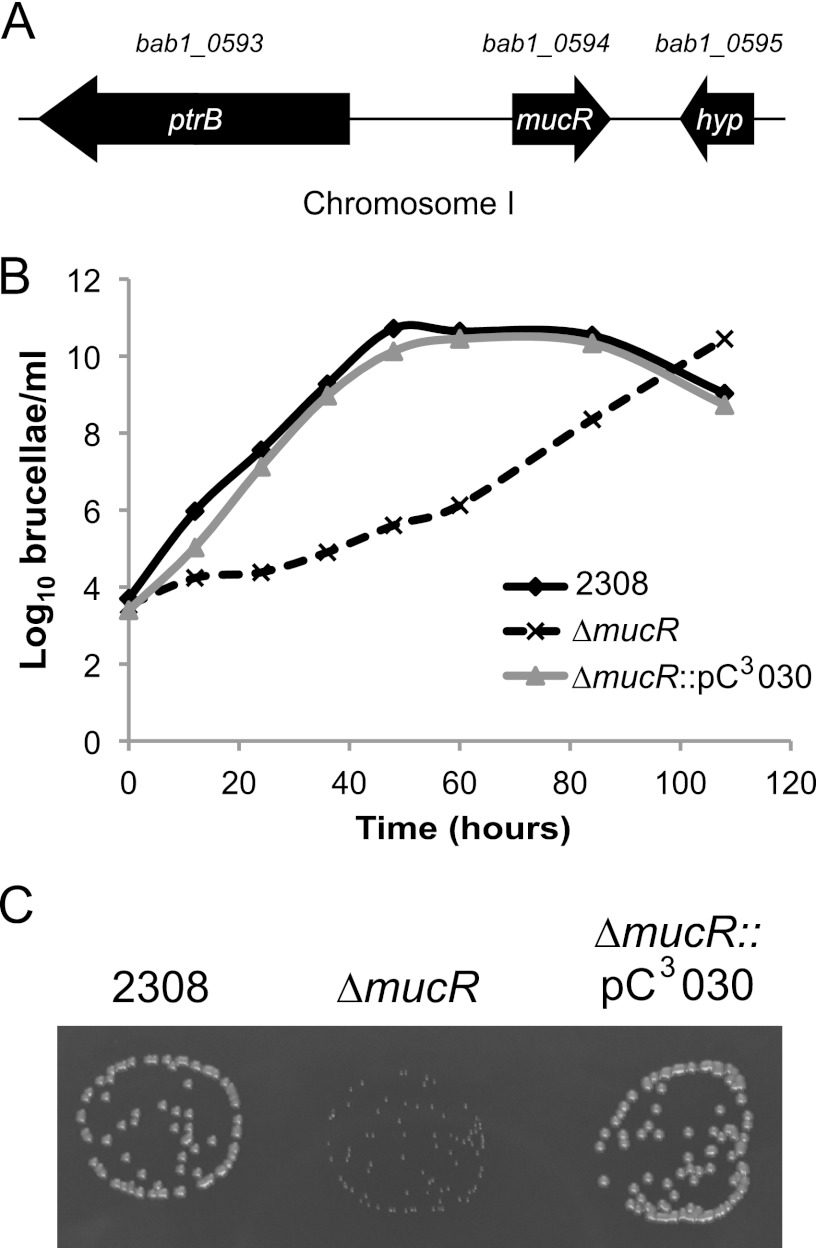
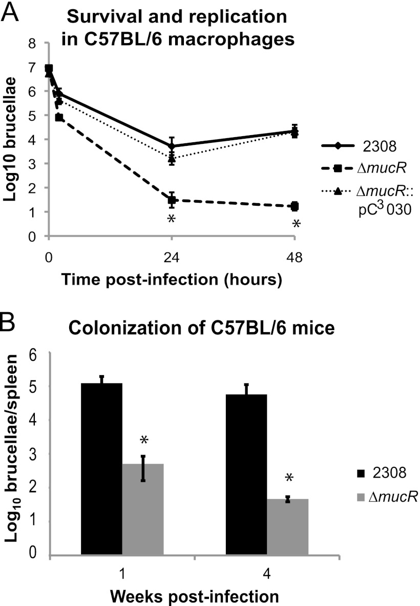
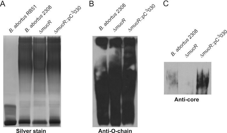
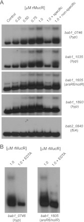
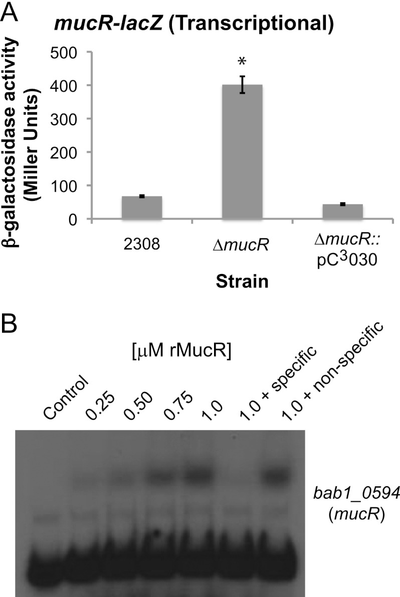
The Ros-type regulator MucR is one of the few transcriptional regulators that have been linked to virulence in Brucella. Here, we show that a Brucella abortus in-frame mucR deletion strain exhibits a pronounced growth defect during in vitro cultivation and, more importantly, that the mucR mutant is attenuated in cultured macrophages and in mice. The genetic basis for the attenuation of Brucella mucR mutants has not been defined previously, but in the present study the genes regulated by MucR in B. abortus have been elucidated using microarray analysis and real-time reverse transcription-PCR (RT-PCR). In B. abortus 2308, MucR regulates a wide variety of genes whose products may function in establishing and maintaining cell envelope integrity, polysaccharide biosynthesis, iron homeostasis, genome plasticity, and transcriptional regulation. Particularly notable among the MucR-regulated genes identified is arsR6 (nolR), which encodes a transcriptional regulator previously linked to virulence in Brucella melitensis 16 M. Importantly, electrophoretic mobility shift assays (EMSAs) determined that a recombinant MucR protein binds directly to the promoter regions of several genes repressed by MucR (including arsR6 [nolR]), and in Brucella, as in other alphaproteobacteria, MucR binds to its own promoter to repress expression of the gene that encodes it. Overall, these studies have uncovered the diverse genetic regulon of MucR in Brucella, and in doing so this work has begun to define the MucR-controlled genetic circuitry whose misregulation contributes to the virulence defect of Brucella mucR mutants.


Figures

Fig 1
Fig 1
Deletion of mucR in Brucella abortus 2308 results in a growth deficiency. (A) Genetic organization of the mucR locus. The mucR gene is found on chromosome I and is designated bab1_0594 in the B. abortus 2308 genome sequence. mucR is flanked by ptrB (bab1_0593), encoding oligopeptidase B, and by a small, hypothetical-protein-encoding gene. (B) Growth curves of Brucella abortus strains in a rich medium. B. abortus 2308, the isogenic mucR mutant (ΔmucR), and the complemented mucR mutant (ΔmucR::pC3030) strains were grown in brucella broth, and the growth of each culture was monitored by plating serial dilutions on SBA to determine the number of CFU/ml. (C) Photograph of Brucella abortus colonies on blood agar after 60 h of growth.
Fig 2
Fig 2
A Brucella abortus mucR mutant is significantly attenuated in the mouse model. (A) Brucella abortus 2308, the mucR isogenic mutant strain (ΔmucR), and the complemented mucR mutant strain (ΔmucR::pC3030) were tested for survival and replication in primary murine macrophages. Macrophages were isolated from the peritoneal cavities of C57BL/6 mice, and the cells were infected with opsonized Brucella strains. Extracellular bacteria were killed by gentamicin treatment. At the indicated times, the macrophages were lysed, and serial dilutions were plated on blood agar to determine the number of viable intracellular brucellae. The asterisks indicate significant differences in survival and replication between parental strain 2308 and the mucR mutant strain (t test; P < 0.05). (B) Spleen colonization of mice experimentally infected with Brucella strains. C57BL/6 mice were infected intraperitoneally with approximately 5 × 10⁴ CFU of B. abortus 2308 or the mucR mutant. At 1 and 4 weeks postinfection, the mice were sacrificed, and the serial dilutions of spleen homogenates were plated on blood agar to determine the number of brucellae colonizing the spleen. The asterisks indicate significant differences in spleen colonization between parental strain 2308 and the mucR mutant strain (t test; P < 0.05).
Fig 3
Fig 3
The lipopolysaccharide (LPS) core is modified in the B. abortus mucR mutant. Brucella strains were grown in brucella broth to stationary phase, and LPS was isolated by hot phenol extraction as described previously (33). Purified LPS from the specified Brucella strains was separated on an SDS-PAGE gel containing 3 M urea, and the LPS was then visualized by silver staining (A), Western blot analysis with an anti-O-chain monoclonal antibody (B), and Western blot analysis with an anticore monoclonal antibody (C).
Fig 4
Fig 4
The Brucella abortus mucR mutant exhibits a cell envelope defect and an iron acquisition defect in vitro. (A) Brucella strains were tested in a disk diffusion assay for their comparative susceptibilities to polymyxin B and sodium dodecyl sulfate (SDS). The results are plotted as the average diameters (±standard deviations) of the zones of inhibition around disks containing the indicated stress agents, and the results are from single experiment that was repeated in triplicate. Asterisks denote statistically significant differences (t test; P < 0.05) between a mutant strains and parental strain 2308. (B) Brucella abortus 2308, the mucR mutant strain (ΔmucR), and the complemented mucR mutant strain (ΔmucR::pC3030) were tested for their ability to utilize ferric (Fe3+) or ferrous (Fe2+) iron in a disk diffusion assay. Iron sources (50 mM FeCl3 or 50 mM ferrous ammonium sulfate) were applied to sterile Whatman paper disks on plates containing the chelator EDDHA, and following incubation at 37°C for 72 h, the diameter (in millimeters) of the zone of bacterial growth around each filter disk was measured. The data are represented as the average and standard deviation of the zones of growth recorded for each strain in triplicate, and asterisks denote statistically significant differences (t test; P < 0.05) between the mucR mutant strain and parental strain 2308.
Fig 5
Fig 5
Brucella abortus MucR binds directly to the promoter regions of MucR-regulated genes. (A) Recombinant MucR (rMucR) protein was tested for binding to the promoter regions of MucR-regulated genes using an electrophoretic mobility shift assay (EMSA). Increasing concentrations of rMucR were incubated with radiolabeled DNA corresponding to the promoter regions of several different genes, and in some binding reactions, unlabeled specific and nonspecific competitor DNA fragments were included as controls. The binding reactions were resolved in 6% native polyacrylamide gels, and the reaction products were visualized by autoradiography. (B) Effect of EDTA on the binding of MucR to the promoter regions of MucR-regulated genes. EMSAs were performed as described for panel A, but here, 0.5 mM EDTA was included in some binding reactions. hyp, hypothetical.
Fig 6
Fig 6
Autoregulation of mucR expression in Brucella abortus 2308. (A) β-Galactosidase activity produced by a mucR-lacZ transcriptional fusion. The activity of a mucR-lacZ transcriptional fusion was tested in B. abortus 2308, the mucR isogenic mutant strain (ΔmucR), and the mucR mutant complemented in transmucR::pC3030). β-Galactosidase activity is shown as average Miller units ± standard deviations, and the results shown are from a single experiment that was repeated in triplicate. The asterisk indicates a significant difference in β-galactosidase activity between the parental strain 2308 and the mucR mutant strain (t test; P < 0.05). (B) Recombinant MucR (rMucR) protein was tested for binding to the mucR promoter region using an EMSA. Similar to the EMSA experiments in Fig. 3A, increasing concentrations of rMucR were incubated with radiolabeled DNA corresponding to the mucR promoter region, and in some binding reactions, unlabeled specific or nonspecific competitor DNA fragments were included as controls. The binding reactions were resolved in 6% native polyacrylamide gels and visualized by autoradiography.

References

    1. Pappas G, Papadimitriou P, Akritidis N, Christou L, Tsianos EV. 2006. The new global map of human brucellosis. Lancet Infect. Dis. 6:91–99 - PubMed
    1. Pappas G, Akritidis N, Bosilkovski M, Tsianos E. 2005. Brucellosis. N. Engl. J. Med. 352:2325–2336 - PubMed
    1. Moreno E, Stackebrandt E, Dorsch M, Wolters J, Busch M, Mayer H. 1990. Brucella abortus 16S rRNA and lipid A reveal a phylogenetic relationship with members of the alpha-2 subdivision of the class Proteobacteria. J. Bacteriol. 172:3569–3576 - PMC - PubMed
    1. Batut J, Andersson SG, O'Callaghan D. 2004. The evolution of chronic infection strategies in the alpha-proteobacteria. Nat. Rev. Microbiol. 2:933–945 - PubMed
    1. Close TJ, Tait RC, Kado CI. 1985. Regulation of Ti plasmid virulence genes by a chromosomal locus of Agrobacterium tumefaciens. J. Bacteriol. 164:774–781 - PMC - PubMed

Publication types

MeSH terms

Associated data