. 2013 Mar 22;288(12):8391-8404.
doi: 10.1074/jbc.M112.408179. Epub 2013 Jan 14.

Identification and cytoprotective function of a novel nestin isoform, Nes-S, in dorsal root ganglia neurons

Affiliations

Identification and cytoprotective function of a novel nestin isoform, Nes-S, in dorsal root ganglia neurons

Peng-Han Su et al. J Biol Chem. .

Abstract

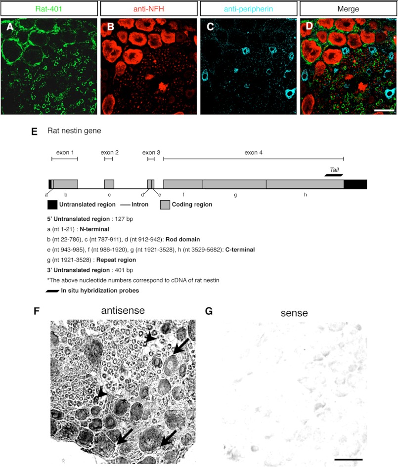
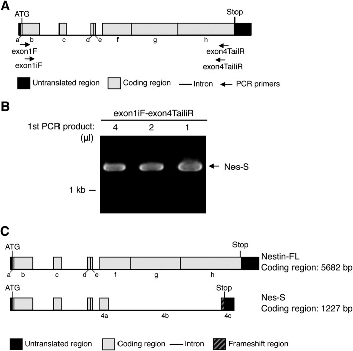
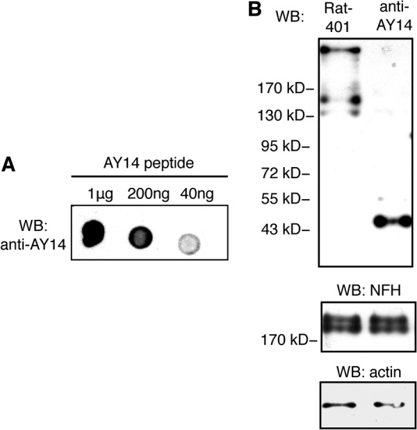
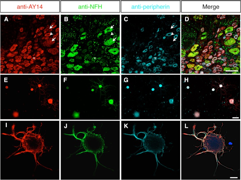
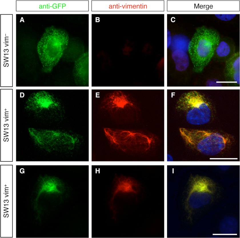
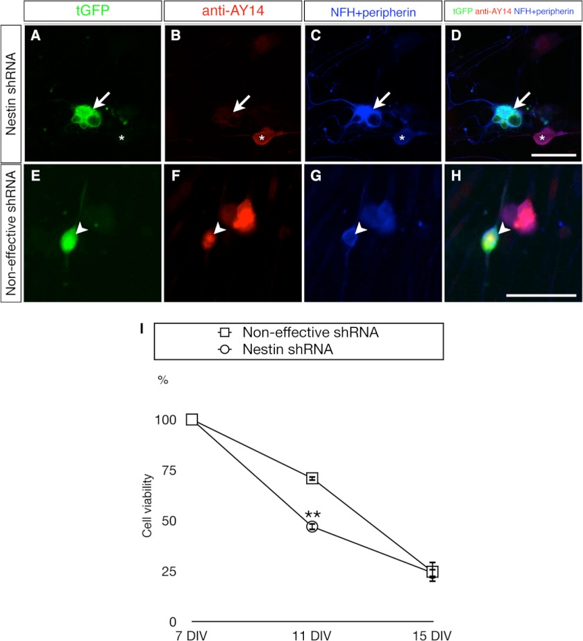
In this study, the first nestin isoform, Nes-S, was identified in neurons of dorsal root ganglia (DRG) of adult rats. Nes-S cannot form filaments by itself in cytoplasmic intermediate filament-free SW13 cells. Instead, it co-assembles into filaments with vimentin when transfected into vimentin(+) SW13 cells, and with peripherin and neurofilament proteins when transfected into N2a cells. In primary DRG neurons, endogenous Nes-S co-assembles with peripherin and neurofilament proteins. The expression of Nes-S first appears in DRG at postnatal day 5 and persists to adulthood. Among the adult tissues we examined, the expression of Nes-S is restricted to the sensory and motor neurons. Finally, exogenous Nes-S enhances viability when transfected into N2a cells, and knockdown of endogenous Nes-S impairs the survival of DRG neurons in primary cultures. Taken together, Nes-S is a new neuronal intermediate filament protein that exerts a cytoprotective function in mature sensory and motor neurons.


Figures

FIGURE 1.
FIGURE 1.
The nestin transcript variant in DRG neurons of adult rats. A–D, triple labeling of DRG sections of adult rats with Rat-401 (A), anti-NFH (B), and anti-peripherin (C). Although satellite cells and Schwann cells were Rat-401+, all of the DRG neurons were Rat-401. D shows merged images. E, schematic diagram of the gene structure of rat nestin and the location of the in situ probe Tail (nt 5178–5682). The coding regions of rat nestin gene were designated as regions from a to h. F and G, in situ hybridization of DRG sections of adult rats with the antisense probe Tail. Specific signals were identified in neurons, satellite cells (arrows), and cytoplasm of Schwann cells surrounding the neuronal processes (arrowheads) (F). The sense probe of Tail was used as the negative control (G). Scale bar: 50 μm.
FIGURE 2.
FIGURE 2.
Identification of the Nes-S transcript variant in DRG neurons by RT-PCR. A, schematic diagram showing locations of the primers used to identify the complete cDNA of Nes-S. The coding regions of rat nestin gene were designated as regions from a to h. B, nested RT-PCR of DRG neuron RNA. The nested RT-PCR was performed using the primer set exon1F-exon4TailR and followed with the primer set exon1iF-exon4TailiR. A specific 1.3-kb band was produced. C, upper panel, schematic diagram of the nestin-FL. Lower panel, schematic diagram of the splicing pattern of the 1.3-kb band, which was named as Nes-S.
FIGURE 3.
FIGURE 3.
Identification of Nes-S protein by immunoblotting of IF-enriched preparations of DRG with anti-AY14. A, dot blotting of the synthesized AY14 peptide with anti-AY14. The specificity of anti-AY14 was confirmed. WB, Western blotting. B, immunoblotting of IF-enriched preparations of DRG of adult rats with Rat-401 and anti-AY14. Although Rat-401 recognized the band corresponding to nestin-FL, anti-AY14 specifically recognized the 45.9-kDa band of Nes-S protein. Immunoblotting with anti-actin and immunoblotting with anti-NFH were used as loading controls.
FIGURE 4.
FIGURE 4.
Immunofluorescence localization of Nes-S protein in DRG neurons of adult rats. A–D, triple labeling of DRG tissue sections of adult rats with anti-AY14 (A), anti-NFH (B), and anti-peripherin (C). All three populations of the DRG neurons, including NFH+/peripherin large neurons (arrows), NFH+/peripherin+ medium neurons (asterisks), and NFH/peripherin+ small neurons (arrowheads), were AY14+. D shows merged images. Scale bar: 50 μm. E–H, triple labeling of primary DRG neurons of adult rats with anti-AY14 (E), anti-NFH (F), and anti-peripherin (G). Anti-AY14-IR was observed in all of the neurons. H shows merged images. Scale bar: 50 μm. I–L, triple labeling of primary DRG neurons of adult rats with anti-AY14 (I), anti-NFH (J), and anti-peripherin (K) by high magnification confocal microscopy. The IFs formed a cage-like cytostructure at the periphery of neurons and extended into the neurites. L shows merged images. Scale bar: 15 μm.
FIGURE 5.
FIGURE 5.
Filament assembly properties of Nes-S. A–I, double labeling of pEGFP-NestS-transfected SW13 cells with anti-GFP (A, D, and G) and anti-vimentin (B, E, and H). Nes-S presented a dispersed pattern in vimentin-free SW13 cells (A–C). The majority of the Nes-S protein co-localized with vimentin IF in the few SW13 cells that expressed vimentin (D–I). Scale bar: 15 μm.
FIGURE 6.
FIGURE 6.
Postnatal expression and tissue distribution of Nes-S. A, immunoblotting of IF-enriched preparations of DRG from rats of different postnatal days, as well as the adult rats, with anti-AY14 and Rat-401. Nes-S expression was not detected until P5, and its intensity reached the highest point at adult. B, immunoblotting of IF-enriched preparations of various tissues of adult rats with anti-AY14 (left) and Rat-401 (right). Nes-S was expressed in DRG, TriG, SCG, and thoracic spinal cord, whereas a smaller amount of Nes-S was detected in sciatic nerve. C–V, triple labeling of TriG, SCG, spinal cord, and sciatic nerve of adult rats with anti-AY14, anti-NFH, and anti-peripherin. In TriG (C–F) and SCG (G–J), all neurons were AY14+. In spinal cord, the preganglionic sympathetic neurons (K–N), and motor neurons (O–R) were AY14+. In sciatic nerve, a trace amount of anti-AY14-IR was detected in the axoplasm of neurites (S–V). IML, intermediate lateral horn; VH, ventral horn. Scale bar: 50 μm.
FIGURE 7.
FIGURE 7.
The cytoprotective effect of Nes-S in N2a neuroblastoma cells. A, promitotic function of Nes-S, as revealed by time-course MTT assays of N2a/NestS and N2a/vector cells. These cells were passaged 12 h before being cultured in either serum-containing (S) or serum-free (SF) medium. The time of medium change was designated as 0 h. The cell number at each time point was normalized to that of N2a/vector cells cultured in serum-containing medium at 0 h. The N2a/NestS cells showed an ∼2-fold proliferation rate when cultured in serum-containing medium, as compared with N2a/vector cells (n = 4, **, p < 0.01, two-tailed t test). Both cell types did not proliferate in serum-free medium. Error bars indicate S.E. B, prosurvival function of Nes-S, as determined by MTT assays of N2a/NestS and N2a/vector cells, as well as untransfected N2a cells (N2a/wt). The survival rates of N2a/NestS cells upon 250 and 325 μm H2O2 treatments (95.8 ± 5.3 and 64.5 ± 6.0%, respectively) were significantly higher than those of N2a/vector cells (56.7 ± 4.2 and 23.2 ± 5.9%, respectively) and N2a/wt cells (44.9 ± 1.7 and 19.4 ± 6.8%, respectively). There was no significant difference in survival rates between N2a/vector cells and N2a/wt cells (n = 4, **, p < 0.01, ***, p < 0.001, two-tailed t test). Error bars indicate S.E. C, caspase-3 activation of N2a/NestS and N2a/vector cells. N2a cells transiently expressing either pEGFP (N2a/vector) or pEGFP-NestS (N2a/NestS) were subjected to serum starvation for 12 h and treated with 250 μm H2O2, and their whole cell extractions were collected at various time points. The results showed that expression of EGFP-tagged Nes-S protein attenuated the activation of caspase-3. D, co-immunoprecipitation of Cdk5 and EGFP-tagged Nes-S protein. N2a cells transiently expressing either pEGFP (N2a/vector) or pEGFP-NestS (N2a/NestS) were subjected to serum starvation for 24 h before harvesting. The lysates were immunoprecipitated with anti-GFP and immunoblotted (WB) with anti-Cdk5. Total lysates were also immunoblotted with anti-Cdk5 as a positive control. Cdk5 was detected in the immunoprecipitates (IP) from N2a/NestS lysates, but not in that from N2a/vector. Arrowhead, Cdk5. Asterisks, IgG bands. E, effect of Thr-316 point mutation on cytoprotective function of Nes-S. The viability of N2a/NestS-T316A cells was significantly higher than that of N2a/NestS cells (87.5 ± 2.3 and 64.5 ± 6.0%, respectively). The viability of N2a/NestS-T316D cells was significantly lower than that of N2a/NestS cells (36.5 ± 6.4 and 64.5 ± 6.0%, respectively). No significant difference in survival rates was observed among N2a/NestS-T316D, N2a/vector, and N2a/wt cells (n = 4, *, p < 0.05, **, p < 0.01, two-tailed t test). Error bars indicate S.E. UT, untreated.
FIGURE 8.
FIGURE 8.
Impaired cell survival in primary DRG neurons of adult rats upon RNAi knockdown of Nes-S. A–D, reduced expression of Nes-S by nestin shRNA expression vector. The transfected neurons in the 7 DIV cultures were recognized by their tGFP expression (A) and morphological features, i.e. a rounded soma and elongated neurites. Triple labeling with anti-AY14 (B) and anti-NFH and anti-peripherin (C, their IRs were recorded in the same channel) showed that anti-AY14-IR intensity was reduced in DRG neurons expressing the tGFP reporter gene of shRNA vector (arrow). The anti-AY14-IR remained in the untransfected neurons (asterisk). D shows merged images. E–H, expression level of Nes-S was not altered by noneffective shRNA control vector. The tGFP+ neurons (E, arrowhead) present AY14-IR (F) as well as NFH- and peripherin-IR (G). H shows merged images. Scale bar: 50 μm. I, reduced cell survival rate in Nes-S knockdown DRG neurons during prolonged culture. Live cell images of tGFP+ neurons were taken by an inverted fluorescence microscope at 7, 11, and 15 DIV. At each day, the number of tGFP-expressing neurons was counted. The viability of Nes-S knockdown neurons was significantly decreased at 11 DIV (n = 3, **, p < 0.01, two-tailed t test). Neurons transfected with the noneffective shRNA vector were used as control. The survival rate of each DIV was calculated using the number of tGFP+ cells of the same plate at 7 DIV as the denominator. For the number of neurons of both transfected groups at each day, see supplemental Table S2. Error bars indicate S.E.

Similar articles

Cited by

References

    1. Lendahl U., Zimmerman L. B., McKay R. D. (1990) CNS stem cells express a new class of intermediate filament protein. Cell 60, 585–595 - PubMed
    1. Yang H. Y., Lieska N., Goldman A. E., Goldman R. D. (1992) Colchicine-sensitive and colchicine-insensitive intermediate filament systems distinguished by a new intermediate filament-associated protein, IFAP-70/280 kD. Cell Motil. Cytoskeleton 22, 185–199 - PubMed
    1. Steinert P. M., Chou Y. H., Prahlad V., Parry D. A., Marekov L. N., Wu K. C., Jang S. I., Goldman R. D. (1999) A high molecular weight intermediate filament-associated protein in BHK-21 cells is nestin, a type VI intermediate filament protein: limited co-assembly in vitro to form heteropolymers with type III vimentin and type IV α-internexin. J. Biol. Chem. 274, 9881–9890 - PubMed
    1. Chou Y. H., Khuon S., Herrmann H., Goldman R. D. (2003) Nestin promotes the phosphorylation-dependent disassembly of vimentin intermediate filaments during mitosis. Mol. Biol. Cell 14, 1468–1478 - PMC - PubMed
    1. Guérette D., Khan P. A., Savard P. E., Vincent M. (2007) Molecular evolution of type VI intermediate filament proteins. BMC Evol. Biol. 7, 164. - PMC - PubMed

Publication types

MeSH terms

LinkOut - more resources