. 2013 Feb 22;288(8):5886-95.
doi: 10.1074/jbc.M112.434852. Epub 2013 Jan 14.

Analysis of Ca2+ signaling motifs that regulate proton signaling through the Na+/H+ exchanger NHX-7 during a rhythmic behavior in Caenorhabditis elegans

Affiliations

Analysis of Ca2+ signaling motifs that regulate proton signaling through the Na+/H+ exchanger NHX-7 during a rhythmic behavior in Caenorhabditis elegans

Erik Allman et al. J Biol Chem. .

Abstract

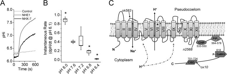
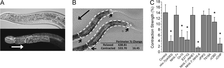
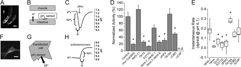
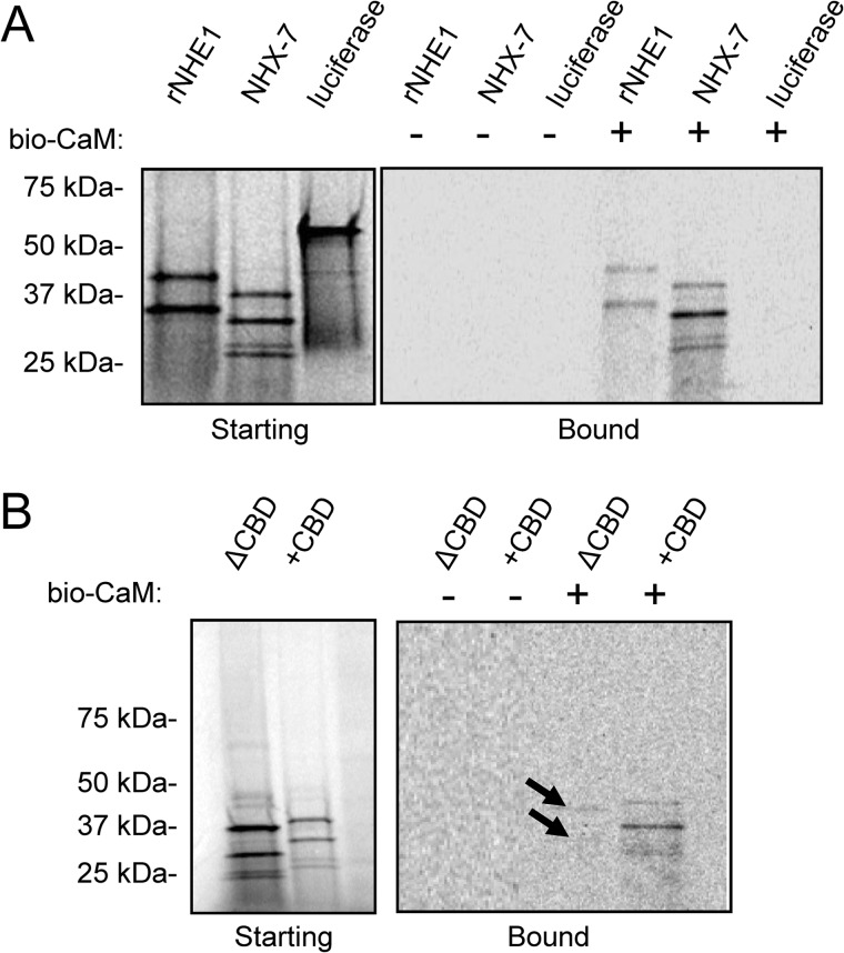
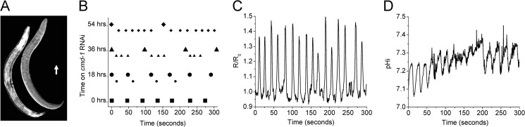
Membrane proton transporters contribute to pH homeostasis but have also been shown to transmit information between cells in close proximity through regulated proton secretion. For example, the nematode intestinal Na(+)/H(+) exchanger NHX-7 causes adjacent muscle cells to contract by transiently acidifying the extracellular space between the intestine and muscle. NHX-7 operates during a Ca(2+)-dependent rhythmic behavior and contains several conserved motifs for regulation by Ca(2+) input, including motifs for calmodulin and phosphatidylinositol 4,5-bisphosphate binding, protein kinase C- and calmodulin-dependent protein kinase type II phosphorylation, and a binding site for calcineurin homologous protein. Here, we tested the idea that Ca(2+) input differentiates proton signaling from pH housekeeping activity. Each of these motifs was mutated, and their contribution to NHX-7 function was assessed. These functions included pH recovery from acidification in cells in culture expressing recombinant NHX-7, extracellular acidification measured during behavior in live moving worms, and muscle contraction strength as a result of this acidification. Our data suggest that multiple levels of Ca(2+) input regulate NHX-7, whose transport capacity normally exceeds the minimum necessary to cause muscle contraction. Furthermore, extracellular acidification limits NHX-7 proton transport through feedback inhibition, likely to prevent metabolic acidosis from occurring. Our findings are consistent with an integrated network whereby both Ca(2+) and pH contribute to proton signaling. Finally, our results obtained by expressing rat NHE1 in Caenorhabditis elegans suggest that a conserved mechanism of regulation may contribute to cell-cell communication or proton signaling by Na(+)/H(+) exchangers in mammals.


Figures

FIGURE 1.
FIGURE 1.
Physiologic characterization of recombinant NHX-7 activity. AP-1 cells, a CHO cell derivative that lacks NHE activity, were used for transient expression and fluorescence-based pH measurements of recombinant Na+/H+ activity. A, representative traces of Na+-dependent pH recovery following acidification in cells expressing C. elegans NHX-7 or rat NHE1 as indicated. The negative control was the pcDNA3.1 vector alone. B, pHe dependence of NHX-7. To calculate instantaneous recovery rate data, a best line fit equation was calculated for a 1-min window following the re-addition of Na+ of a plot of dpHi/dt versus pH and extrapolated to pH 6.1. Each data point represents three to four replicate experiments (>10 cells per experiment). The mean and median are designated by the small interior box and horizontal line, respectively. Error (large box) is S.E., whiskers represent the maximum and minimum values, and asterisks indicate p < 0.05 versus pH 7.6 via analysis of variance. C, schematic of NHX-7 protein: domain organization and Ca2+ regulatory motifs. Transmembrane domains are labeled with Roman numerals. Relevant nhx-7 mutant alleles are listed in italics, with their positions denoted by arrows. ok583 is a deletion allele that acts as a null, whereas n2568 and ox10 result in truncated proteins. Motifs targeted for mutagenesis were based upon homology with rat NHE1 (supplemental Fig. S1) and are denoted as well. The E271Q pore mutation is indicated by an asterisk in transmembrane domain VII. Other targets are marked schematically and by amino acid location within the nhx-7 C-terminal coding sequence, including CHP, PIP2, and CBD motifs and the single potential CaMKII phosphorylation site at Thr-618. Fusions between the N terminus of NHX-7 and the C terminus of other Na+/H+ exchangers occur in a conserved sequence in transmembrane domain XII.
FIGURE 2.
FIGURE 2.
In vivo structure-function analysis of NHX-7. A transgenic construct coding for an nhx-7 minigene fused to a cDNA for the red fluorescent protein mCherry was mutated at potential Ca2+ regulatory motifs or replaced with a homologous region from rNHE1 or NHX-6. The ability of these constructs to restore pBoc strength in an nhx-7(ok585) null mutant was assessed. A, confocal micrograph of pnhx-7::NHX-7::mCherry rescue construct expression in intestinal rings 7–9 (lower) and differential interference contrast (DIC) (upper). The white arrow points from posterior to anterior and is scaled to 50 μm. B, representative contraction assay using perimeter analysis. Values were calculated post hoc from a DIC video by measuring the perimeter (dashed lines) of the worm at both maximal relaxation (lower) and contraction (upper). Arrows indicate the vulva and anus, which were used as anterior and posterior reference points, respectively. An example calculation is shown in the inset. C, mutant pBoc strength. Transgenic constructs are denoted as follows: NHX-7+ is the WT transgene; ox10 recapitulates a loss-of-function mutation identified through a forward genetic screen (deletion/frameshift starting approximately A702); E271Q is a pore mutation; NHX-7::rNHE1 and NHX-7::NHX-6 are fusions between the transmembrane domain of NHX-7 and the cytoplasmic domain of the indicated NHEs; ΔPIP2 is a mutation of the PIP2-binding site (K569A/K570A); T618A is a mutation of a CaMKII recognition site; ΔCBD is a deletion of the CBD from amino acids 695 to 723; ΔCHP is a mutation of the PBO-1-binding site (amino acids 541–545, MVQHL to RRQHR). Error is S.D. for between 7 and 27 worms per strain. Asterisks denote p < 0.05 versus the wild-type control via analysis of variance.
FIGURE 3.
FIGURE 3.
Analysis of in vivo/in vitro exchanger activity. A genetically encoded fluorescent pH sensor was targeted to the extracellular side of the posterior intestinal basolateral membrane to measure regional changes in pseudocoelomic pH during defecation in transgenic strains expressing WT and mutant nhx-7 minigenes. To measure the basal activity of recombinant proteins, pHi was monitored in transfected AP-1 cells in culture, and the rate of Na+-dependent pH recovery following acidification was calculated. A, confocal micrograph of transgenic worms expressing the extracellular pH sensor at the basolateral membrane of intestinal rings 7–9. The white arrow denotes posterior-to-anterior orientation of the worm and is scaled to 50 μm. B, schematic of the extracellular pH sensor with protons moving from the intestine into the space between cells. C, representative trace of extracellular (pseudocoelomic) pH fluctuations that occur between the intestine and body wall muscle relative to pBoc. Dynamic fluorescent measurements of pHe in freely moving worms were used to calculate the initial rate of acidification (dpH/dt), which is a function of NHX-7 activity. D, extracellular acidification rates resulting from expression of the mutant constructs are represented as normalized to the control strain. The resting pH of the pseudocoelom was not significantly different between strains, and the overall extent of acidification was proportional to the initial rate of acidification. Error is S.E. for three independent trials, with more than five worms per trial. Asterisks indicate p < 0.05 compared with the wild-type control via analysis of variance. E, instantaneous Na+/H+ exchange rate data for mutant NHX-7 constructs expressed in AP-1 cells in vitro. To calculate instantaneous recovery rate data, a best line fit equation was calculated for a 1-min window following the re-addition of Na+ of a plot of dpHi/dt versus pH and extrapolated to pH 6.1. Values were obtained from between three and nine independent experiments, with >10 cells imaged per experiment. The mean and median are designated by the small interior box and horizontal line, respectively. Error (large box) is S.E., whiskers represent the maximum and minimum values, and asterisks indicate p < 0.05 compared with cells expressing the wild-type NHX-7 via analysis of variance. F, confocal micrograph of an AP-1 cell expressing recombinant V5 epitope-tagged NHX-7 that has been visualized using anti-V5 primary and fluorescent Alexa Fluor 488-conjugated anti-mouse secondary antibodies. Scale bar = 10 μm. G, schematic of pHi measurements in cell culture with protons moving out of the cell into the medium. H, representative trace of pHi during Na+-dependent pH recovery following acidification. The recovery rate was calculated based upon a plot of dpHi/dt versus pHi, with a best fit line extrapolated to pH 6.1.
FIGURE 4.
FIGURE 4.
Analysis of in vitro calmodulin binding by NHX-7. The cytoplasmic C termini of both NHX-7 and rNHE1 were expressed in vitro as radiolabeled proteins, and their ability to bind to biotinylated bovine calmodulin (bio-CaM; 98% identical to worm CMD-1) was assessed by streptavidin fractionation, gel electrophoresis of the starting and bound fractions, and autoradiographic detection. A, autoradiograms of gel-fractionated 35S-labeled proteins. The starting products from in vitro transcription/translation reactions (left) and bound fractions (right) are shown. An equivalent fraction of each was analyzed. Luciferase was used as a negative control. Multiple products are likely due to the use of PCR-amplified templates for in vitro transcription and do not interfere with the conclusion. Predicted molecular masses are as follows: rNHE1, ∼36 kDa; NHX-7, ∼31 kDa; and luciferase, 61 kDa. B, the CBD (identical to ΔCBD) was deleted, and the ability of the mutant ΔCBD protein to bind CaM was assessed as described above. Arrows denote slight residual CaM binding. The predicted molecular mass of ΔCBD is ∼28 kDa.
FIGURE 5.
FIGURE 5.
The calmodulin gene cmd-1 is required for normal defecation signaling. RNAi was used to reduce cmd-1 expression specifically in the intestine. Worms were placed onto cmd-1 or control RNAi plates as L3 larva, and measurements were made following 0, 18, 36, and 54 h of RNAi. The fluorescent biosensors D3cpv and pHluorin were used to follow intestinal Ca2+ and pHi oscillations, respectively, as cmd-1 expression was reduced over time. A, composite image of a transgenic worm expressing a transcriptional fusion of the cmd-1 promoter and the fluorescent protein mCherry (left) and the corresponding DIC image (right). The arrow indicates posterior-to-anterior orientation and is scaled to 50 μm. B, dot plots representing pBoc timing in individual worms following exposure to cmd-1 RNAi for the time periods indicated (0, 18, 36, and 54 h). Strong contractions are plotted as symbols, whereas shadows or weak reiterative contractions are plotted as smaller symbols of like type placed lower on the y axis. C, representative Ca2+ oscillations in a cmd-1 RNAi worm at 54 h. The oscillatory period shown here is similar to that of the behavioral reiterations shown in B. D, representative intestinal pHi oscillations in a cmd-1 RNAi worm at 54 h. Note that this trace represents fluctuations in pHi rather than pHe and that the period is similar to that of both Ca2+ oscillations and pBoc.

Similar articles

Cited by

References

    1. Casey J. R., Grinstein S., Orlowski J. (2010) Sensors and regulators of intracellular pH. Nat. Rev. Mol. Cell Biol. 11, 50–61 - PubMed
    1. Alper S. L. (2002) Genetic diseases of acid-base transporters. Annu. Rev. Physiol. 64, 899–923 - PubMed
    1. Garbern J. Y., Neumann M., Trojanowski J. Q., Lee V. M., Feldman G., Norris J. W., Friez M. J., Schwartz C. E., Stevenson R., Sima A. A. (2010) A mutation affecting the sodium/proton exchanger, SLC9A6, causes mental retardation with tau deposition. Brain 133, 1391–1402 - PMC - PubMed
    1. Martínez-Zaguilán R., Seftor E. A., Seftor R. E., Chu Y. W., Gillies R. J., Hendrix M. J. (1996) Acidic pH enhances the invasive behavior of human melanoma cells. Clin. Exp. Metastasis 14, 176–186 - PubMed
    1. Ludwig M. G., Vanek M., Guerini D., Gasser J. A., Jones C. E., Junker U., Hofstetter H., Wolf R. M., Seuwen K. (2003) Proton-sensing G-protein-coupled receptors. Nature 425, 93–98 - PubMed

Publication types

LinkOut - more resources