. 2013 Feb 22;288(8):5364-73.
doi: 10.1074/jbc.M112.414060. Epub 2013 Jan 14.

The MLK-related kinase (MRK) is a novel RhoC effector that mediates lysophosphatidic acid (LPA)-stimulated tumor cell invasion

Affiliations

The MLK-related kinase (MRK) is a novel RhoC effector that mediates lysophosphatidic acid (LPA)-stimulated tumor cell invasion

Olga Korkina et al. J Biol Chem. .

Abstract

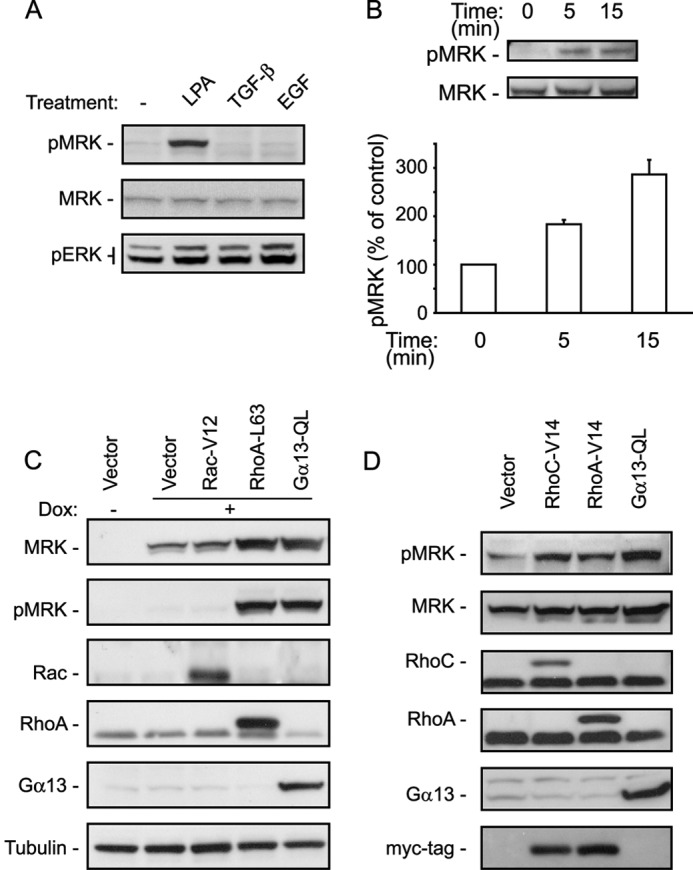
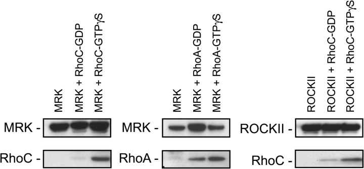
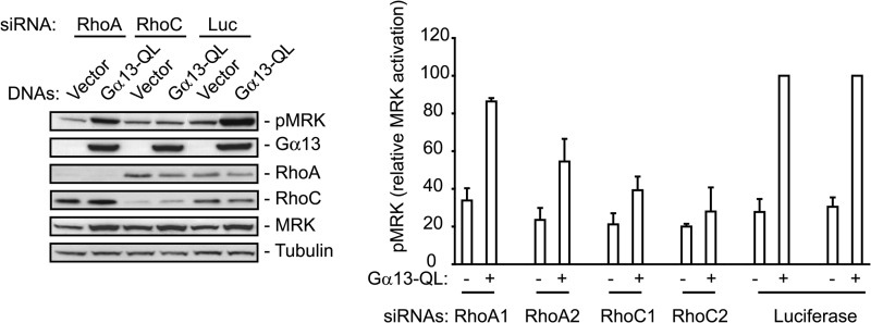
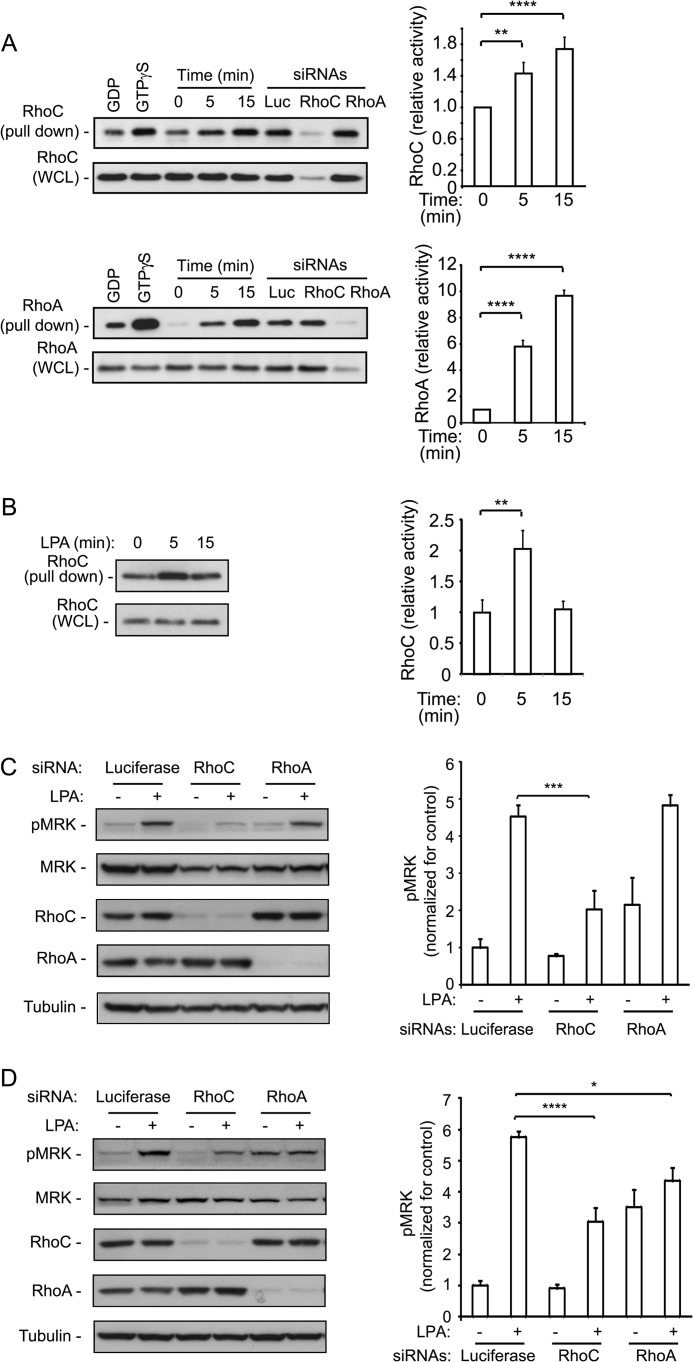
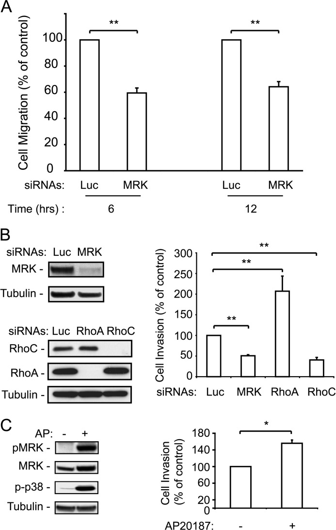
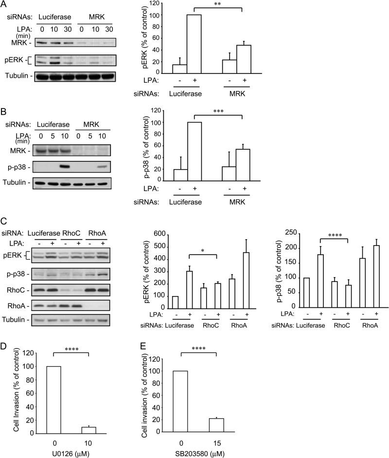
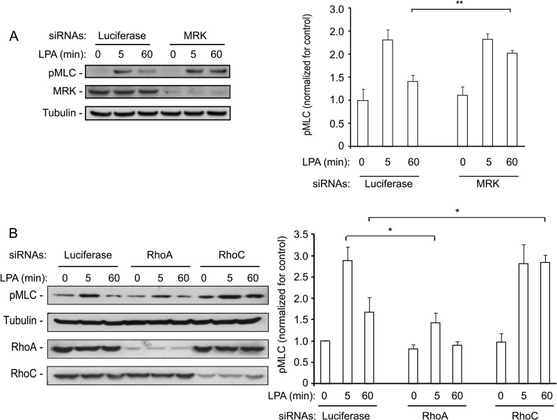
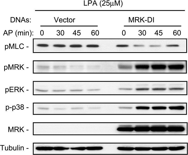
The small GTPase RhoC is overexpressed in many invasive tumors and is essential for metastasis. Despite its high structural homology to RhoA, RhoC appears to perform functions that are different from those controlled by RhoA. The identity of the signaling components that are differentially regulated by these two GTPases is only beginning to emerge. Here, we show that the MAP3K protein MRK directly binds to the GTP-bound forms of both RhoA and RhoC in vitro. However, siRNA-mediated depletion of MRK in cells phenocopies depletion of RhoC, rather than that of RhoA. MRK depletion, like that of RhoC, inhibits LPA-stimulated cell invasion, while depletion of RhoA increases invasion. We also show that active MRK enhances LPA-stimulated invasion, further supporting a role for MRK in the regulation of invasion. Depletion of either RhoC or MRK causes sustained myosin light chain phosphorylation after LPA stimulation. In addition, activation of MRK causes a reduction in myosin light chain phosphorylation. In contrast, as expected, depletion of RhoA inhibits myosin light chain phosphorylation. We also present evidence that both RhoC and MRK are required for LPA-induced stimulation of the p38 and ERK MAP kinases. In conclusion, we have identified MRK as a novel RhoC effector that controls LPA-stimulated cell invasion at least in part by regulating myosin dynamics, ERK and p38.


Figures

FIGURE 1.
FIGURE 1.
MRK is activated by LPA, Gα13 and Rho proteins. A, UMDI cells, expressing recombinant MRK fused to a drug-binding domain of FKBP12, were treated for 5 min with the following growth factors: 25 μm LPA, 10 ng/ml TGF-β1, and 50 μg/ml EGF, or left untreated (−). Cell lysates were tested by Western blotting for MRK activation using the P-MRK specific antibody. The P-ERK antibody was used to test for cellular stimulation by the growth factors. B, M28 cells that express KT3-tagged MRK after treatment with doxycyclin, were stimulated with LPA for the indicated times and tested for MRK activation as in A. Blots were scanned and the band intensities were quantified using ImageJ. Histograms are the means + S.E. of three independent experiments. C, M28 cells were transfected with vector control, Rac-V12, RhoA-L63, or Gα13-QL constructs. Forty-eight hours later they were treated overnight with doxycyclin (Dox) to induce expression of KT3-MRK and then tested for MRK activity by Western blotting with the P-MRK specific antibody. D, UMDI cells were transfected with the indicated DNA plasmids, 48 h later cells were harvested and tested for MRK activity as in C.
FIGURE 2.
FIGURE 2.
MRK interacts with Rho proteins in vitro. KT3-MRK was immunoprecipitated with KT3 antibodies from M28 cells grown in serum-free medium and incubated in vitro with purified RhoC or RhoA proteins loaded with GDP or GTP-γS. ROCKII protein was used as a positive control for RhoC-binding. Rho proteins were detected using the X-Press antibody that recognizes their tag.
FIGURE 3.
FIGURE 3.
Rho proteins are necessary for Gα13-QL-induced stimulation of MRK activation. M28 cells were co-transfected with vector control or Gα13-QL plasmids and either RhoA, RhoC, or luciferase (L) siRNAs. After 24 h they were treated with doxycyclin for 18 h to induce MRK expression and then harvested. Whole cell extracts were probed with the indicated antibodies. Two different siRNAs targeting RhoA and RhoC were tested. Histograms are means + S.E. of five independent experiments.
FIGURE 4.
FIGURE 4.
LPA-stimulated MRK activation is inhibited by Rho proteins down-regulation. LPA stimulates activation of RhoA and RhoC proteins. U2OS cells (A) or EMDI cells (B) were treated with 25 μm LPA for the indicated times and then tested for activation of Rho proteins using pull-down assay with GST-Rhotekin-RBD. The last 3 lanes of each blot in A are pull-downs from steady-state lysates of cells transfected with the individual siRNAs that were used to control for band specificity. Each set of experiments was repeated 3–5 times; bands were quantified with the ImageJ program and expressed as means + S.E. in the corresponding histograms. C, UMDI cells, or D, EMDI cells, were transfected with control luciferase, RhoC or RhoA targeting siRNAs and then treated with 25 μm LPA for 5 min. Cell lysates were immunoblotted with the pMRK specific antibody to test for MRK activity, and with the indicated antibodies for controls. The pMRK bands were quantified with ImageJ. Histograms are means + S.E. of three independent experiments. p values representing Student's t test were *, p < 0.05; **, p < 0.01; ***, p < 0.005; ****, p < 0.001.
FIGURE 5.
FIGURE 5.
MRK and RhoC positively control cell invasion, whereas RhoA negatively regulates it. A, ES-2 ovarian cancer cells were transfected with luciferase (Luc) or MRK siRNAs and then tested for migration in the wound healing assay for 6 and 12 h. B, ES-2 cells, transfected with the indicated siRNAs, were tested for LPA-stimulated cell invasion through Matrigel chambers as described under “Experimental Procedures.” Blots show depletion of the respective proteins by RNA interference. C, enhancement of LPA-stimulated cell invasion by active MRK induced by forced homodimerization. Detection by immunoblotting of MRK activated by the homodimerizing drug AP20187 in the EMDI cells and cell invasion through matrigel in response to LPA (1 μm) and AP20187 (2 μm). Histograms are means + S.E. from 3–6 independent experiments. *, p < 0.005; **, p < 0.001.
FIGURE 6.
FIGURE 6.
MRK and RhoC down-regulation inhibit LPA-stimulated ERK and p38 activation. UMDI cells were transfected with the MRK siRNA (A and B) for 36 h and with the Rho proteins specific siRNAs (C). Cells were starved overnight and treated with 25 μm LPA for 15 min. Cell lysates were blotted with the indicated antibodies. The pERK and p-p38 bands were quantified using ImageJ. Histograms represent the means + S.E. from three independent experiments. D and E, inhibition of ERK or p38 activity blocks invasion. ES-2 cells were processed for the Matrigel invasion assay as in Fig. 5 in the presence of the ERK inhibitor (D) and the p38 inhibitor (E). Histograms are the means + S.E. from 3–5 independent experiments. *, p < 0.05; **, p < 0.02; ***, p < 0.01; ****, p < 0.001.
FIGURE 7.
FIGURE 7.
MRK and RhoC down-regulation cause sustained LPA-induced MLC activation. ES-2 cells were transfected with the indicated siRNAs and 24 h later they were starved overnight. Cells were then stimulated with 25 μm LPA for the indicated time, harvested directly in SDS-containing sample buffer, and processed for Western blot analysis to test for P-MLC. Histograms represent the means + S.E. of 3–4 independent experiments. *, p < 0.05; **, p < 0.02.
FIGURE 8.
FIGURE 8.
Active MRK reduces LPA-stimulated MLC phosphorylation. EMDI cells were starved overnight, they were treated with the homodimerizing drug AP20187 (100 nm) for 1 h and then stimulated with LPA (10 μm) for 10 min. Cells were harvested directly in SDS-containing sample buffer and processed for Western blot with the indicated antibodies. Blot is representative of three independent experiments.
FIGURE 9.
FIGURE 9.
Down-regulation of MRK or RhoC increases focal adhesion number and length as well as cell area. ES-2 cells were transfected with the indicated siRNAs, 48 h later they were plated on coverslips and 24 h later processed for immunofluorescence. Histograms are means + S.E. of data from 15–30 cells and are representative of two independent experiments. L: luciferase; A: RhoA; C: RhoC; M: MRK. *, p < 0.02; **, p < 0.001.

Similar articles

Cited by

References

    1. Sahai E., Marshall C. J. (2002) RHO-GTPases and cancer. Nat. Rev. Cancer 2, 133–142 - PubMed
    1. Karlsson R., Pedersen E. D., Wang Z., Brakebusch C. (2009) Rho GTPase function in tumorigenesis. Biochim. Biophys. Acta 1796, 91–98 - PubMed
    1. Kamai T., Arai K., Tsujii T., Honda M., Yoshida K. (2001) Overexpression of RhoA mRNA is associated with advanced stage in testicular germ cell tumour. BJU. Int. 87, 227–231 - PubMed
    1. Suwa H., Ohshio G., Imamura T., Watanabe G., Arii S., Imamura M., Narumiya S., Hiai H., Fukumoto M. (1998) Overexpression of the rhoC gene correlates with progression of ductal adenocarcinoma of the pancreas. Br. J. Cancer 77, 147–152 - PMC - PubMed
    1. van Golen K. L., Davies S., Wu Z. F., Wang Y., Bucana C. D., Root H., Chandrasekharappa S., Strawderman M., Ethier S. P., Merajver S. D. (1999) A novel putative low-affinity insulin-like growth factor-binding protein, LIBC (lost in inflammatory breast cancer), and RhoC GTPase correlate with the inflammatory breast cancer phenotype. Clin. Cancer Res. 5, 2511–2519 - PubMed

Publication types

MeSH terms

LinkOut - more resources