. 2013 Jan 21;200(2):219-33.
doi: 10.1083/jcb.201207036. Epub 2013 Jan 14.

Perlecan regulates bidirectional Wnt signaling at the Drosophila neuromuscular junction

Affiliations

Perlecan regulates bidirectional Wnt signaling at the Drosophila neuromuscular junction

Keisuke Kamimura et al. J Cell Biol. .

Abstract

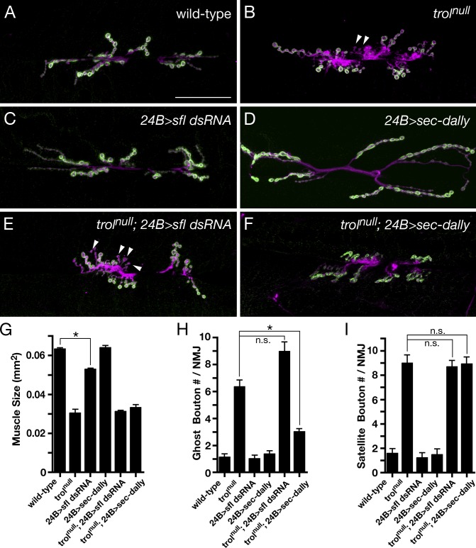
Heparan sulfate proteoglycans (HSPGs) play pivotal roles in the regulation of Wnt signaling activity in several tissues. At the Drosophila melanogaster neuromuscular junction (NMJ), Wnt/Wingless (Wg) regulates the formation of both pre- and postsynaptic structures; however, the mechanism balancing such bidirectional signaling remains elusive. In this paper, we demonstrate that mutations in the gene of a secreted HSPG, perlecan/trol, resulted in diverse postsynaptic defects and overproduction of synaptic boutons at NMJ. The postsynaptic defects, such as reduction in subsynaptic reticulum (SSR), were rescued by the postsynaptic activation of the Frizzled nuclear import Wg pathway. In contrast, overproduction of synaptic boutons was suppressed by the presynaptic down-regulation of the canonical Wg pathway. We also show that Trol was localized in the SSR and promoted postsynaptic accumulation of extracellular Wg proteins. These results suggest that Trol bidirectionally regulates both pre- and postsynaptic activities of Wg by precisely distributing Wg at the NMJ.


Figures

Figure 1.
Figure 1.
trol mutations affect both muscle size and NMJ morphology. (A–D) Type I boutons of wild-type (A) and trolnull mutant (B–D) larvae were stained with anti-HRP (magenta) and anti-DLG (green) antibodies. trol mutants showed ghost (arrowheads in B and C) and satellite boutons (B, arrow; and D). (E–H) Area of muscle 6/7 (E), total number of boutons (F), and numbers of ghost (G) and satellite (H) boutons in the NMJs of the first-, second-, and third-instar larvae of wild-type and trol (trolnull and trol7) mutants were measured. Error bars represent SEM (n = 10; *, P < 0.05; **, P < 0.01; and ***, P < 0.001 relative to wild type). Bars: (A) 30 µm; (C) 12 µm.
Figure 2.
Figure 2.
Larval locomotion and electrophysiological analyses of trolnull mutants. (A) Larval locomotion was quantified as the number of muscular body contractions per minute during crawling of first- and third-instar larvae of wild-type and trolnull mutants. (B) Representative traces of mEJP and eEJP showed that trolnull mutants had defective synaptic transmission. (C–E) trolnull mutants displayed decreased frequency (C) and amplitude (D) of mEJP and decreased eEJP amplitude (E). Error bars represent SEM (*, P < 0.05 and **, P < 0.005 relative to wild type). Sample numbers: (A) n = 10; (C–E) n = 11.
Figure 3.
Figure 3.
Trol is localized at postsynaptic SSR and functions postsynaptically. (A–C) Area of muscle 6/7 (A) and the numbers of ghost (B) and satellite boutons (C) in the NMJs of 24B>trol dsRNA, elav>trol dsRNA, and repo>trol dsRNA animals, in which Trol was knocked down specifically in muscle, neuron, and glia, respectively, were quantified. Muscle-specific trol knockdown caused reduction of muscle area and increase of ghost and satellite boutons. Error bars represent SEM (n = 20; *, P < 0.01 and **, P < 0.001 relative to wild type). (D and E) Immunoelectron microscopy was performed for the synaptic bouton regions of the trol FlyTrap line. Localization of Trol was analyzed by immunostaining with the anti-GFP antibody. Arrows indicate Trol signals in the SSR. (E) White arrowheads indicate the localization of Trol signals at the perisynaptic region between pre- and postsynaptic membranes. (D) Trol signals were also detected on basement membranes (black arrowheads). Bars: (D) 1 µm; (E) 300 nm. B, synaptic bouton.
Figure 4.
Figure 4.
Both the core protein and HS moieties of Trol are required for NMJ formation. (A–F) Larval NMJs of wild type (A), trolnull (B), 24B>sfl dsRNA (C), 24B>sec-dally (D), trolnull; 24B>sfl dsRNA (E), and trolnull; 24B>sec-dally (F) were stained with anti-HRP (magenta) and anti-DLG (green) antibodies. Arrowheads in B and E indicate ghost boutons. Bars, 50 µm. (G–I) Area of muscle 6/7 (G) and the numbers of ghost (H) and satellite (I) boutons in animals of indicated genotypes were measured. Muscle-specific expression of sfl dsRNA reduced muscle size (24B>sfl dsRNA). The ghost bouton phenotype of the trolnull mutant was rescued by muscle-specific expression of sec-dally (trolnull; 24B>sec-dally) but not by sfl dsRNA (trolnull; 24B>sfl dsRNA). Expression of neither sfl dsRNA nor sec-dally affected the satellite bouton phenotype of the trolnull mutant. Error bars represent SEM (n = 18; *, P < 0.01).
Figure 5.
Figure 5.
Trol is required for formation of postsynaptic structures and glutamate receptor localization at postsynaptic sites. (A–F) Electron micrographs of wild-type (A, C, and E) and trolnull mutant (B, D, and F) tissues are presented. In trolnull mutants, the cell surface of muscle is located closer to the basement membrane (arrowheads in B) than in the wild type (A). Mutations in trol caused small SSRs (D) and abnormally enlarged pockets in the postsynaptic region apposed to active zones (asterisks in D and F). The structure of T bar appeared normal in the trolnull mutant (arrowheads in E and F). Arrowheads in A indicate the cell surface of the muscle. An asterisk in E indicates the normal postsynaptic area apposed to the active zone. (G–I) Morphometric analyses of type Ib boutons showed that the SSR area was decreased (G), and the number of enlarged pockets increased (H) in trolnull mutants. On the other hand, the number of synaptic vesicles was not altered in trolnull mutants (I). To count the number of synaptic vesicles, we selected those within 250 nm of the active zones. Error bars represent SEM (n = 15; *, P < 0.0001). (J–L) Synaptic regions of wild-type (J) and trolnull mutants (K) were stained with the antibodies against active zone marker BRP and GluRIIA. (J) BRP and GluRIIA clusters were located on apposed membranes at the synaptic boutons in wild-type larvae. (K) In trolnull mutants, the levels of GluRIIA, but not BRP, were dramatically decreased compared with those of wild-type animals. (L) Bar graphs show the mean staining intensity for GluRIIA and BRP at the NMJ. Error bars represent SEM (n = 20; **, P < 0.001). Bars: (A, B, E, and F) 500 nm; (C and D) 1.5 µm; (J and K) 15 µm. AZ, active zone.
Figure 6.
Figure 6.
Trol regulates Wg-mediated postsynaptic development. (A–C) Muscle 6 in wild-type (A) and trolnull (B) animals was stained with antibodies to the C terminus of Dfz2 (green) and α-Tubulin (magenta). Immunoreactive puncta in the nuclei (arrowheads in dashed circles) were markedly reduced in trolnull mutants. DFz2-C localization in the synaptic boutons was not affected by mutations in trol (arrows). (C) The numbers of DFz2-C nuclear puncta in third-instar larval muscles of wild-type and trolnull animals were counted. (D–F) Electron micrographs of synaptic type I boutons of wild-type (D), trolnull (E), and trolnull; 24B>DFz2-C–NLS (F) animals are presented. The SSR region is colored brown. (G–J) Expression of DFz2-C–NLS in trolnull muscles rescued their ghost bouton (G), small SSR (H), and enlarged postsynaptic pocket (I) phenotypes. However, DFz2-C–NLS expression did not rescue the abnormal localization of GluRIIA at trolnull synaptic boutons (J). Error bars represent SEM (n = 20; **, P < 0.01; ***, P < 0.001; and ****, P < 0.0001). Bars: (A) 15 µm; (D) 1 µm. AZ, active zone.
Figure 7.
Figure 7.
Reduction of presynaptic Wg signaling suppresses satellite bouton phenotype of trol mutants. (A) Activation of FNI pathway in the muscles (trolnull; 24B>DFz2-C–NLS) did not rescue the satellite bouton phenotype of trolnull mutants. (B–H) The numbers of satellite boutons in muscle 6/7 of the indicated genotypes were counted (B). Larval NMJs of wild type (C), trolnull (D), trolnull; wgCX4/+ (E), trolnull; gbb1/+ (F), trolnull; OK6>sggact (G), and trolnull; OK6>dshDIX (H) were stained with anti-HRP (magenta) and anti-DLG (green) antibodies. Heterozygous mutation of wg (E), but not that of gbb (F), suppressed the satellite bouton phenotype of trolnull animals (B). Presynaptic suppression of canonical Wg signaling (G and H) rescued the satellite bouton phenotype (B), but not the ghost bouton phenotype, of trolnull animals. Arrows and arrowheads indicate the satellite and ghost boutons, respectively. (I–K) The NMJs of wild-type (I) and trolnull (J) animals were stained with anti-HRP (magenta) and anti-Futsch (green) antibodies. (K) Bar graph shows the percentage of boutons with Futsch loops and with unbundled Futsch in wild-type and trolnull animals. In the NMJs of trolnull mutants, synaptic boutons with Futsch loops significantly increased compared with those in wild-type animals (arrowheads in I and J). Error bars represent SEM (n = 20; *, P < 0.05; **, P < 0.001). Bars, 50 µm.
Figure 8.
Figure 8.
Trol regulates the extracellular distribution of Wg. (A–F) The NMJs of OK6>GFP-wg (A and D) and trolnull; OK6>GFP-wg (B and E) larvae, which expressed Wg-GFP specifically in the motor neurons, were stained using a protocol detecting extracellular Wg (Ext-Wg). The immunostainings of presynaptic membrane (HRP) and total Wg-GFP are also shown. The majority of the trolnull boutons showed greatly reduced levels of extracellular Wg-GFP protein (arrowheads in B) compared with the wild type (A). (C) Bar graph shows the mean staining intensity of extracellular Wg-GFP at the NMJ. (D) In the wild type, extracellular Wg-GFP proteins were diffusely spread into the postsynaptic region (arrowheads). (E) In the trolnull boutons, extracellular Wg-GFP proteins were closely associated with the presynaptic membranes with little spreading into postsynaptic regions (arrowheads). Dotted curved lines trace the outlines of presynaptic terminals. (F) Plot profiles of the intensities of HRP and extracellular Wg labeling along a line crossing a bouton of wild type and trolnull are shown. The data shown are from a single representative experiment out of three repeats (n = 13). (G–I) OK6>GFP-wg larval NMJs were incubated with (H) or without (G) heparitinase I followed by extracellular staining of Wg-GFP. Removal of HS did not affect the total levels of Wg-GFP but significantly reduced the extracellular levels of Wg-GFP. (I) Bar graphs show the mean staining intensities of total (left) and extracellular Wg-GFP (right) levels at the NMJ. Hep, heparitinase. Error bars represent SEM (n = 25; *, P < 0.05). Bars: (A and G) 10 µm; (D) 1 µm.
Figure 9.
Figure 9.
Proposed model for Trol function in Wg pathway at pre- and postsynaptic cells. In the wild type (left), presynaptically released Wg sends its signal to both presynaptic and postsynaptic cells, which is required for proliferation of boutons and formation of postsynaptic SSR, respectively. Trol binds and sequesters Wg on the surface of SSR. In trol mutants (right), stability of Wg at the postsynaptic site is decreased, and therefore, Wg might preferably bind its receptor DFz2 on the presynaptic membrane, inducing overproliferation of boutons. Thus, Trol regulates bidirectional activity of Wg signaling at the NMJ.

Similar articles

Cited by

References

    1. Arikawa-Hirasawa E., Rossi S.G., Rotundo R.L., Yamada Y. 2002. Absence of acetylcholinesterase at the neuromuscular junctions of perlecan-null mice. Nat. Neurosci. 5:119–123 10.1038/nn801 - DOI - PubMed
    1. Ataman B., Ashley J., Gorczyca D., Gorczyca M., Mathew D., Wichmann C., Sigrist S.J., Budnik V. 2006. Nuclear trafficking of Drosophila Frizzled-2 during synapse development requires the PDZ protein dGRIP. Proc. Natl. Acad. Sci. USA. 103:7841–7846 10.1073/pnas.0600387103 - DOI - PMC - PubMed
    1. Ataman B., Ashley J., Gorczyca M., Ramachandran P., Fouquet W., Sigrist S.J., Budnik V. 2008. Rapid activity-dependent modifications in synaptic structure and function require bidirectional Wnt signaling. Neuron. 57:705–718 10.1016/j.neuron.2008.01.026 - DOI - PMC - PubMed
    1. Axelrod J.D., Miller J.R., Shulman J.M., Moon R.T., Perrimon N. 1998. Differential recruitment of Dishevelled provides signaling specificity in the planar cell polarity and Wingless signaling pathways. Genes Dev. 12:2610–2622 10.1101/gad.12.16.2610 - DOI - PMC - PubMed
    1. Bayat V., Jaiswal M., Bellen H.J. 2011. The BMP signaling pathway at the Drosophila neuromuscular junction and its links to neurodegenerative diseases. Curr. Opin. Neurobiol. 21:182–188 10.1016/j.conb.2010.08.014 - DOI - PMC - PubMed

Publication types

MeSH terms

LinkOut - more resources