. 2013 Jan 29;110(5):1680-5.
doi: 10.1073/pnas.1222130110. Epub 2013 Jan 14.

Frustration in the energy landscapes of multidomain protein misfolding

Affiliations

Frustration in the energy landscapes of multidomain protein misfolding

Weihua Zheng et al. Proc Natl Acad Sci U S A. .

Abstract

Frustration from strong interdomain interactions can make misfolding a more severe problem in multidomain proteins than in single-domain proteins. On the basis of bioinformatic surveys, it has been suggested that lowering the sequence identity between neighboring domains is one of nature's solutions to the multidomain misfolding problem. We investigate folding of multidomain proteins using the associative-memory, water-mediated, structure and energy model (AWSEM), a predictive coarse-grained protein force field. We find that reducing sequence identity not only decreases the formation of domain-swapped contacts but also decreases the formation of strong self-recognition contacts between β-strands with high hydrophobic content. The ensembles of misfolded structures that result from forming these amyloid-like interactions are energetically disfavored compared with the native state, but entropically favored. Therefore, these ensembles are more stable than the native ensemble under denaturing conditions, such as high temperature. Domain-swapped contacts compete with self-recognition contacts in forming various trapped states, and point mutations can shift the balance between the two types of interaction. We predict that multidomain proteins that lack these specific strong interdomain interactions should fold reliably.


Conflict of interest statement

The authors declare no conflict of interest.

Figures

Fig. 1.
Fig. 1.
The fraction of misfolded domains found upon simulated annealing is plotted against the sequence identity between the two domains in the fused construct. Titin I27 was fused with five different proteins via a four-residue GLY linker to form two-domain proteins, as indicated in the plot. Forty annealing simulations were run for each fused protein from totally extended conformations. All proteins were studied with the same annealing schedule. A domain is considered folded when the fraction of native contacts within the domain formula image
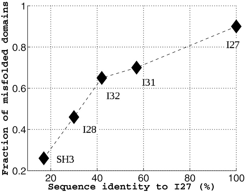
, i = 1, 2 at the end of all of the simulations. As the sequence identity increases, the fraction of misfolded domains increases accordingly.
Fig. 2.
Fig. 2.
Misfolded structures and their contact maps. (Left) A domain-swapped misfolded structure from simulated annealing simulations of the fused dimer SH3-SH3. (Right) A misfolded structure with a significant amount of self-recognition contacts from simulations of the fused dimer I27-I27. The different levels of frustration in the tertiary contacts as determined by the frustratometer analysis (28) are illustrated in the structures. In the structures, the two domains are in blue and yellow color, respectively. Minimally frustrated interactions are shown in green lines, and frustrated interactions are in red. The swapped contacts formed at the domain interface of SH3-SH3 are minimally frustrated, as expected from the principle of minimal frustration for native contacts. The self-recognition contacts formed at the domain interface of I27-I27 are also minimally frustrated, indicating that these contacts are stronger than random contacts. Within the set of four contact maps for each fused dimer, the lower left and upper right contact maps are for each domain, respectively. Formed native contacts are represented by black dots, formed nonnative contacts are in red, and the native contacts that are not formed are in yellow. The upper left contact map is the interdomain contact map. Black dots stand for formed swapped contacts. Red dots represent other types of interdomain contacts. In the case of I27-I27, we observe a different type of contact, the self-recognition contacts (in red), along the diagonal of the map. The lower left contact map is a summary contact map showing the total number of occurrences of each formed contact from the end structures of 40 annealing simulations. For SH3-SH3, the swapped contacts that are formed between the residues near the linker position appear more frequently than those that are more distantly connected through the sequence. For I27-I27, the self-recognition contacts formed between residue indexes 56 and 61 (HILILH) appear in almost all of the simulations.
Fig. 3.
Fig. 3.
Energy and free-energy surfaces for I27-I27 at its folding temperature. Nself and Nswap are the number of self-recognition contacts and the number of domain-swapped contacts, respectively. The trapped states I have higher energies than the native states N, as shown in the z axis, but have similar free energies to those of the native states, as shown by the color coding of the free energy, with scale indicated in the side bar. We see that the ensemble I states are entropically favored. As temperature increases, the intermediate ensemble will become more stable than the native ensemble.
Fig. 4.
Fig. 4.
Competing roles of the swapped contacts and self-recognition contacts in the “wild-type” (WT) fused dimer I27-I27, SH3-SH3, and their mutants (MT) I27*-I27* and SH3*-SH3*. Forty annealing simulations were carried out for all dimers. 〈Ncontacts〉 is the number of contacts averaged over all 40 simulations. Nintercontact is the number of interdomain contacts. The interdomain contacts include the swapped contacts, self-recognition contacts, and other types of interdomain contacts. (A) Single-point mutation introduces a pair of strong self-recognition contacts in SH3*-SH3*, suppressing the formation of swapped contacts. Significant misfolding with formation of self-recognition contacts occurs after the mutation. (B) Two point mutations eliminated two pairs of the strongest self-recognition contacts in I27; therefore the swapped contacts play a more dominant role.
Fig. 5.
Fig. 5.
Comparisons of stability (energy per residue) among various zero-temperature structures and ensembles of thermally sampled structures of I27 (A and C) and SH3 (B). The stability for the native monomeric structure (green vertical line) is calculated from the AWSEM energy function. The strongest nonnative hexamer pairing possible in the monomer (magenta bar) is significantly less stable than the native structure, indicating that misfolding by inappropriate pairing of strands will be unlikely during the folding of the monomer for both I27 and SH3. In A and B, the blue bars represent the distribution of the stability of all of the self-recognition hexamer pairs, calculated from the AWSEM-Amylometer (SI Text). If the stability of the strongest self hexamer pair is competitive with the native structure, as in the case of I27-I27, the particular self pair becomes responsible for the misfolding of the fused protein in our simulation and potentially, would trigger further aggregation in solution. For SH3-SH3, B predicts that fused protein should fold as well as the monomer in the simulation, because all self hexamer pairings are weaker than the most stable nonnative hexamer pairing in the monomer. In C, the stability distributions of various ensembles of structures collected from the simulations of I27-I27 are shown. The native ensemble (green) is energetically more stable than both the domain-swapped ensemble (blue) and the self-recognition ensemble (red). Nevertheless, the local interactions between the self-pairing hexamers from the self-recognition ensembles, shown in cyan, are even stronger than typical energies in the native folded ensemble.

Similar articles

Cited by

References

    1. Wolynes PG. Energy landscapes and solved protein-folding problems. Philos Trans R Soc A. 2005;363:453–467. - PubMed
    1. Selkoe DJ. Folding proteins in fatal ways. Nature. 2003;426(6968):900–904. - PubMed
    1. Chiti F, Dobson CM. Protein misfolding, functional amyloid, and human disease. Annu Rev Biochem. 2006;75:333–366. - PubMed
    1. Silow M, Tan Y-J, Fersht AR, Oliveberg M. Formation of short-lived protein aggregates directly from the coil in two-state folding. Biochemistry. 1999;38(40):13006–13012. - PubMed
    1. Yang S, et al. Domain swapping is a consequence of minimal frustration. Proc Natl Acad Sci USA. 2004;101(38):13786–13791. - PMC - PubMed

Publication types

LinkOut - more resources