. 2013 Apr 1;73(7):2170-80.
doi: 10.1158/0008-5472.CAN-12-2481. Epub 2013 Jan 14.

FOXP3 regulates sensitivity of cancer cells to irradiation by transcriptional repression of BRCA1

Affiliations

FOXP3 regulates sensitivity of cancer cells to irradiation by transcriptional repression of BRCA1

Weiquan Li et al. Cancer Res. .

Abstract

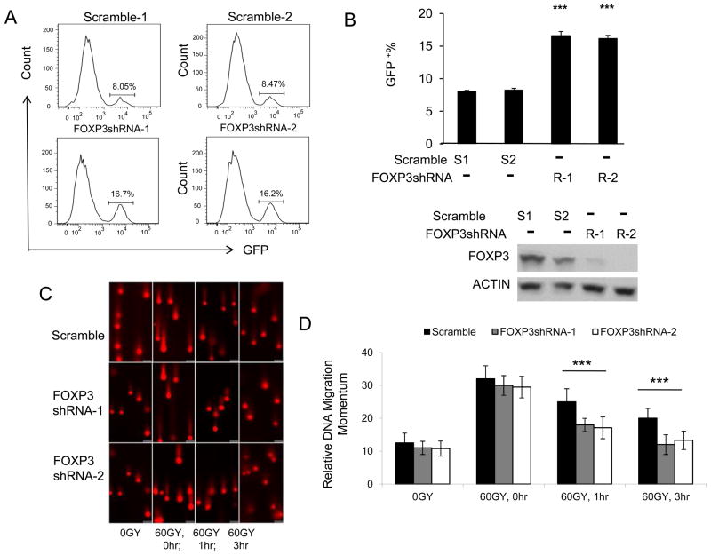
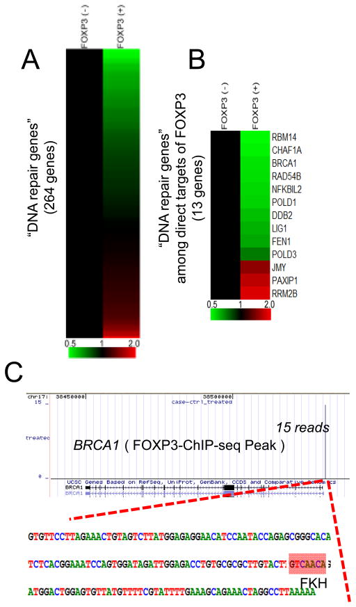
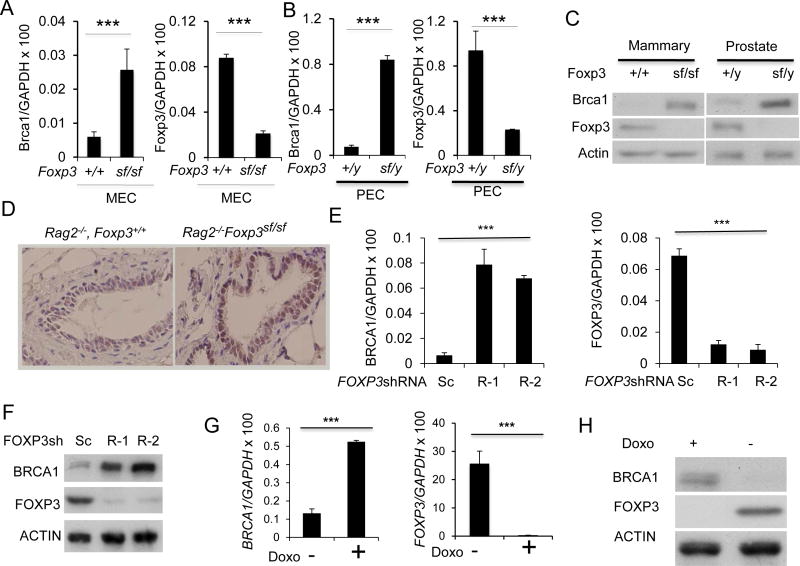
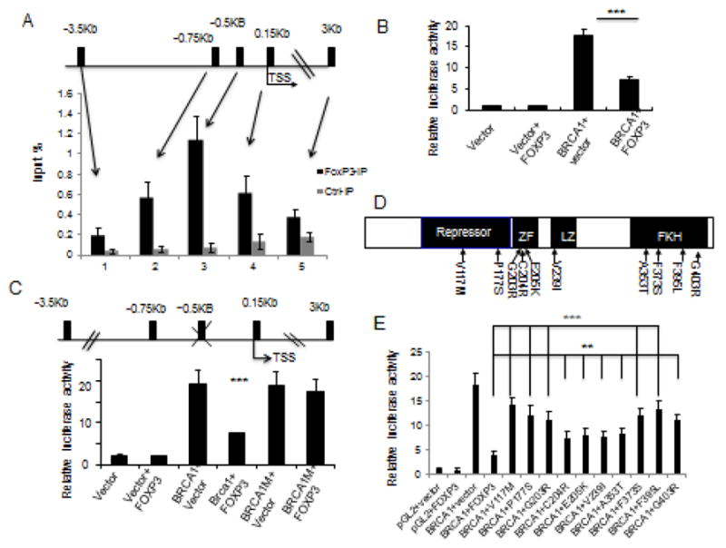
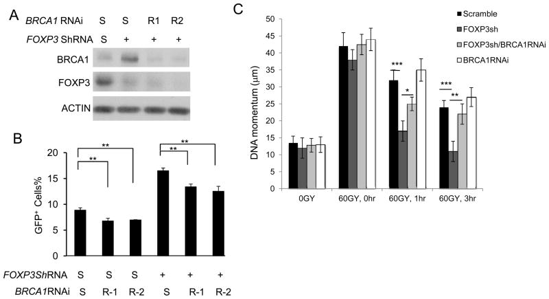
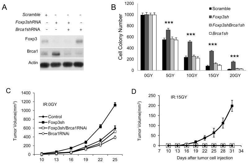
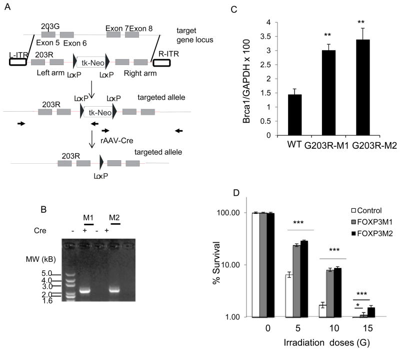
FOXP3 is an X-linked tumor suppressor gene and a master regulator in T regulatory cell function. This gene has been found to be mutated frequently in breast and prostate cancers and to inhibit tumor cell growth, but its functional significance in DNA repair has not been studied. We found that FOXP3 silencing stimulates homologous recombination-mediated DNA repair and also repair of γ-irradiation-induced DNA damage. Expression profiling and chromatin-immunoprecipitation analyses revealed that FOXP3 regulated the BRCA1-mediated DNA repair program. Among 48 FOXP3-regulated DNA repair genes, BRCA1 and 12 others were direct targets of FOXP3 transcriptional control. Site-specific interaction of FOXP3 with the BRCA1 promoter repressed its transcription. Somatic FOXP3 mutants identified in breast cancer samples had reduced BRCA1 repressor activity, whereas FOXP3 silencing and knock-in of a prostate cancer-derived somatic FOXP3 mutant increased the radioresistance of cancer cells. Together our findings provide a missing link between FOXP3 function and DNA repair programs.


Conflict of interest statement

The authors have no conflict of interest in this study.

Figures

Figure 1
Figure 1
FOXP3 represses DNA repairs. A. Representative histograms depicting distribution of GFP signals. U2OS cells with integrated DR-GFP reporter were stably transduced with scrambled or FOXP3 shRNA lentiviral vectors. Then the cells were infected with adenovirus carried restriction enzyme I-Sce I. Since the HR DNA repair corrects the GFP open-reading frame, the repair efficiency was measured by GFP+ cells recorded with flow cytometry.. B. Upper panel shows summary data on DNA repairing efficiency as measured by GFP+ cells. Data shown are means +/− S.D. from 3 independent experiments. *** P < 0.001. Lower panel shows FOXP3 protein expressions detected by Western blot, as shown in the lower panel. C. Comet assay of DNA damage response. U2OS cells stably transduced with scramble or FOXP3 shRNA were irradiated for 60GY. The cells were cultured for the given periods and then subject to agarose gel electrophoresis. The photographs show the momentum of the nuclear DNA movement. D. Summary data of momentum of cellular DNA. Data shown are means +/− S.D. from three independent experiments. *** P< 0.001.
Figure 2
Figure 2
Regulation of DNA damage response by FOXP3. A. Gene expression profile of DNA repair-related genes. DNA repair related genes were selected by UniProt database and their gene expression levels are shown as a heat map. Gene expression levels without FOXP3 induction were normalized to 1.0. Color scale is indicated at the bottom of the figure. Using P<0.001 as statistical endpoint, 48/247 DNA repair genes are affected by FOXP3. See Table S1 and Figures S1 for details. B. Gene expression profile of DNA repair-related genes among direct targets of FOXP3 revealed by ChIP-seq. Gene names are indicated at the right side of the heat map. See Table S2 and Figure S2 for details. C. FOXP3 binding site at the BRCA1 promoter identified by a FOXP3-ChIP-seq analysis. A single peak is identified with 15 reads in ChIP-seq. DNA sequence around the FOXP3-binding site is shown as a bottom panel. “GTCAACA” marked as a red box is a known Forkhead binding motif (FKH).
Figure 3
Figure 3
Foxp3 repressed Brca1 expression in mammary and prostate epithelial cells in the mice. A. Mouse mammary epithelial cells (MEC) were isolated from Rag2−/− Foxp3+/+ and Rag2−/− FoxP3sf/sf mammary glands. Brca1(left panel) and Foxp3 (right panel) mRNA levels were determined by real-time RT-PCR. Data shown were means+/−S.D. of mRNA, presented as %GAPDH and have been repeated three times. ***, P< 0.001. B. Mouse prostate epithelial cells (PEC) were isolated from Rag2−/− Foxp3+/y and Rag2−/− Foxp3sf/y prostates. Brca1(left panel) and Foxp3 (right panel) mRNA levels were determined by real-time RT-PCR. Data shown were means+/−S.D. of mRNA, presented as % Gapdh. The results have been repeated three times. C. The left panel shows Brca1 and Foxp3 expression in MEC by Western blot, while the right panel shows those in PEC. D. Immunohistochemical analysis for Brca1protein in benign mammary tissue from WT and Foxp3sf/sf mice. E–H. FOXP3 represses the BRCA1 gene in normal and malignant human mammary epithelial cells. E. Silencing FOXP3 in MCF10A cells up-regulates BRCA1 mRNA level. The levels of BRCA1 mRNA (left panel) and FOXP3 (right panel) in two scrambled or two FOXP3 siRNA transfected cells as determined by real-time RT-PCR. Data shown are means+/−S.D. of three independent experiments. F. Under the same conditions as in E, MCF10A cells were lysed for Western blot to detect BRCA1 and FOXP3 protein expression as indicated. G. Reduction of BRCA1 mRNA by FOXP3 in MCF-7 Tet-off expression system. MCF-7 cells with Tet-off expression of either GFP control or GFP in conjunction with FOXP3 were cultured for 48 hours in the presence or absence of doxycyclin. The BRCA1 mRNA was quantified by real-time PCR(left panel). The induction of FOXP3 mRNA was shown in the right panel. Data shown are means+/−S.D. of three independent experiments. ***: p value< 0.001. H. BRCA1 and FOXP3 protein were detected by Western blot as shown.
Figure 4
Figure 4
Identification of a functional FOXP3 binding site in the BRCA1 gene. A. The top panel shows a diagram of the potential forkhead binding motif in BRCA1 promoter, as marked black bars, while the lower panel shows that the % of input DNA precipitated by anti-FOXP3 or control IgG. Data shown are means+/−S.D. of % input DNA, from three independent experiments. TSS: transcriptional start site. B. Relative luciferase activity assay of BRCA1 promoter. 3.5 kB DNA upstream of BRCA1 TSS was cloned into luciferase reporter vector pGL2 transfected into 293 cells. Luciferase activity was assayed at 48 hours after transfection. Data shown are means+/−S.D. of three independent experiments. C. Identification of FOXP3 responsive element by mutational analysis. The FKH at −0.5 kB of BRCA1 TSS (GTCAACA) was mutated to GTCGGCA. The constructs used are diagrammed on the upper panel, while the transcriptional activity in the presence or absence of FOXP3 is presented in the lower panel. Data shown are means+/−S.D. of three independent analyses. D. Diagram depicting the position of somatic FOXP3 mutants from breast cancer samples used in this study. E. FOXP3 mutations abrogated its repression of BRCA1 promoter. Somatic mutations of FOXP3 from human breast and prostate cancer samples were tested for their repression of BRCA1 promoter using a luciferase reporter assay. Data shown are means+/−S.D. of triplicate sample analyses. **P<0.01, ***P<0.001.
Figure 5
Figure 5
FOXP3-BRCA1 interaction regulates DNA repair in U2OS cells. A, B. U2OS cells integrated with DR-GFP reporter were infected either with scrambled or FOXP3 shRNA. Transfectants were selected by puromycin for two weeks. The U2OS cells with scramble and FOXP3 shRNA were treated with scrambled RNAi (S) or BRCA1 RNAi. A. FOXP3 and BRCA1 expression. B. Repair of DSB as measured by GFP positive cells with flow cytometry. Data shown in B are means +/− S.D. from 3 independent experiments. C. Comet assay to measure repair of γ-ray-induced DNA damages. U2OS cells with scramble and FOXP3 shRNA, FOXP3 and/or BRCA1 ShRNAs were irradiated for 60 Gy. The cells were cultured for given periods and used for comet assays. Data showed were +/−S.D. means from three independent experiments. *P<0.05, **P<0.01, ***P<0.001.
Figure 6
Figure 6
Foxp3-Brca1 interaction regulates survival and tumorigenicity of mouse mammary tumor cell line TSA. A. Efficacy of ShRNA silencing of Foxp3 and siRNA silencing of Brca1, as detected by Western blot. B. Foxp3-Brca1 interaction determines TSA resistance to irradiation, as measured by colony formation assay. TSA with ShRNA silencing of Foxp3 and/or Brca1 received 0, 5, 10, 15, or 20Gy of γ–irradiation. The irradiated cell lines were cultured for a week before the colonies were counted. The data shown are from those from triplicate cultures. ***: P <0.001. C and D. Foxp3-Brca1 interaction regulates tumorigencity of γ-irradiated TSA. TSA cell lines that were stably transfected with scrambled, Foxp3 shRNA and/or Brca1shRNA were either unirradiated (C) or irradiated with 15Gy of γ–ray (D) were transplanted into mammary fatpads of syngeneic BALB/c mice. The tumor sizes were monitored over 5 weeks. Data shown are means+/−S.D. of tumor volumes and have been repeated three times.
Figure 7
Figure 7
Cancer-related somatic mutation of FOXP3 increases cancer cell resistance to irradiation. Somatic missense mutation uncovered from prostate cancer tissue (203G>R) were knocked into colon cancer cell line, as diagramed in A. The position of primers used for both screening of homologous recombinant and for Cre-mediated excision of Neo cassette is marked with bold arrows. Red color is used for vector sequence, while the black color is used for sequence of endogenous locus. B. Confirmation of homologous recombination and excision of Neo cassette by PCR. Two homologous recombination clones were identified from two 96-well-plates of G418 resistant single clones. The neomycin resistance gene was excised by Cre recombinase carried by adenovirus. Similar data were obtained for the homologous recombination in the right arm. C. Knockin of mutant FOXP3 increase BRCA1 transcripts as determined by real-time PCR. D. Survival of cancer cells after given doses of γ-irradiation. Data shown are means and S.D. of triplicated culture. *P<0.05, ***P<0.001.

Similar articles

Cited by

References

    1. Begg AC, Stewart FA, Vens C. Strategies to improve radiotherapy with targeted drugs. Nat Rev Cancer. 2011;11:239–53. - PubMed
    1. Delaney G, Jacob S, Featherstone C, Barton M. The role of radiotherapy in cancer treatment: estimating optimal utilization from a review of evidence-based clinical guidelines. Cancer. 2005;104:1129–37. - PubMed
    1. Bhide SA, Nutting CM. Recent advances in radiotherapy. BMC Med. 2010;8:25. - PMC - PubMed
    1. Baumann M, Krause M, Hill R. Exploring the role of cancer stem cells in radioresistance. Nat Rev Cancer. 2008;8:545–54. - PubMed
    1. Bao S, Wu Q, McLendon RE, Hao Y, Shi Q, Hjelmeland AB, et al. Glioma stem cells promote radioresistance by preferential activation of the DNA damage response. Nature. 2006;444:756–60. - PubMed

Publication types

MeSH terms