. 2012:2012:620834.
doi: 10.1155/2012/620834. Epub 2012 Dec 18.

Conditional Inactivation of Pten with EGFR Overexpression in Schwann Cells Models Sporadic MPNST

Affiliations

Conditional Inactivation of Pten with EGFR Overexpression in Schwann Cells Models Sporadic MPNST

Vincent W Keng et al. Sarcoma. 2012.

Abstract

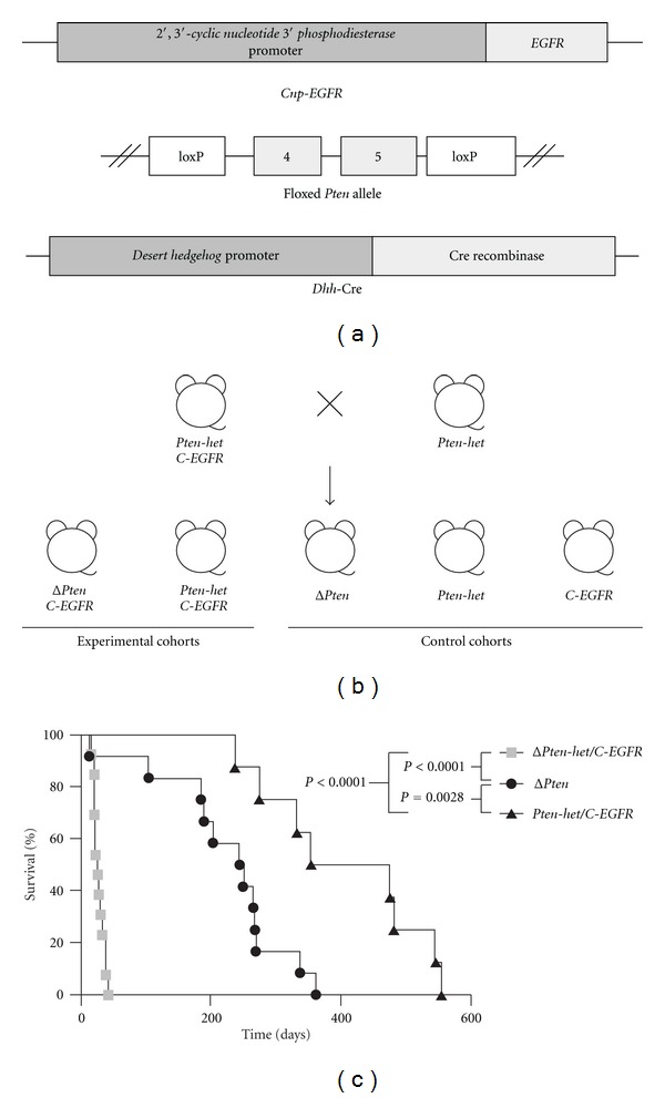
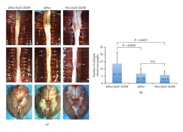
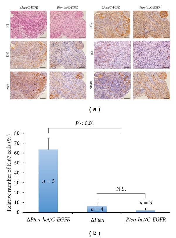
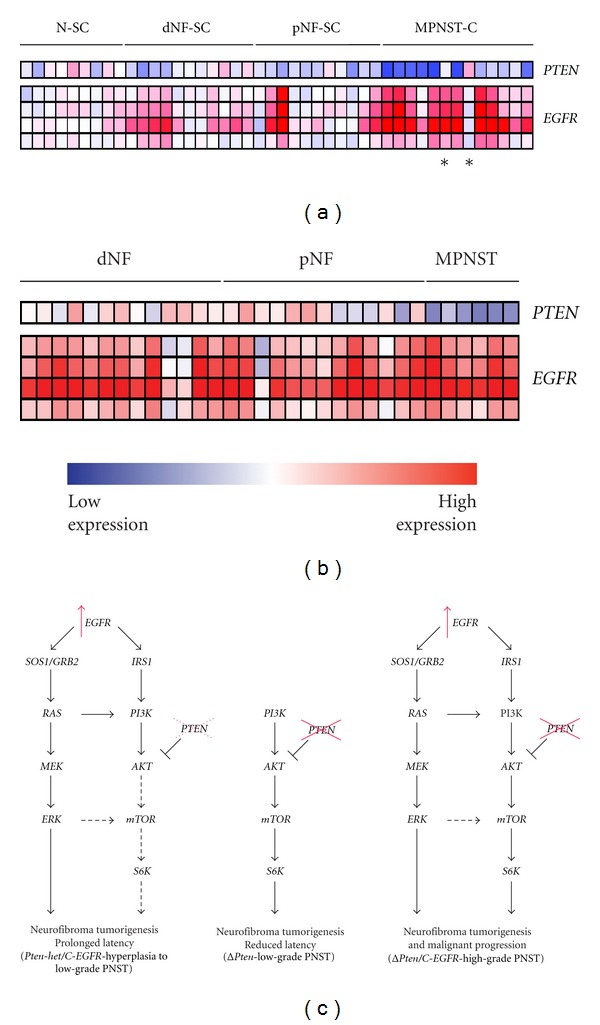
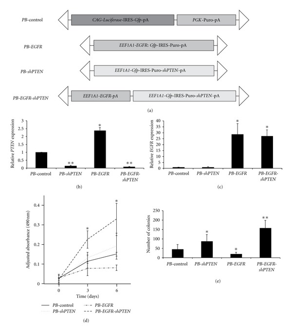
The genetic mechanisms involved in the transformation from a benign neurofibroma to a malignant sarcoma in patients with neurofibromatosis-type-1- (NF1-)associated or sporadic malignant peripheral nerve sheath tumors (MPNSTs) remain unclear. It is hypothesized that many genetic changes are involved in transformation. Recently, it has been shown that both phosphatase and tensin homolog (PTEN) and epidermal growth factor receptor (EGFR) play important roles in the initiation of peripheral nerve sheath tumors (PNSTs). In human MPNSTs, PTEN expression is often reduced, while EGFR expression is often induced. We tested if these two genes cooperate in the evolution of PNSTs. Transgenic mice were generated carrying conditional floxed alleles of Pten, and EGFR was expressed under the control of the 2',3'-cyclic nucleotide 3'phosphodiesterase (Cnp) promoter and a desert hedgehog (Dhh) regulatory element driving Cre recombinase transgenic mice (Dhh-Cre). Complete loss of Pten and EGFR overexpression in Schwann cells led to the development of high-grade PNSTs. In vitro experiments using immortalized human Schwann cells demonstrated that loss of PTEN and overexpression of EGFR cooperate to increase cellular proliferation and anchorage-independent colony formation. This mouse model can rapidly recapitulate PNST onset and progression to high-grade PNSTs, as seen in sporadic MPNST patients.


Figures

Figure 1
Figure 1
Establishing a novel sporadic peripheral nerve sheath tumor (PNST) progression mouse model. (a) Transgenes used to establish the novel sporadic PNST mouse model. Cnp-EGFR consists of the Cnp regulatory elements driving the EGFR gene expression specifically in Schwann cells and/or their precursor cells. Floxed Pten allele consists of the essential exons 4 and 5 of the Pten gene floxed with loxP sites. Dhh-Cre consists of the Dhh regulatory elements driving Cre recombinase to remove the loxP sites and allow for the inactivation of the floxed Pten alleles specifically in Schwann cells and/or their precursor cells. (b) Breeding strategy for generating experimental and control animals. Transgenic mice each carrying a single transgene was bred to obtain doubly transgenic mice Dhh-Cre; Pten flox/+ mice (Pten-het). Doubly transgenic mice were then bred with remaining transgene to obtain triple transgenic Dhh-Cre; Pten flox/+; Cnp-EGFR mice (Pten-het/C-EGFR). Finally, Pten-het mice were bred with Pten-het/C-EGFR mice to obtain the required experimental and control cohorts. Dhh-Cre; Pten flox/flox; Cnp-EGFRPten/C-EGFR) and Dhh-Cre; Pten flox/+; Cnp-EGFR (Pten-het/C-EGFR) experimental cohorts. Dhh-Cre; Pten flox/floxPten), Pten-het and Cnp-EGFR (C-EGFR) control cohorts. (c) Kaplan-Meier survival curves of various experimental and control cohorts. Pten dosage augmented the peripheral nervous system phenotype in the context of EGFR overexpression in Schwann cell and/or their precursor cells, resulting in decreased survival. P: log-rank test.
Figure 2
Figure 2
Pten dosage with EGFR overexpression affected enlarged dorsal root ganglia tumor multiplicity. (a) Left: representative of an early-onset peripheral nervous system phenotype observed in a 38-day Dhh-Cre; Pten flox/flox; Cnp-EGFRPten/C-EGFR) experimental mouse. Brachial plexus: majority of the dorsal root ganglia and trigeminal nerves were enlarged. Middle: representative of a late-onset peripheral nervous system phenotype observed in a 104-day Dhh-Cre; Pten flox/floxPten) control mouse. Brachial plexus: several dorsal root ganglia and trigeminal nerves were enlarged. Right: representative of a late-onset peripheral nervous system phenotype observed in a 274-day Dhh-Cre; Pten flox/+; Cnp-EGFR (Pten-het/C-EGFR) control mouse. Brachial plexus: several dorsal root ganglia and trigeminal nerves were enlarged. Top panels: brachial plexi; middle panels: dorsal root ganglia; bottom panels: brain with trigeminal nerves; arrows indicate peripheral nervous system phenotype; scale bars, 2 mm. (b) Statistically significant differences in the number of enlarged dorsal root ganglia isolated from ΔPten/C-EGFR experimental cohort compared with control cohorts (ΔPten and Pten-het/C-EGFR). Mean ± standard deviation; P: unpaired t-test; n: number of mice evaluated in each cohort; N.S.: nonsignificant.
Figure 3
Figure 3
Histological analyses of peripheral nervous system phenotype. Standard hematoxylin-eosin staining (HE) and immunohistochemical (IHC) staining were performed on all peripheral nervous system tissue sections. (a) Representative HE and IHC staining of peripheral nerves taken from a Dhh-Cre; Pten flox/flox; Cnp-EGFRPten/C-EGFR) experimental and Dhh-Cre; Pten flox/+; Cnp-EGFR (Pten-het/C-EGFR) control mice using antibodies against the proliferative marker (Ki67), Schwann cell/oligodendrocyte lineage marker (S100ß), activated Ras/Mapk/Erk signaling by phospho-Erk1/2 (pErk), activated Pi3k/Akt signaling by phospho-Akt (pAkt) detection, and activated mTor signaling by phospho-S6 (pS6). Negative controls, sections incubated without the primary antibody gave no significant signal above background (data not shown). (b) Semiquantitative analysis of proliferative peripheral nerve cells in various control and experimental cohorts. Representative peripheral nerves were isolated from each cohort and IHC stained using the Ki67 proliferative marker. The number of Ki67-positive peripheral nerve cells was counted and shown as a percentage of total cells per counted field of view at 20x magnification (mean ± standard deviation). Peripheral nerves were taken from ΔPten/C-EGFR experimental mice, Dhh-Cre; Pten flox/floxPten) and Pten-het/C-EGFR control mice. No Ki67-positive cells were detected in sciatic nerves isolated from FVB/N mice (Supplementary Figure 1). n, number of mice from each cohort; N.S.: nonsignificance between indicated cohorts; P: unpaired t-test.
Figure 4
Figure 4
Expression microarray analysis of PTEN and EGFR in human peripheral nerve tumors. (a) Purified human Schwann cells from normal sciatic nerve (N-SC), dermal neurofibroma cell lines (dNF-SC), and plexiform neurofibroma cell lines (pNF-SC). Transformed cells from malignant peripheral nerve sheath cell lines (MPNST-C). Asterisks indicate sporadic MPNST samples. (b) Solid dermal neurofibromas (dNF), plexiform neurofibromas (pNF), and malignant peripheral nerve sheath tumors (MPNST). Four different probes for EGFR were used. Red, increase in red intensity as expression increases; Blue, increase in blue intensity as expression decreases. (c) Conditional inactivation of Pten and EGFR overexpression in Schwann cells resulted in high-grade PNST initiation and/or progression due to the upregulation of both Ras/Mapk/Erk and Pi3k/Akt/mTor signaling pathways (right). Inactivation of Pten alone resulted in reduced latency with low-grade PNST tumorigenesis at low penetrance (middle). Conditional inactivation of Pten alone can result in low-grade PNST tumorigenesis via the upregulation of the Pi3k/Akt/mTor signaling pathway. Partial conditional inactivation of Pten in the context of EGFR overexpression in Schwann cells resulted in prolonged latency with hyperplasia to low-grade PNST tumorigenesis at low penetrance (left), resulting in upregulation of Ras/Mapk/Erk and slight upregulation of the Pi3k/Akt/mTor signaling pathways. SOS1: son of sevenless homolog 1; GRB2: growth factor receptor-bound protein 2; IRS2: insulin receptor substrate 2; Dhh-Cre; Pten flox/flox; EGFRPten/C-EGFR), Dhh-Cre; Pten flox/+; EGFR (Pten-het/C-EGFR) and Dhh-Cre; Pten flox/floxPten) animals.
Figure 5
Figure 5
Knockdown of PTEN and overexpression of EGFR cooperate in vitro to oncogenically transform immortalized human Schwann cells. (a) PiggyBac (PB) constructs used to knock down PTEN (PB-shPTEN) and/or overexpress EGFR (PB-EGFR) in HSC2λ immortalized human Schwann cells. CAG: cytomegalovirus early enhancer element and chicken beta-actin promoter; PGK: phosphoglycerate kinase; EEF1A1: eukaryotic translation elongation factor 1 alpha 1 promoter; IRES: internal ribosome entry site; Gfp: green fluorescent protein; pA: polyadenylation signal; Puro: puromycin resistance gene; triangles: PB-specific inverted terminal repeat sequences. Quantitative PCR analysis demonstrating that PTEN mRNA levels are reduced (b) and EGFR mRNA levels are increased (c) when these constructs are stably transfected into HSC2λ cells. (d) MTS proliferation assay shows that PTEN knockdown or EGFR overexpression alone do not change the rate of proliferation compared to control transfected cells, but when combined significantly increase cellular proliferation. (e) Soft agar colony formation assay demonstrates that PTEN knockdown moderately increases colony formation, but in the context of EGFR overexpression, reduction in PTEN significantly increases the number of colonies formed. *P < 0.05 and **P < 0.0001, unpaired t-test; mean ± standard deviation.

Similar articles

Cited by

References

    1. Boyd KP, Korf BR, Theos A. Neurofibromatosis type 1. Journal of the American Academy of Dermatology. 2009;61(1):1–14. - PMC - PubMed
    1. Friedman JM. Epidemiology of neurofibromatosis type 1. American Journal of Medical Genetics. 1999;89(1):1–6. - PubMed
    1. Rosenfeld A, Listernick R, Charrow J, Goldman S. Neurofibromatosis type 1 and high-grade tumors of the central nervous system. Child’s Nervous System. 2010;26(5):663–667. - PubMed
    1. Beert E, Brems H, Daniels B, et al. Atypical neurofibromas in neurofibromatosis type 1 are premalignant tumors. Genes Chromosomes Cancer. 2011;50(12):1021–1032. - PubMed
    1. Perrone F, Da Riva L, Orsenigo M, et al. PDGFRA, PDGFRB, EGFR, and downstream signaling activation in malignant peripheral nerve sheath tumor. Neuro-Oncology. 2009;11(6):725–736. - PMC - PubMed

LinkOut - more resources