

Effects of immediate orthodontic and orthopedic forces on peri-miniscrew bones: histomorphologic and histomorphometric assessment in dogs
- PMID: 23319949
- PMCID: PMC3540708
- DOI: 10.1155/2012/851740
Effects of immediate orthodontic and orthopedic forces on peri-miniscrew bones: histomorphologic and histomorphometric assessment in dogs
Abstract
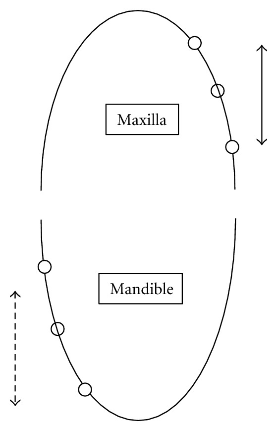
Aim. The aim of this study was histomorphologic and histomorphometric evaluation of peri-screw bone after immediate orthodontic and orthopedic forces and comparing them with a control group. Materials and Methods. 18 dual-top miniscrews were inserted in the premolar region of three Iranian dogs. Screws were divided into three groups: (1) the first group undergoing immediate orthodontic force of 300 cN, (2) the second group undergoing immediate orthopedic force of 600 cN and (3) a control group. Screws were explanted with adequate amount of surrounding bone after three months. Bone-screw contact (BSC), percentage of lamellar bone, and percentage of woven bone were evaluated. Wilcoxon and Man Whitney tests were used to analyze the data using SPSS software ver. 15 (α = 0.05). Results. There was no significant difference among the groups in terms of bone-screw contact (P value = 0.42), percentage of lamellar bone (P value = 0.83), and percentage of woven bone (P value = 0.88). Conclusion. By applying orthodontic and orthopedic forces to mini-screws the quality of surrounding bone and osseointegration will not be affected. Clinical Significance. Application of force to mini-screws is helpful in orthodontic-screw therapies.
Figures

Similar articles
-
Application of Orthodontic Immediate Force on Dental Implants: Histomorphologic and Histomorphometric Assessment.Ann Maxillofac Surg. 2017 Jan-Jun;7(1):11-17. doi: 10.4103/ams.ams_35_15. Ann Maxillofac Surg. 2017. PMID: 28713730 Free PMC article.
-
Osseointegration of orthodontic micro-screws after immediate and early loading.Angle Orthod. 2010 Mar;80(2):354-60. doi: 10.2319/021909-106.1. Angle Orthod. 2010. PMID: 19905862 Free PMC article.
-
Histomorphometric evaluation of cortical bone thickness surrounding miniscrew for orthodontic anchorage.Clin Implant Dent Relat Res. 2011 Sep;13(3):197-205. doi: 10.1111/j.1708-8208.2009.00197.x. Epub 2009 May 12. Clin Implant Dent Relat Res. 2011. PMID: 19438949
-
Tapered orthodontic miniscrews induce bone-screw cohesion following immediate loading.Eur J Orthod. 2006 Dec;28(6):541-6. doi: 10.1093/ejo/cjl044. Eur J Orthod. 2006. PMID: 17142258
-
Effects of orthodontic forces on bone turnover biomarkers in peri-miniscrew crevicular fluid: A systematic review.Int Orthod. 2020 Sep;18(3):403-411. doi: 10.1016/j.ortho.2020.03.005. Epub 2020 Apr 22. Int Orthod. 2020. PMID: 32335125
Cited by
-
Temporary anchorage devices and the forces and effects on the dentition and surrounding structures during orthodontic treatment: a scoping review.Eur J Orthod. 2023 May 31;45(3):324-337. doi: 10.1093/ejo/cjac072. Eur J Orthod. 2023. PMID: 36763546 Free PMC article.
-
Revisiting the Complications of Orthodontic Miniscrew.Biomed Res Int. 2022 Aug 1;2022:8720412. doi: 10.1155/2022/8720412. eCollection 2022. Biomed Res Int. 2022. PMID: 35958810 Free PMC article. Review.
-
Dental extrusion with orthodontic miniscrew anchorage: a case report describing a modified method.Case Rep Dent. 2015;2015:909314. doi: 10.1155/2015/909314. Epub 2015 Feb 2. Case Rep Dent. 2015. PMID: 25713739 Free PMC article.
References
-
- Huang LH, Shotwell JL, Wang HL. Dental implants for orthodontic anchorage. American Journal of Orthodontics and Dentofacial Orthopedics. 2005;127(6):713–722. - PubMed
-
- Graber TM, Vanarsdall RL. Orthodontics: current Principles and Techniques. Rio de Janeiro, Brazil edition. Guanabara Koogan: 1999.
-
- Melsen B, Petersen JK, Costa A. Zygoma ligatures: an alternative form of maxillary anchorage. Journal of Clinical Orthodontics. 1998;32(3):154–158. - PubMed
-
- Roberts WE, Smith RK, Zilberman Y, Mozsary PG, Smith RS. Osseous adaptation to continuous loading of rigid endosseous implants. American Journal of Orthodontics. 1984;86(2):95–111. - PubMed
-
- Wehrbein H, Merz BR, Diedrich P, Glatzmaier J. The use of palatal implants for orthodontic anchorage: design and clinical application of the orthosystem. Clinical Oral Implants Research. 1996;7(2):410–416. - PubMed
LinkOut - more resources
Full Text Sources
