. 2012:2012:732578.
doi: 10.1155/2012/732578. Epub 2012 Dec 18.

Caffeic Acid phenethyl ester inhibits oral cancer cell metastasis by regulating matrix metalloproteinase-2 and the mitogen-activated protein kinase pathway

Affiliations

Caffeic Acid phenethyl ester inhibits oral cancer cell metastasis by regulating matrix metalloproteinase-2 and the mitogen-activated protein kinase pathway

Chih-Yu Peng et al. Evid Based Complement Alternat Med. 2012.

Erratum in

Abstract

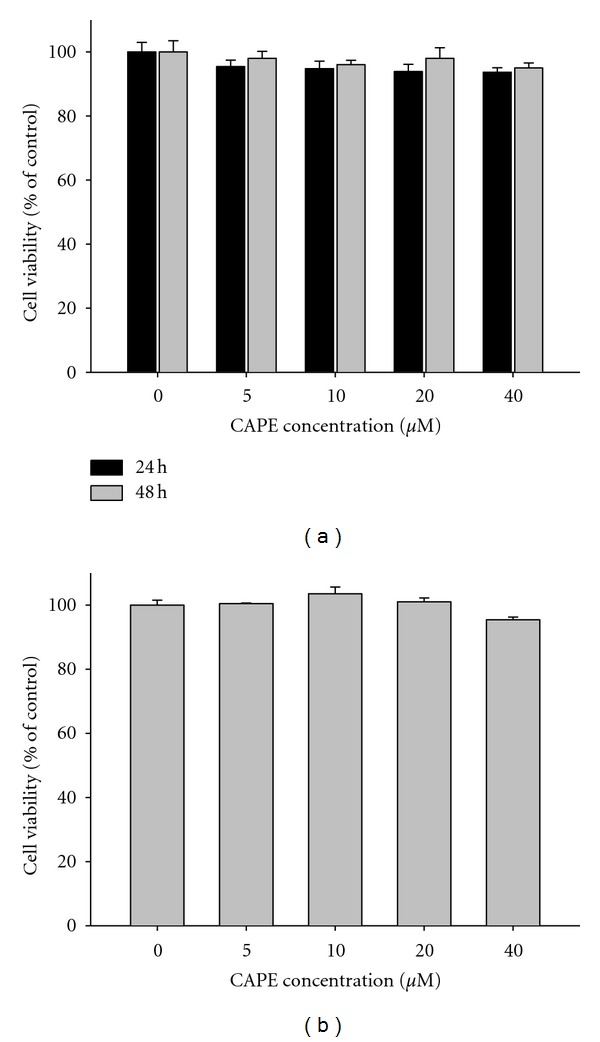
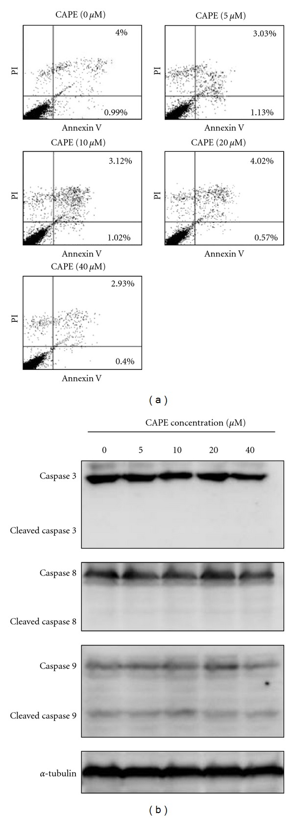
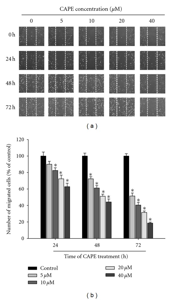
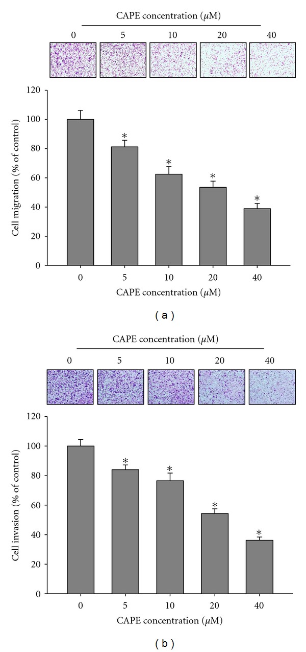
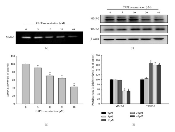
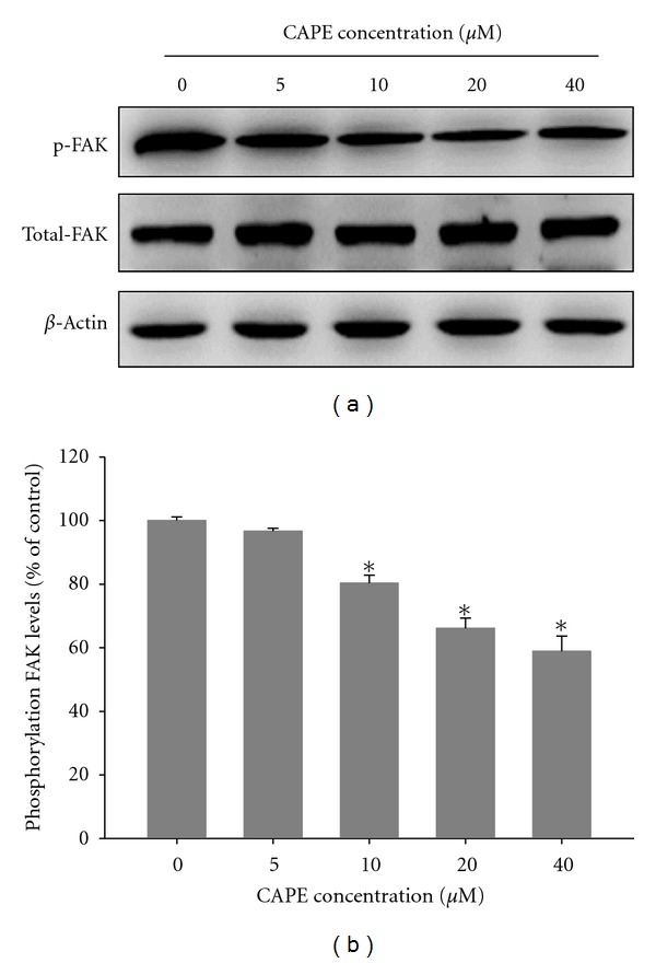
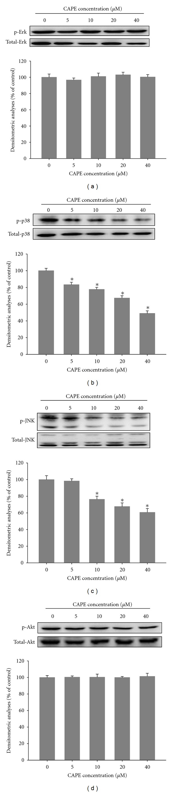
Caffeic acid phenethyl ester (CAPE), an active component extracted from honeybee hives, exhibits anti-inflammatory and anticancer activities. However, the molecular mechanism by which CAPE affects oral cancer cell metastasis has yet to be elucidated. In this study, we investigated the potential mechanisms underlying the effects of CAPE on the invasive ability of SCC-9 oral cancer cells. Results showed that CAPE attenuated SCC-9 cell migration and invasion at noncytotoxic concentrations (0 μM to 40 μM). Western blot and gelatin zymography analysis findings further indicated that CAPE downregulated matrix metalloproteinase-2 (MMP-2) protein expression and inhibited its enzymatic activity. CAPE exerted its inhibitory effects on MMP-2 expression and activity by upregulating tissue inhibitor of metalloproteinase-2 (TIMP-2) and potently decreased migration by reducing focal adhesion kinase (FAK) phosphorylation and the activation of its downstream signaling molecules p38/MAPK and JNK. These data indicate that CAPE could potentially be used as a chemoagent to prevent oral cancer metastasis.


Figures

Figure 1
Figure 1
Effect of CAPE on cell viability. (a) SCC-9 cells were treated with CAPE (0, 5, 10, 20, and 40 μM) for 24 h and 48 h before being subjected to an MTT assay for cell viability. (b) Normal gingival fibroblast cells were treated with CAPE (0, 5, 10, 20, and 40 μM) for 24 h before being subjected to an MTT assay for cell viability. The values represented the means ± SD of at least three independent experiments.
Figure 2
Figure 2
Effects of CAPE on the Annexin V flipping and caspase cleavage in SCC-9 cells. SCC-9 cells were treated with CAPE (0–40 μM) for 24 h and then subjected to Annexin V and PI double-stained flow cytometry (a) or Western blotting to analyze the protein levels of caspase 3, 8, and 9 (b).
Figure 3
Figure 3
Effect of CAPE on in vitro wound closure in oral cancer cells. (a) SCC-9 cells were wounded and then treated with vehicle (DMSO) or CAPE (0, 5, 10, 20, and 40 μM) for 0 h, 24 h, 48 h, and 72 h in 0.5% FBS-containing medium. At 0, 24 h, 48 and 72 h, phase-contrast pictures of the wounds at three different locations were taken. (b) Cells migrating into the wound area were counted using the dashed line as time zero. A quantitative assessment of the mean number of cells in the denuded zone is the mean ± SD (n = 3).
Figure 4
Figure 4
Effect of CAPE on cell migration and invasion in SCC-9 cells. (a) The cell migration and (b) cell invasion were measured using a Boyden chamber for 16 h and 24 h with polycarbonate filters, respectively. The migration and invasion abilities of SCC-9 cells were quantified by counting the number of cells that invaded to the underside of the porous polycarbonate as described in Section 2. The values represented the means ± SD of at least three independent experiments. *P < 0.05 as compared with the vehicle group.
Figure 5
Figure 5
Effects of CAPE on the activity and protein level of MMP-2 and the protein level of the endogenous inhibitor TIMP-2. (a-b) SCC-9 cells were treated with CAPE (0, 5, 10, 20, and 40 μM) for 24 h and then subjected to gelatin zymography to analyze the activity of MMP-2. (c-d) SCC-9 cells were treated with CAPE (0, 5, 10, 20, and 40 μM) for 24 h and then subjected to western blotting to analyze the protein levels of MMP-2 and TIMP-2. Quantitative results of MMP-2 and TIMP-2 protein levels which were adjusted with β-actin protein level. The values represented the means ± SD of at least three independent experiments. *P < 0.05 as compared with the vehicle group.
Figure 6
Figure 6
Effects of CAPE on the phosphorylation level of FAK. (a) SCC-9 cells were treated with CAPE (0, 5, 10, 20, and 40 μM) for 24 h and then subjected to western blotting to analyze the levels of FAK. (b) Quantitative results of phosphorylation level of FAK which were adjusted with total FAK protein level. The values represented the means ± SD of at least three independent experiments. *P < 0.05 as compared with the vehicle group.
Figure 7
Figure 7
Effects of CAPE on the MAPKs pathway and Akt signalings. SCC-9 cells were cultured in various concentrations of CAPE (0, 5, 10, 20, and 40 μM) for 24 hours, and then the cell lysates were subjected to SDS-PAGE followed by western blots with (a) anti-ERK1/2, (b) anti-p38, (c) anti-JNK, and (d) anti-Akt (total and phosphorylated) antibodies as described in Section 2. Determined activities of these proteins were subsequently quantified by densitometric analyses with that of control being 100% as shown just after the gel data. The values represented the means ± SD of at least 3 independent experiments. *P < 0.05 as compared with the vehicle group.

References

    1. Spiro RH, Alfonso AE, Farr HW, Strong EW. Cervical node metastasis from epidermoid carcinoma of the oral cavity and oropharynx. A critical assessment of current staging. American Journal of Surgery. 1974;128(4):562–567. - PubMed
    1. Stetler-Stevenson WG, Liotta LA, Kleiner DE. Extracellular matrix 6: role of matrix metalloproteinases in tumor invasion and metastasis. FASEB Journal. 1993;7(15):1434–1441. - PubMed
    1. Chien MH, Ying TH, Hsieh YS, et al. Dioscorea nipponica Makino inhibits migration and invasion of human oral cancer HSC-3 cells by transcriptional inhibition of matrix metalloproteinase-2 through modulation of CREB and AP-1 activity. Food and Chemical Toxicology. 2012;50:558–566. - PubMed
    1. Yang SF, Chen MK, Hsieh YS, et al. Antimetastatic effects of Terminalia catappa L. on oral cancer via a down-regulation of metastasis-associated proteases. Food and Chemical Toxicology. 2010;48(4):1052–1058. - PubMed
    1. Egeblad M, Werb Z. New functions for the matrix metalloproteinases in cancer progression. Nature Reviews Cancer. 2002;2(3):161–174. - PubMed