

A critical HA1 neutralizing domain of H5N1 influenza in an optimal conformation induces strong cross-protection
- PMID: 23320093
- PMCID: PMC3539987
- DOI: 10.1371/journal.pone.0053568
A critical HA1 neutralizing domain of H5N1 influenza in an optimal conformation induces strong cross-protection
Abstract
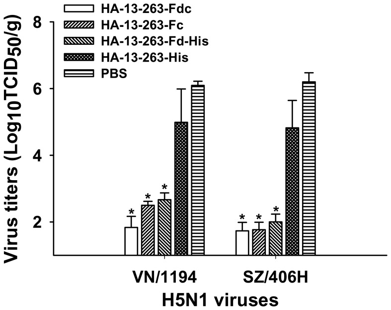
The highly pathogenic avian influenza (HPAI) H5N1 viruses, especially the laboratory-generated H5N1 mutants, have demonstrated the potential to cross the species barrier and infect mammals and humans. Consequently, the design of an effective and safe anti-H5N1 vaccine is essential. We previously demonstrated that the full-length hemagglutinin 1 (HA1) could induce significant neutralizing antibody response and protection. Here, we intended to identify the critical neutralizing domain (CND) in an optimal conformation that can elicit strong cross-neutralizing antibodies and protection against divergent H5N1 strains. We thus constructed six recombinant proteins covering different regions of HA1 of A/Anhui/1/2005(H5N1), each of which was fused with foldon (Fd) and Fc of human IgG. We found that the critical fragment fused with Fd/Fc (HA-13-263-Fdc, H5 numbering) that could elicit the strongest neutralizing antibody response is located in the N-terminal region of HA1 (residues 13-263), which covers the receptor-binding domain (RBD, residues 112-263). We then constructed three additional recombinants fused with Fd plus His tag (HA-13-263-Fd-His), Fc only (HA-13-263-Fc), and His tag only (HA-13-263-His), respectively. We found that the HA-13-263-Fdc, which formed an oligomeric conformation, induced the strongest neutralizing antibody response and cross-protection against challenges of two tested H5N1 virus strains covering clade 1: A/VietNam/1194/2004 (VN/1194) or clade 2.3.4: A/Shenzhen/406H/06 (SZ/406H), while HA-13-263-Fc dimer and HA-13-263-Fd-His trimer elicited higher neutralizing antibody response and protection than HA-13-263-His monomer. These results suggest that the oligomeric form of the CND containing the RBD can be further developed as an effective and safe vaccine for cross-protection against divergent strains of H5N1 viruses.
Conflict of interest statement
Figures
Similar articles
-
Intranasal vaccination of recombinant H5N1 HA1 proteins fused with foldon and Fc induces strong mucosal immune responses with neutralizing activity: Implication for developing novel mucosal influenza vaccines.Hum Vaccin Immunother. 2015;11(12):2831-8. doi: 10.1080/21645515.2015.1074363. Hum Vaccin Immunother. 2015. PMID: 26260706 Free PMC article.
-
A recombinant vaccine of H5N1 HA1 fused with foldon and human IgG Fc induced complete cross-clade protection against divergent H5N1 viruses.PLoS One. 2011 Jan 27;6(1):e16555. doi: 10.1371/journal.pone.0016555. PLoS One. 2011. PMID: 21304591 Free PMC article.
-
Unmasking Stem-Specific Neutralizing Epitopes by Abolishing N-Linked Glycosylation Sites of Influenza Virus Hemagglutinin Proteins for Vaccine Design.J Virol. 2016 Sep 12;90(19):8496-508. doi: 10.1128/JVI.00880-16. Print 2016 Oct 1. J Virol. 2016. PMID: 27440889 Free PMC article.
-
Potential cross-species transmission of highly pathogenic avian influenza H5 subtype (HPAI H5) viruses to humans calls for the development of H5-specific and universal influenza vaccines.Cell Discov. 2023 Jun 16;9(1):58. doi: 10.1038/s41421-023-00571-x. Cell Discov. 2023. PMID: 37328456 Free PMC article. Review.
-
The Role of Fc Gamma Receptors in Broad Protection against Influenza Viruses.Vaccines (Basel). 2018 Jun 29;6(3):36. doi: 10.3390/vaccines6030036. Vaccines (Basel). 2018. PMID: 29966222 Free PMC article. Review.
Cited by
-
A recombinant receptor-binding domain of MERS-CoV in trimeric form protects human dipeptidyl peptidase 4 (hDPP4) transgenic mice from MERS-CoV infection.Virology. 2016 Dec;499:375-382. doi: 10.1016/j.virol.2016.10.005. Epub 2016 Oct 15. Virology. 2016. PMID: 27750111 Free PMC article.
-
Designing a multi-epitope vaccine to provoke the robust immune response against influenza A H7N9.Sci Rep. 2021 Dec 29;11(1):24485. doi: 10.1038/s41598-021-03932-2. Sci Rep. 2021. PMID: 34966175 Free PMC article.
-
A safe and convenient pseudovirus-based inhibition assay to detect neutralizing antibodies and screen for viral entry inhibitors against the novel human coronavirus MERS-CoV.Virol J. 2013 Aug 26;10:266. doi: 10.1186/1743-422X-10-266. Virol J. 2013. PMID: 23978242 Free PMC article.
-
Nanodiamond enhances immune responses in mice against recombinant HA/H7N9 protein.J Nanobiotechnology. 2017 Oct 5;15(1):69. doi: 10.1186/s12951-017-0305-2. J Nanobiotechnology. 2017. PMID: 28982373 Free PMC article.
-
Identification of an ideal adjuvant for receptor-binding domain-based subunit vaccines against Middle East respiratory syndrome coronavirus.Cell Mol Immunol. 2016 Mar;13(2):180-90. doi: 10.1038/cmi.2015.03. Epub 2015 Feb 2. Cell Mol Immunol. 2016. PMID: 25640653 Free PMC article.
References
Publication types
MeSH terms
Substances
Grants and funding
LinkOut - more resources
Full Text Sources
Other Literature Sources
Medical
Research Materials
