. 2013;8(1):e53596.
doi: 10.1371/journal.pone.0053596. Epub 2013 Jan 8.

Neurogenic and neurotrophic effects of BDNF peptides in mouse hippocampal primary neuronal cell cultures

Affiliations

Neurogenic and neurotrophic effects of BDNF peptides in mouse hippocampal primary neuronal cell cultures

Maria del Carmen Cardenas-Aguayo et al. PLoS One. 2013.

Abstract

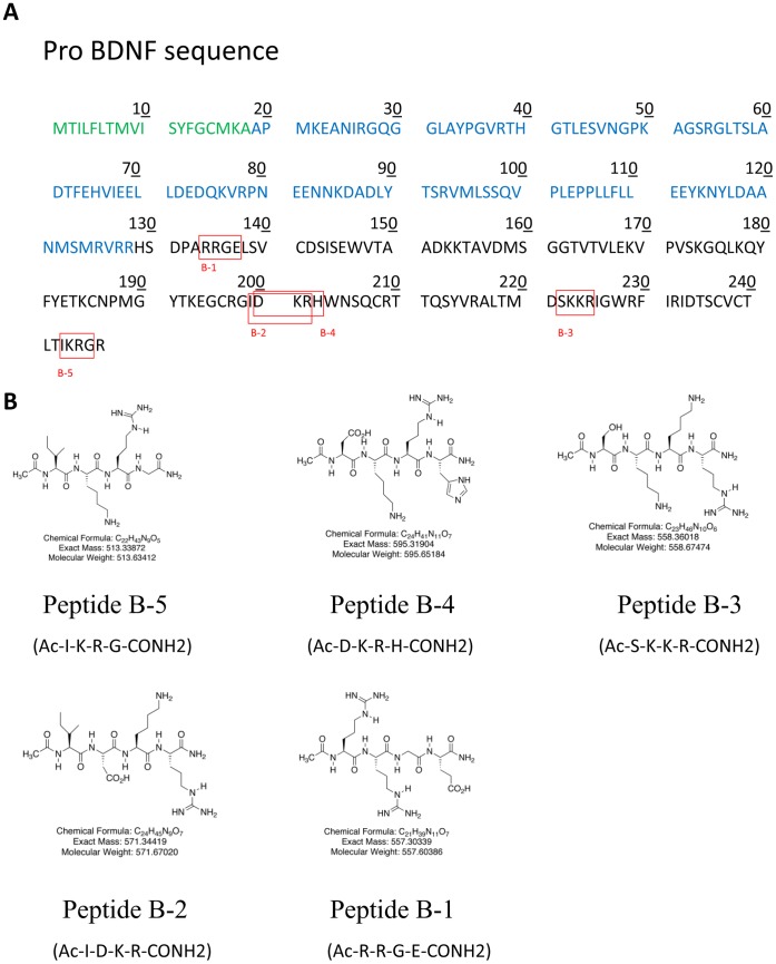
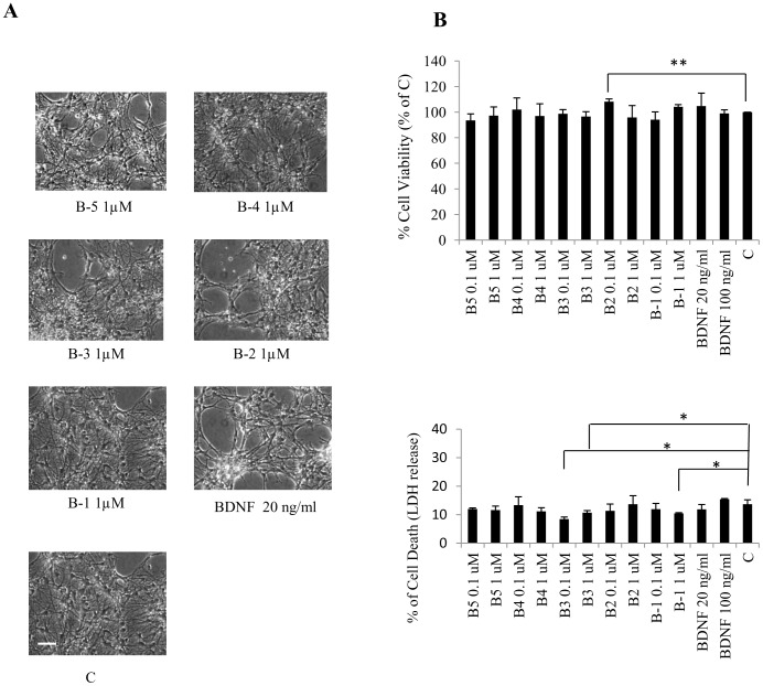
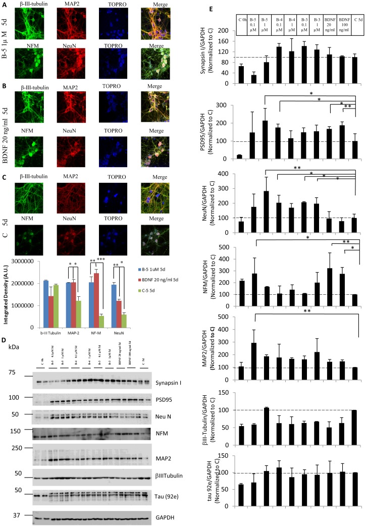
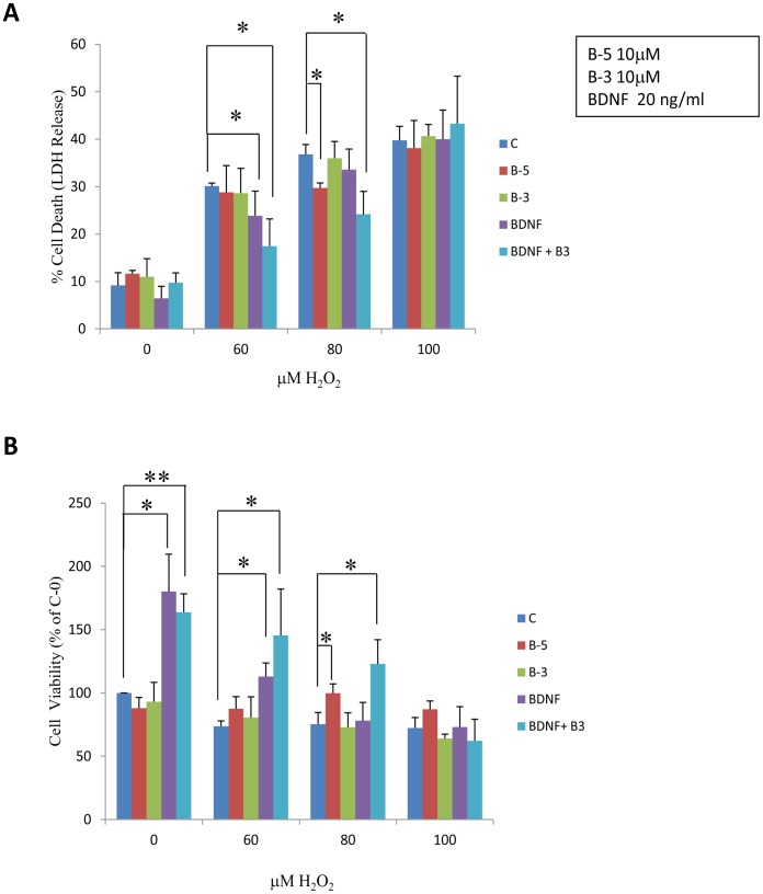
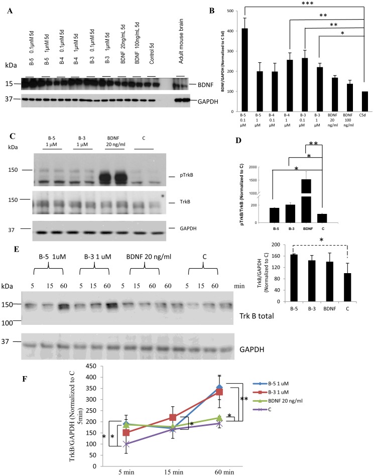
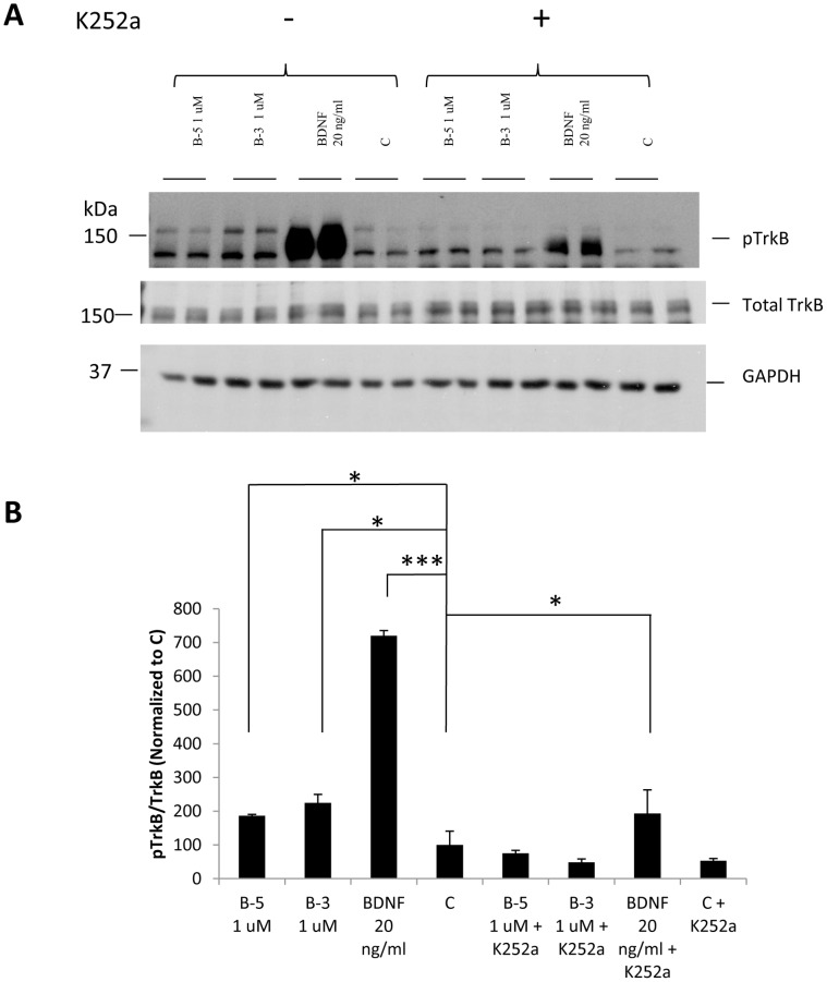
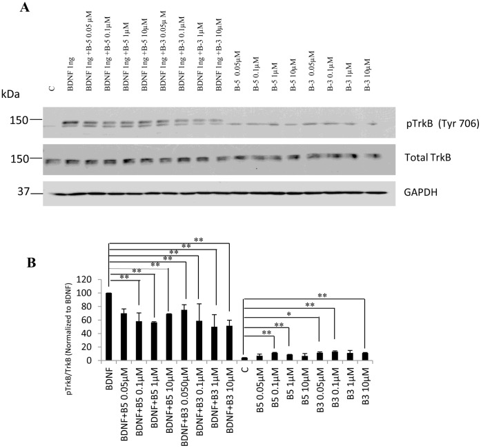
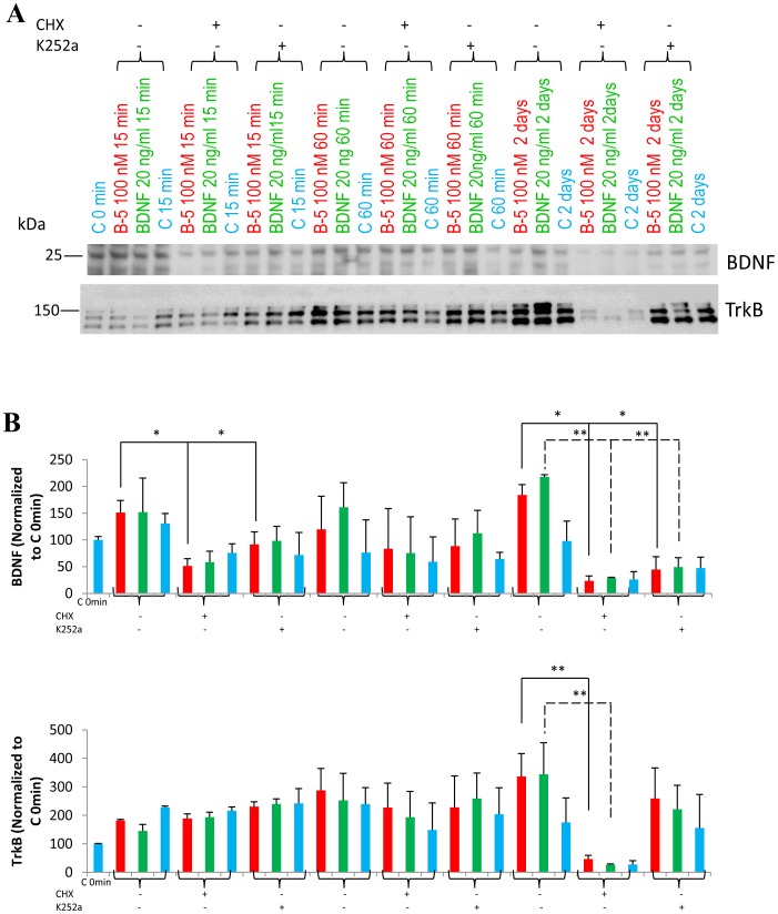
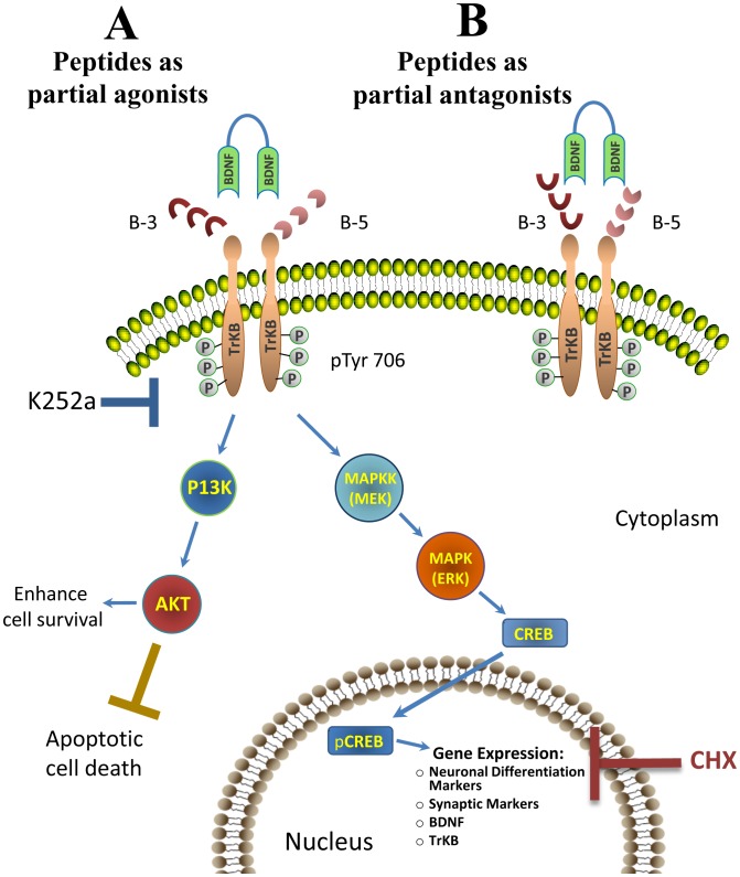
The level of brain-derived neurotrophic factor (BDNF), a member of the neurotrophin family, is down regulated in Alzheimer's disease (AD), Parkinson's disease (PD), depression, stress, and anxiety; conversely the level of this neurotrophin is increased in autism spectrum disorders. Thus, modulating the level of BDNF can be a potential therapeutic approach for nervous system pathologies. In the present study, we designed five different tetra peptides (peptides B-1 to B-5) corresponding to different active regions of BDNF. These tetra peptides were found to be non-toxic, and they induced the expression of neuronal markers in mouse embryonic day 18 (E18) primary hippocampal neuronal cultures. Additionally, peptide B-5 induced the expression of BDNF and its receptor, TrkB, suggesting a positive feedback mechanism. The BDNF peptides induced only a moderate activation (phosphorylation at Tyr 706) of the TrkB receptor, which could be blocked by the Trk's inhibitor, K252a. Peptide B-3, when combined with BDNF, potentiated the survival effect of this neurotrophin on H(2)O(2)-treated E18 hippocampal cells. Peptides B-3 and B-5 were found to work as partial agonists and as partial antagonists competing with BDNF to activate the TrkB receptor in a dose-dependent manner. Taken together, these results suggest that the described BDNF tetra peptides are neurotrophic, can modulate BDNF signaling in a partial agonist/antagonist way, and offer a novel therapeutic approach to neural pathologies where BDNF levels are dysregulated.


Conflict of interest statement

Competing Interests: Studies described in this article were supported in part by the New York State Office of Peoplewith Developmental Disabilities, and a research grant from EVER NeuroPharma GmbH, Unterach, Austria. This does not alter the authors’ adherence to all the PLOS ONE policies on sharing data and materials.

Figures

Figure 1
Figure 1. BDNF tetrapeptides used in this study.
(A) Amino acid sequence of human BDNF. The signal peptide (18 amino acids, positions 1–18) is shown in green, the propeptide (110 amino acids, positions 19–128) in blue, and the BDNF sequence, (119 amino acids, positions 129–247, molecular weight 26 kDa) in black. The sequences of the 5 tetra peptides used in this study (B-1 to B-5) are boxed (as red squares). (B) Chemical structures of the BDNF peptides used in this study.
Figure 2
Figure 2. BDNF peptides B-1 to B-5 are non-toxic to E18 mouse primary hippocampal neuronal cells.
(A) Phase contrast photomicrographs of hippocampal neurons vehicle-treated or treated with BDNF (20 ng/ml) or the indicated peptides (at 1 µM) for 5 days. Scale bar represents 20 µm. (B) Effect of BDNF peptides on neuronal survival. Lactate dehydrogenase (LDH) assay showing the percentages of cell viability and cell death after 5 days of treatment with the peptides (at 0.1 or 1 µM) or BDNF (at 20 and 100 ng/ml; 0.79 and 3.95 nM respectively). Counts were normalized to survival achieved with vehicle treatment for 5 days (control, C). Values for each concentration derived from 3 independent experiments. *p<0.05, **p<0.01. ANOVA with post-hoc test and/or Student’s t test.
Figure 3
Figure 3. BDNF peptides B-5, B-4, and B-3 are neurogenic and neurotrophic.
(A–C) Peptide B-5 induces the expression of the neuronal markers β-III-tubulin, MAP2, NFM, and NeuN in E18 primary hippocampal cell cultures. Representative confocal images illustrating double immunolabeling of β–III-tubulin (green) and MAP2 (red) and NFM (green) and NeuN (red) in cells treated for 5 days with (A) Peptide B-5 at 1 µM, (B) with BDNF, 20 ng/ml (0.79 nM) for 5 days, and,(C) vehicle only (control, C). TOPRO-3 (blue) was used to stain the nuclei. Magnification bar = 10 µm. The bottom panel shows the level of fluorescence intensity using Image J software to measure the level of expression. (D, E) BDNF peptides (B-5, B-4, and B-3) induce the expression of neuronal markers in E18 primary hippocampal neurons. (D) Representative Western blots showing an increase in the expression of the neuronal markers PSD95, NeuN, NFM, and MAP-2 in cells treated with the peptides (B5, B-4 and B-3) at concentrations of 0.1 µM and 1 µM or BDNF at concentrations of 20 or 100 ng/ml (0.79 or 3.95 nM respectively). GPADH was used as a loading control. (E) Quantification of the Western blots of neuronal markers shown in D. The integrated density value of the bands in Western blots was determined using densitometry (Fuji software, Multi Gauge, Version 3.0), and data was normalized to GAPDH and to control (medium treated cells for 5 days, C 5d). Data are shown as mean ± standard deviation, n = 3. 10% SDS-PAGE gels. *p<0.05, **p<0.01, ***p<0.001. One-way ANOVA/post-hoc test/Student’s t test.
Figure 4
Figure 4. BDNF peptides potentiate the effect of BDNF in rescuing H2O2-induced neurotoxicity.
(A) LDH cytotoxicity assay showing the percentage of cell death in hippocampal cells treated with increasing concentrations of H2O2 i.e. 0, 60, 80, and 100 µM for 6 h, and then after changing the medium, exposed to B-5, B-3, BDNF or BDNF+B-3 for 24 h. Peptide B-5 significantly reduced cell death caused by 80 µM H2O2 and Peptide B-3 significantly potentiated the neuroprotective effect of BDNF. (B) Percent viability of hippocampal cells by LDH assay. The cells were treated in the same way as in A. Peptide B-3 significantly increased the viability when combined with BDNF in cells not treated or treated with 60 or 80 µM of H2O2. Data were normalized to control (vehicle treated cells). * p<0.05, **p<0.01. ANOVA and/or Student’s t test, n = 3.
Figure 5
Figure 5. (A) BDNF peptides B-5, B-4 and B-3 induce the expression of BDNF.
Western blot analyses of cells treated with peptides B-5, B-4 and B-3, or with BDNF, or vehicle for 5 days, showed an increase in BDNF expression in cells treated with the peptides. A sample of adult mouse brain was included as a control for the migration of the bands corresponding to pro-BDNF and BDNF. (B) Densitometric quantitation of the Western blots developed with anti-BDNF. Data was normalized to GAPDH as loading control and then to 5 days control vehicle treated cells, C 5d. (C) Peptides B-5 and B-3 activated TrkB receptor in primary E18 hippocampal cells. Western blots showing phosphorylation of TrkB at Tyr 706 on treatment with Peptide B-5 (1 µM), Peptide B-3 (1 µM) or BDNF (20 ng/ml, 0.79 nM) for 1 h as compared to control treated cells, C. Lower blots show the levels of TrkB receptor and the levels of GAPDH as a loading control. (D) Densitometric analysis of the Western-blots for pTrkB normalized to TrkB, and TrkB normalized to GAPDH. Control was taken as 100 percent in each case. (E) BDNF peptides (B-5 and B-3) increased the expression of TrkB. Increase in expression of TrkB in TrkB stably-expressing NIH-3T3 fibroblast cells, as a function of time. Cells were treated for 5, 15 or 60 min with B-5 (1 µM), B-3 (1 µM), BDNF (20 ng/ml), or vehicle only (Control, C). Western blots of total TrkB, and GAPDH included as a loading control. (F) Densitometric quantitation of the Western blots for TrkB normalized to GAPDH. Data are shown as mean ± standard deviation, n = 3. *p<0.05, **p<0.01, ***p<0.001. One-way ANOVA/post-hoc test/Student’s t test. Dashed line in (D) denotes that B-5 induction of TrkB expression almost approaches significance (p = 0.057, one-way ANOVA).
Figure 6
Figure 6. Activation of the TrkB receptor by BDNF peptides B-5 and B-3 can be blocked by the TrkB inhibitor, K252a.
(A) Cells were pretreated with or without K252a for 1 h and then exposed to Peptide B-5 or Peptide B-3 at 1 µM or 20 ng/ml BDNF for 5 min. Western blots of pTrkB (Tyr 706), total TrkB and GAPDH included as loading control. (B) Densitometric quantitation of the Western-blots for pTrkB normalized to TrkB. Data are shown as mean ± standard deviation, n = 3. *p<0.05,**p<0.01, ***p<0.001. One-way ANOVA/post-hoc test/Student’s t-test.
Figure 7
Figure 7. (A, B) BDNF peptides act as partial agonists and antagonists of BDNF.
Competition experiments showing inhibition of the activation (pTrkB) of the TrkB receptor when the TrkB receptor stably expressing NIH-3T3 fibroblasts were treated with increasing concentrations of peptides B-5 or B-3 in the presence or absence of BDNF for 15 min. (A) Western blots of pTrkB (Tyr 706) and of total TrkB. GAPDH is included as a loading control. (B) Densitometric quantitation of Western blots for pTrkB normalized to TrkB (after normalizing TrkB to GAPDH) and shown as a percentage of BDNF. Data are shown as mean ± standard deviation, n = 3. *p<0.05, **p<0.01. One-way ANOVA/post-hoc test/Student’s t test.
Figure 8
Figure 8. (A,B) The increase in expression of BDNF in primary hippocampal cells caused by BDNF peptides or BDNF after two days of treatment can be blocked by pre-exposure (1 h before adding the peptides) to protein synthesis inhibitor, cycloheximide (CHX) or to TrkB inhibitor, K252a.
(A) Western blots of BDNF, and total TrkB, of hippocampal cells treated with 0.1 µM peptide B-5 or 20 ng/ml BDNF or vehicle treated, and when indicated, also pretreated with CHX or K252a (B) Densitometric quantitation of the Western-blots for BDNF and TrkB normalized to the corresponding control cells (control, C) in each condition. Data are shown as mean ± standard deviation, n = 3. *p<0.05, **p<0.01. One-way ANOVA/post-hoc test/Student’s t test.
Figure 9
Figure 9. Proposed mechanism of action of BDNF peptides.
BDNF peptides B-5 and B-3 may interact with or compete for the binding site of BDNF to its transmembrane receptor TrkB. Depending on the concentration or the cellular state or condition (i.e. during stress, like in the presence of H2O2), the peptides could act as partial agonists or partial antagonists. Fig. 8A shows the partial agonistic role of the peptides. In this case, the peptides favor the activation of the TrkB receptor, and in the presence of BDNF, they synergize with it. Once the TrkB receptor gets activated, it is dimerized and autophosphorylated (one of the residues that gets phosphorylated is the Tyr 706) and the signal is transduced. The cascades that could be activated by the peptides include the differentiation pathway through MAPK and pCREB regulating gene expression of markers of neuronal phenotype and plasticity, and also the expression of BDNF and TrkB, giving the possibility of a feedback mechanism. The other cascade that could be activated by the peptides is the survival one, in which PI3K and AKT participate to enhance survival and inhibit cell death. Fig. 8B represents the partial antagonistic role of the peptides where the peptides compete with BDNF for the activation of the receptor blocking the TrkB activation by BDNF and its signal transduction pathway. The sites where the TrkB inhibitor K252a and the protein synthesis inhibitor CHX can block the pathway are shown with a grey and red bar, respectively.

Similar articles

Cited by

References

    1. Kaplan DR, Miller FD (2000) Neurotrophin signal transduction in the nervous system. Curr Opin Neurobiol 10: 381–391. - PubMed
    1. Chao MV (2003) Neurotrophins and their receptors: a convergence point for many signalling pathways. Nat Rev Neurosci 4: 299–309. - PubMed
    1. Li N, Liu GT (2010) The novel squamosamide derivative FLZ enhances BDNF/TrkB/CREB signaling and inhibits neuronal apoptosis in APP/PS1 mice. Acta Pharmacol Sin 31: 265–272. - PMC - PubMed
    1. Counts SE, Mufson EJ (2005) The role of nerve growth factor receptors in cholinergic basal forebrain degeneration in prodromal Alzheimer disease. J Neuropathol Exp Neurol 64: 263–272. - PubMed
    1. Tapia-Arancibia L, Aliaga E, Silhol M, Arancibia S (2008) New insights into brain BDNF function in normal aging and Alzheimer disease. Brain Res Rev 59: 201–220. - PubMed

Publication types

MeSH terms