. 2013;8(1):e53940.
doi: 10.1371/journal.pone.0053940. Epub 2013 Jan 8.

Katanin localization requires triplet microtubules in Chlamydomonas reinhardtii

Affiliations

Katanin localization requires triplet microtubules in Chlamydomonas reinhardtii

Jessica M Esparza et al. PLoS One. 2013.

Erratum in

Abstract

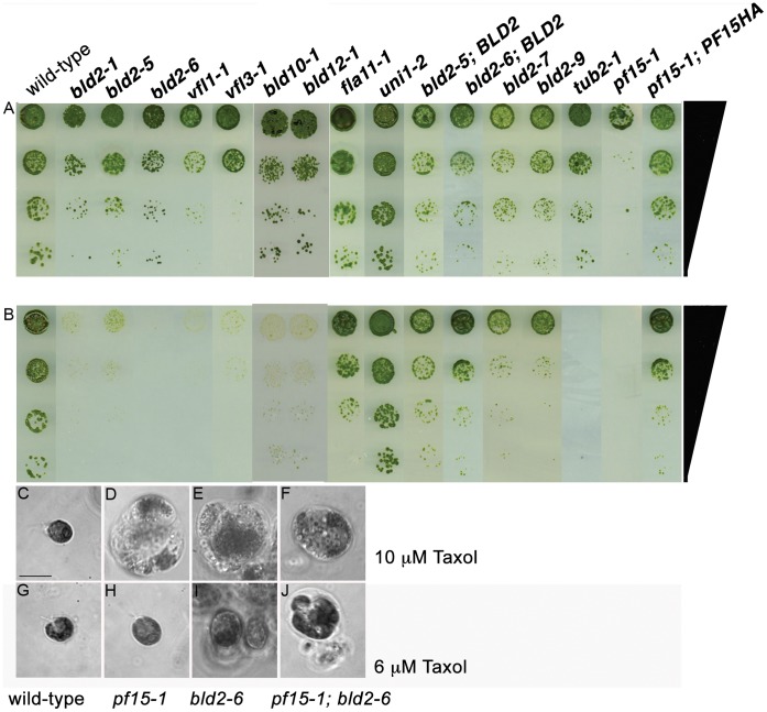
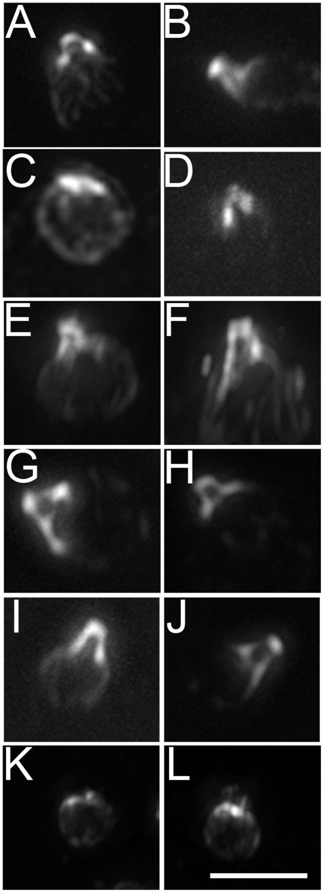
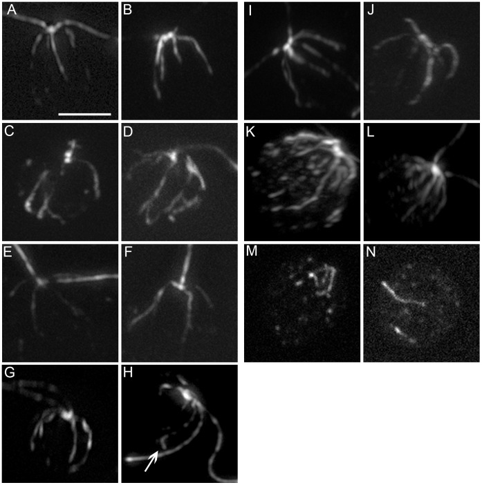
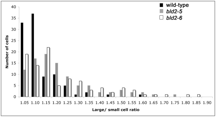
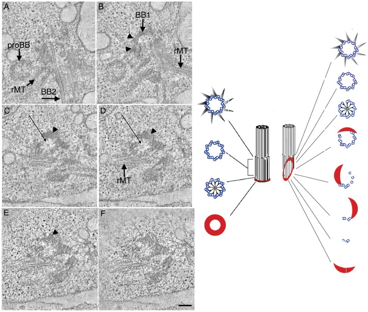
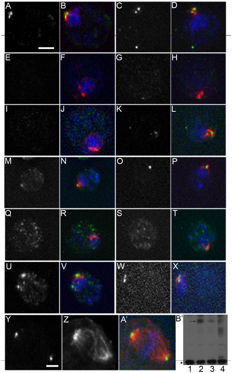
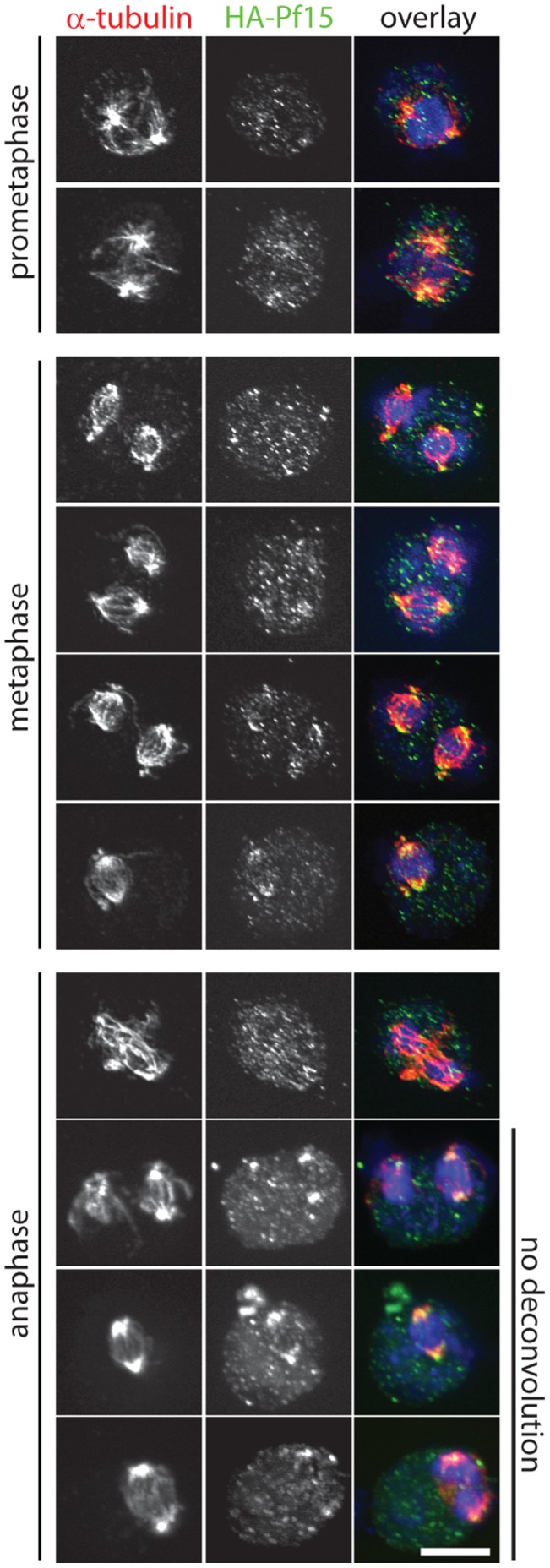
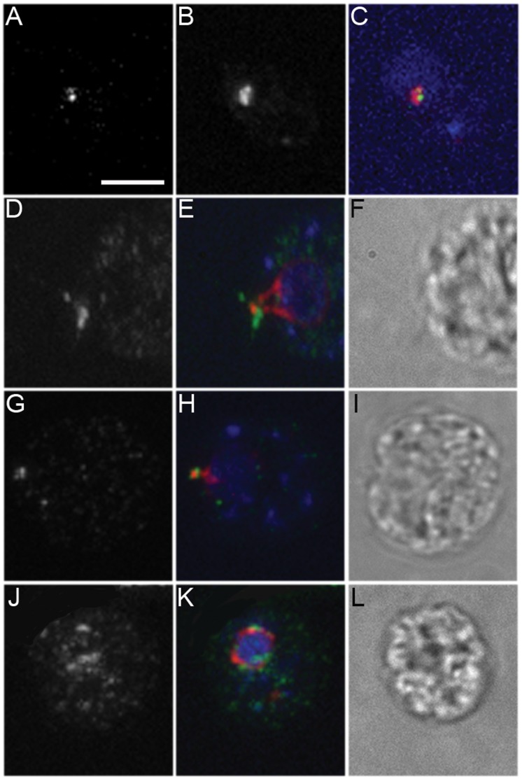
Centrioles and basal bodies are essential for a variety of cellular processes that include the recruitment of proteins to these structures for both centrosomal and ciliary function. This recruitment is compromised when centriole/basal body assembly is defective. Mutations that cause basal body assembly defects confer supersensitivity to Taxol. These include bld2, bld10, bld12, uni3, vfl1, vfl2, and vfl3. Flagellar motility mutants do not confer sensitivity with the exception of mutations in the p60 (pf19) and p80 (pf15) subunits of the microtubule severing protein katanin. We have identified additional pf15 and bld2 (ε-tubulin) alleles in screens for Taxol sensitivity. Null pf15 and bld2 alleles are viable and are not essential genes in Chlamydomonas. Analysis of double mutant strains with the pf15-3 and bld2-6 null alleles suggests that basal bodies in Chlamydomonas may recruit additional proteins beyond katanin that affect spindle microtubule stability. The bld2-5 allele is a hypomorphic allele and its phenotype is modulated by nutritional cues. Basal bodies in bld2-5 cells are missing proximal ends. The basal body mutants show aberrant localization of an epitope-tagged p80 subunit of katanin. Unlike IFT proteins, katanin p80 does not localize to the transition fibers of the basal bodies based on an analysis of the uni1 mutant as well as the lack of colocalization of katanin p80 with IFT74. We suggest that the triplet microtubules are likely to play a key role in katanin p80 recruitment to the basal body of Chlamydomonas rather than the transition fibers that are needed for IFT localization.


Conflict of interest statement

Competing Interests: The authors have declared that no competing interests exist.

Figures

Figure 1
Figure 1. Basal body mutant strains show supersensitivity to Taxol.
(A) Serial dilution of mutant, rescued, and intragenic revertant strains on control medium and (B) 8 µM Taxol-containing medium. Phase images of cells on media with different Taxol concentrations. (C, G) Wild-type, (D, H) pf15-1, (E, I) bld2-6 and (F, J) bld2-6, pf15-1 double mutant on 10 µM (C–F) or 6 µM Taxol (G–J) containing medium. The bld2-6, pf15-1 double mutant is unable to grow on 6 µM Taxol containing medium compared to the single mutant strains. Scale bar in Panel C equals 10 µm. Panels C–J are at the same magnification.
Figure 2
Figure 2. Centrin localization varies in the bld2-5 and bld2-6 strains.
(A, B) Wild-type cells with an extended pattern of centrin. (C) Approximately 41% of bld2-5 cells have centrin that collapses on the nucleus (n = 70). (D) bld2-5 cells with a wild-type centrin pattern. (E, F) The rescued transformant, bld2-5; BLD2, and (G, H) the pseudorevertants, bld2-7 and (I, J) bld2-9 have extended centrin. (K, L) All bld2-6 cells show centrin collapses on or around the nucleus. Scale bar in panel L equals 10 µm. Panels A–L are at the same magnification.
Figure 3
Figure 3. Rootlet microtubules are disorganized in the bld2-5 and bld2-6 strains.
(A, B) Rootlet microtubules in wild-type cells form a cruicate pattern. (C, D) bld2-5 cells show an aberrant number and placement of rootlet microtubules. (E, F) The bld2-5; BLD2 strain shows a wild-type rootlet microtubule phenotype (N = 15). Pseudorevertants bld2-7 (G, H) and bld2-9 (I, J) have a nearly wild-type rootlet microtubule phenotype but splaying occurs at the ends of the microtubules (arrow). (K, L) The tub2-1 strain has increased acetylated α-tubulin staining. (M, N) The bld2-6 cells have a severe disorganization of rootlet microtubules. Scale bar in Panel A equals 5 µm. Panels A–N are at the same magnification.
Figure 4
Figure 4. The bld2-5 and bld2-6 strains misplace the cleavage furrow.
A. The ratio of the areas of wild-type sister cells is approximately equal to one (black bars), whereas the ratio of the areas of bld2-5 (gray bars) and bld2-6 (white bars) sister cells is equal to or greater than one, which suggests a defect in proper placement of the cleavage furrow . These results are statistically significant compared by a permutation test .
Figure 5
Figure 5. Mature basal bodies in the bld2-5 strain contain defects in microtubule blade organization.
Selected tomographic slices show the progression from the proximal (A) to the distal end (F) of the basal body. (A) Probasal bodies contain a ring of amorphous material at their proximal base. (B–E; arrowheads) Mature basal bodies (BB1) contain amorphous material that extends distally rather than in a proximal ring. (D–F) The assembly of microtubule blades is also incomplete with singlet, doublet and sometimes triplet microtubules present. (C, D; arrows) The cartwheel structure is observed distally. BB1, mature basal body 1; BB2, mature basal body 2; rMT, rootlet microtubules. Scale bar equals 100 nm and Panels A–F are at the same magnification. Schematic representation of the structure of a wild-type basal body and the defects in bld2-5 basal bodies as one moves from the proximal to distal region of the basal body.
Figure 6
Figure 6. Katanin p80 is absent or diffusely localized in the bld2 mutant strains.
(A–D) Katanin p80 localizes as two dots (green) near centrin (red) in wild-type cells. DNA is stained with DAPI (blue). (E–H) bld2-6 cells lack or show diffuse katanin p80 staining. (I–L) in the bld2-5 strain has heterogeneous katanin p80 staining that is correlated with the centrin localization pattern. Katanin p80 localization appears wild-type in bld2-7 (M, N) and bld2-9 (O, P) pseudorevertants. bld10 cells do not localize katanin p80 (Q, R), while uni3 cells show variable staining (S–V). Katanin p80 localization appears wild-type in uni1 cells (W, X). Katanin p80 localizes to the spindle poles during mitosis where the spindle microtubules (red) are stained with an antibody against α-tubulin and DNA (blue) is stained with DAPI (Y–A’). (B’) Immunoblot of katanin p80 (arrow) and PbsA (arrowhead) in lysates from wild-type (1), pf15-1; PF15HA (2), bld2-5; PF15HA (3), and bld2-6; PF15HA (4) cells. Cell lysates of wild-type and mutants show no difference in the level of katanin p80 as standardized by PbsA. Scale bar in Panel A equals 5 µm. Panels A–X are at the same magnification. Scale bar equals 2 µm. All images are at the same magnification.
Figure 7
Figure 7. Katanin localizes to the spindle poles in mitotic cells.
Synchronized cultures were fixed and stained for α-tubulin (red, left panel), HA (green, middle panel), and DNA (blue). Cell cycle stages were determined based on the DNA and tubulin staining patterns. The prometaphase, metaphase, and top row of anaphase are deconvoluted maximum projections of the z-stack. The bottom three rows for anaphase are maximum projections of the z-stack without deconvolution. Scale bar equals 5 µm.
Figure 8
Figure 8. IFT74 localization is aberrant in the bld2 alleles.
(A–C) IFT74 and katanin p80 do not colocalize. (D–F) IFT74 (green) localizes to the base of the flagella in wild-type cell as a band as well as to the proximal region of the flagella and partially colocalizes with centrin (red) in the striated fiber at the distal end of the basal body but not along the nucleo basal body connectors at the proximal end (red). (G–I) bld2-5 cells show staining at the base of the flagella, however the localization appears reduced compared to wild-type cells. (J–L) IFT74 localizes throughout the cytoplasm in bld2-6 cells. In about one-half of the cells examined, IFT74 localizes near the aberrant centrin staining at the nucleus. DNA (blue) is stained with DAPI. Scale bar in Panel A equals 5 µm. Panels A–L are at the same magnification.

Similar articles

Cited by

References

    1. Orr GA, Verdier-Pinard P, McDaid H, Horwitz SB (2003) Mechanisms of Taxol resistance related to microtubules. Oncogene 22: 7280–7295. - PMC - PubMed
    1. Schibler MJ, Huang B (1991) The colR4 and colR15 beta-tubulin mutations in Chlamydomonas reinhardtii confer altered sensitivities to microtubule inhibitors and herbicides by enhancing microtubule stability. J Cell Biol 113: 605–614. - PMC - PubMed
    1. Hartman JJ, Mahr J, McNally K, Okawa K, Iwamatsu A, et al. (1998) Katanin, a microtubule-severing protein, is a novel AAA ATPase that targets to the centrosome using a WD40-containing subunit. Cell 93: 277–287. - PubMed
    1. McNally FJ, Okawa K, Iwamatsu A, Vale RD (1996) Katanin, the microtubule-severing ATPase, is concentrated at centrosomes. J Cell Sci 109 (Pt 3): 561–567. - PubMed
    1. Zhang D, Rogers GC, Buster DW, Sharp DJ (2007) Three microtubule severing enzymes contribute to the “Pacman-flux” machinery that moves chromosomes. J Cell Biol 177: 231–242. - PMC - PubMed

Publication types

MeSH terms

LinkOut - more resources