. 2013;8(1):e54007.
doi: 10.1371/journal.pone.0054007. Epub 2013 Jan 8.

Does changing androgen receptor status during prostate cancer development impact upon cholesterol homeostasis?

Affiliations

Does changing androgen receptor status during prostate cancer development impact upon cholesterol homeostasis?

James Robert Krycer et al. PLoS One. 2013.

Abstract

Background: Recent evidence associates prostate cancer with high cholesterol levels, with cholesterol being an important raw material for cell-growth. Within the cell, cholesterol homeostasis is maintained by two master transcription factors: sterol-regulatory element-binding protein 2 (SREBP-2) and liver X receptor (LXR). We previously showed that the androgen receptor, a major player in prostate cell physiology, toggles these transcription factors to promote cholesterol accumulation. Given that prostate cancer therapy targets the androgen receptor, selecting for cells with altered androgen receptor activity, how would this affect SREBP-2 and LXR activity? Using a novel prostate cancer progression model, we explored how this crosstalk between the androgen receptor and cholesterol homeostasis changes during prostate cancer development.

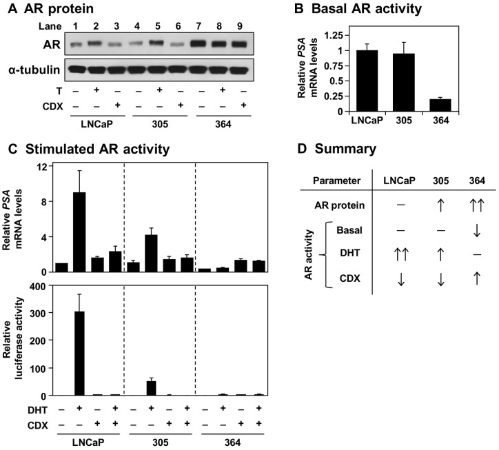
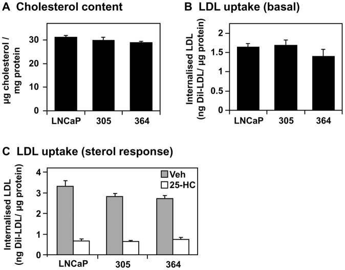
Methodology/principal findings: Firstly, we characterised our progression model, which involved 1) culturing LNCaP cells at physiological testosterone levels to generate androgen-tolerant LNCaP-305 cells, and 2) culturing LNCaP-305 with the anti-androgen casodex to generate castration-resistant LNCaP-364 cells. This progression was accompanied by upregulated androgen receptor expression, typically seen clinically, and a reduction in androgen receptor activity. Although this influenced how SREBP-2 and LXR target genes responded to androgen treatment, cellular cholesterol levels and their response to changing sterol status was similar in all LNCaP sub-lines.

Conclusion/significance: Overall cholesterol homeostasis is unaffected by changing androgen receptor activity in prostate cancer cells. This does not negate the relationship between androgens and cholesterol homeostasis, but rather suggests that other factors compensate for altered androgen receptor activity. Given that cholesterol regulation is maintained during progression, this supports the growing idea that cholesterol metabolism is a suitable target for prostate cancer.


Conflict of interest statement

Competing Interests: The authors have declared that no competing interests exist.

Figures

Figure 1
Figure 1. Growth characteristics of the 305 and 364 cell-lines.
(A) Schematic outlining the development of these LNCaP sub-lines, involving long-term culturing in the presence of either testosterone (T) or casodex (CDX). Details in the text. (B–D) Cells were treated with 10% (v/v) sera and the concentrations of drugs indicated. In (B), this includes FBS (LNCaP) or FBS supplemented with 10 nM T (305) or 10 μM CDX (364). In (C) and (D), this includes FBS or CS-FBS, with T and CDX at concentrations indicated. Cell proliferation was determined as described in the Materials and Methods. (B–D) Data presented as mean + S.E., from three separate experiments per cell-line, each performed with quadruplicate wells per condition. In (D), error bars are contained within the symbols.
Figure 2
Figure 2. Androgen receptor status of the 305 and 364 cell-lines.
(A–B) Cells were grown in Medium A with 10 nM testosterone (T) or 10 μM casodex (CDX). (A) Protein was harvested and subjected to SDS-PAGE and Western blotting against the androgen receptor (AR) and α-tubulin. (B) RNA was harvested and PSA mRNA levels were determined by qRT-PCR, normalised to the LNCaP cells. (C) Top panel: Cells were starved in Medium B for 24 h, before treatment with 1 nM dihydrotestosterone (DHT) and/or 10 μM CDX in Medium B for another 24 h. Following treatment, RNA was harvested and PSA mRNA levels were determined by qRT-PCR, normalised to the vehicle-treated LNCaP cells. Bottom panel: Following transfection, cells were seeded in Medium B. The next day, cells were treated with 1 nM DHT and/or 10 μM CDX in Medium B for another 24 h. Following treatment, cells were assayed for luciferase activity, made relative to the vehicle condition within each cell-line. (D) Summary of the results obtained in (A–C). (A) Blots are representative of four separate experiments. (B–C) Data presented as mean + S.E., from three separate experiments per cell-line, each performed with triplicate wells per condition.
Figure 3
Figure 3. The effect of androgen receptor status on androgen-regulated cholesterol homeostasis.
(A) Schematic outlining the effects of the androgen receptor (AR) on key transcription factors in cholesterol homeostasis. Details in the text. (B–C) Cells were starved in Medium B for 24 h, before treatment with 1 nM dihydrotestosterone (DHT) and/or 10 μM CDX in Medium B for another 24 h. Following treatment, RNA was harvested and (B) LDLR and HMGCR, and (C) ABCG1 and ABCA1, mRNA levels were determined by qRT-PCR, normalised to the vehicle condition in each cell-line. (B–C) Data presented as mean ± S.E., from three separate experiments per cell-line, each performed with triplicate wells per condition.
Figure 4
Figure 4. The effect of androgen receptor status on basal cholesterol homeostasis.
Cells were grown in their basal media: Medium A (LNCaP), supplemented with 10 nM testosterone (305) or 10 μM casodex (364). RNA was harvested and (A) LDLR and HMGCR, and (B) ABCG1 and ABCA1, mRNA levels were determined by qRT-PCR, normalised to the LNCaP cells. (A–B) Data presented as mean + S.E., from three separate experiments per cell-line, each performed with triplicate wells per condition.
Figure 5
Figure 5. The effect of androgen receptor status on cholesterol levels and LDL uptake.
(A) Cells were grown in their basal media: Medium A (LNCaP), supplemented with 10 nM testosterone (305) or 10 μM casodex (364). Cholesterol levels were determined as described in the Materials and Methods. (B) Cells were treated in their basal media, after which LDL uptake was determined. (C) Cells were plated in their basal media, then starved overnight in Medium C. The next days, cells were treated for 6 h with or without 10 μM 25-hydroxycholesterol (25-HC) in Medium C, after which LDL uptake was determined. (A–C) Data presented as mean + S.E., from three separate experiments per cell-line, each performed with triplicate wells per condition.
Figure 6
Figure 6. The effect of androgen receptor status on the response to changing sterol status.
Cells were plated in their basal media: Medium A (LNCaP), supplemented with 10 nM testosterone (305) or 10 μM casodex (364). Cells were starved overnight in Medium C, and then treated for 6 h with 25-hydroxycholesterol (25-HC) in Medium C, at the concentrations indicated. Following treatment, RNA was harvested and LDLR, HMGCR, and ABCG1 levels were determined by qRT-PCR, normalised to the vehicle condition within each cell-line. Data presented as mean ± S.E., from three separate experiments per cell-line, each performed with triplicate wells per condition.

Similar articles

Cited by

References

    1. Huggins C, Hodges CV (1941) Studies on prostatic cancer – I The effect of castration, of estrogen and of androgen injection on serum phosphatases in metastatic carcinoma of the prostate. Cancer Res 1: 293–297. - PubMed
    1. Sedelaar JP, Isaacs JT (2009) Tissue culture media supplemented with 10% fetal calf serum contains a castrate level of testosterone. Prostate 69: 1724–1729. - PMC - PubMed
    1. Olsson M, Ekstrom L, Guillemette C, Belanger A, Rane A, et al. (2011) Correlation between circulatory, local prostatic, and intra-prostatic androgen levels. Prostate 71: 909–914. - PubMed
    1. Sharifi N, Gulley JL, Dahut WL (2010) An update on androgen deprivation therapy for prostate cancer. Endocr Relat Cancer 17: R305–315. - PMC - PubMed
    1. Akaza H (2011) Combined androgen blockade for prostate cancer: review of efficacy, safety and cost-effectiveness. Cancer Sci 102: 51–56. - PubMed

Publication types