Comparative Study
. 2013;8(1):e54038.
doi: 10.1371/journal.pone.0054038. Epub 2013 Jan 8.

Ultrastructure of calcareous dinophytes (Thoracosphaeraceae, Peridiniales) with a focus on vacuolar crystal-like particles

Affiliations
Comparative Study

Ultrastructure of calcareous dinophytes (Thoracosphaeraceae, Peridiniales) with a focus on vacuolar crystal-like particles

Carmen Zinssmeister et al. PLoS One. 2013.

Abstract

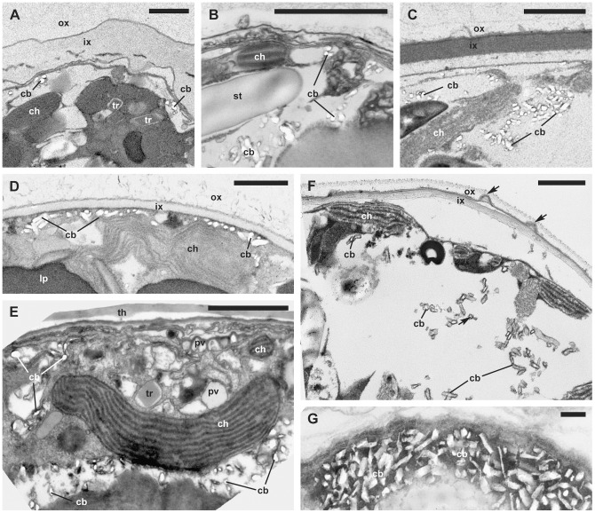
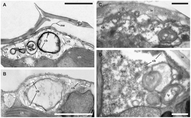
Biomineralization in calcareous dinophytes (Thoracosphaeracaea, Peridiniales) takes place in coccoid cells and is presently poorly understood. Vacuolar crystal-like particles as well as collection sites within the prospective calcareous shell may play a crucial role during this process at the ultrastructural level. Using transmission electron microscopy, we investigated the ultrastructure of coccoid cells at an early developmental stage in fourteen calcareous dinophyte strains (corresponding to at least ten species of Calciodinellum, Calcigonellum, Leonella, Pernambugia, Scrippsiella, and Thoracosphaera). The shell of the coccoid cells consisted either of one (Leonella, Thoracosphaera) or two matrices (Scrippsiella and its relatives) of unknown element composition, whereas calcite is deposited in the only or the outer layer, respectively. We observed crystal-like particles in cytoplasmic vacuoles in cells of nine of the strains investigated and assume that they are widespread among calcareous dinophytes. However, similar structures are also found outside the Thoracosphaeraceae, and we postulate an evolutionarily old physiological pathway (possibly involved in detoxification) that later was specialized for calcification. We aim to contribute to a deeper knowledge of the biomineralization process in calcareous dinophytes.


Conflict of interest statement

Competing Interests: The authors have declared that no competing interests exist.

Figures

Figure 1
Figure 1. Morphological diversity of calcareous coccoid cells.
(SEM). A: Scrippsiella bicarinata (GeoB 411, note the bicarinate tabulation resulting from the fusion of pre- and postcingular plate equivalents). B: Thoracosphaera heimii (CCCM 670, note the small size). C: Scrippsiella trochoidea (GeoB*185, note the spiny surface). D: Calciodinellum operosum (SZN#74, note the holotabulate tabulation). E: Scrippsiella aff. trochoidea (GeoB 283, note the spiny surface). F: Calciodinellum aff. operosum (GeoB 34, note the smooth surface). Scale bar: 10 µm.
Figure 2
Figure 2. General ultrastructure.
(TEM). A. Large thecate cell of Scrippsiella bicarinata (GeoB 411, note the peripheral chloroplast connected to a multiply-stalked pyrenoid). B. Early coccoid cell of Pernambugia tuberosa (GeoB*61, note the numerous peripheral lipid droplets, the two matrices surrounding the cell and the protrusions highlighted by arrows). C. Large thecate cell (longisection) of Scrippsiella bicarinata (GeoB 411, note the multiply-stalked pyrenoids and the numerous mitochondria in the center of the cell). D. Early coccoid cell of Scrippsiella aff. trochoidea (GeoB 283, note the chloroplasts with stalked pyrenoids and the lipid droplets). E. Mature coccoid cell of Leonella granifera (GeoB 38, note the numerous starch grains and lipid droplets and that the cell is surrounded by a single layer containing large, regularly arranged calcareous crystals). Abbreviations: ab, accumulation body; ch, chloroplast; cs, chromosomes; ix, inner matrix; lp, lipid droplet; nu, nucleus; mt, mitochondrion; ox, outer matrix; py, pyrenoid; st, starch grain. Scale bars: 2 µm.
Figure 3
Figure 3. Ultrastructural traits in detail.
(TEM). A. Multiply-stalked pyrenoid covered by a starch shed of Calciodinellum aff. operosum (GeoB 34, note the large, vacuolar crystal-like particles). B. Different chloroplast types of Scrippsiella bicarinata (GeoB 411, note that chloroplasts could be connected to multiply-stalked pyrenoids covered by a starch shed, or have interlamellar pyrenoids, with thylakoid lamellae leading through the pyrenoid. C. Two to four thylakoid lamellae (arrows) of Scrippsiella bicarinata (GeoB 411). D. Mitochondria with tubular cristae of Scrippsiella aff. trochoidea (GeoB 283). Abbreviations: ch, chloroplast; cb, crystal-like particle; me, membrane; mt, mitochondrion; py, pyrenoid; st, starch grain; tr, trichocyst. Scale bars: A and B 1 µm, C and D 0.1 µm.
Figure 4
Figure 4. Ultrastructural traits in detail.
(TEM). A. ‘Apical furrow’ system of Scripsiella trochoidea (GeoB 377, note the numerous vesicles under the cell surface). B. Overlapping thecal plates of Scripsiella trochoidea (GeoB 188, note the outer protrusions of overlapping theca plates). C. Overlapping theca plates in the sulcal region of “Calciodinellum” spec. (tub*2). D. Strand of peripheral microtubules in Scrippsiella bicarinata (GeoB 411, note the multiple membranes under the cell surface). E. Thecal plate boundary of Scripsiella trochoidea (GeoB 377, note the detached outer unit membrane). F. Trichocysts of Scrippsiella trochoidea var. aciculifera (GeoB 228). G. Subcellular organization of Scrippsiella bicarinata (GeoB 411, note the longisection of a trichocyst and the rough endoplasmatic reticulum indicated by an arrow). H. Golgi apparatus of Scrippsiella trochoidea (M34*25/5). J. Pusule of Scrippsiella bicarinata (GeoB 411, note the multiple membranes under the cell surface). Abbreviations: cb, crystal-like particle; ch, chloroplast; cp, cover plate; d, dictyosome, fb, microtubular fiber; gv, Golgi-derived vesicle; me, unit membrane; mt, mitochondrion; po, pore plate; pv, pusular vesicle; py, pyrenoid; re, rough endoplasmatic reticulum; st, starch grain; tr, trichocyst; th, thecal plate. Scale bars: A to C and E to G 1 µm, D, H, J 0.1 µm.
Figure 5
Figure 5. Vacuolar crystal-like particles.
(TEM). A. Scrippsiella trochoidea var. aciculifera (GeoB 228, note the two matrices surrounding the cell, the middle unit membrane is partly disbanded). B. Thoracosphaera heimii (GeoB 211, note the single matrix that will calcify). C. Calcigonellum infula (GeoB*110, note the two matrices surrounding the cell). D. Pernambugia tuberosa (GeoB*61, note the two matrices surrounding the cell). E. Scrippsiella bicarinata (GeoB 411, early coccoid cell, the theca is still attached). F. Calciodinellum aff. operosum (GeoB 34, note the two matrices surrounding the cell and the protrusions between the outer and inner matrix indicated by arrows). G. Calciodinellum operosum (SZN#74, note the large crystal-like particles). Abbreviations: cb, crystal-like particle; ch, chloroplast; ix, inner matrix; ox, outer matrix; pv, pusular vesicle; py, pyrenoid; st, starch grain; tr, trichocyst; th, thecal plate. Scale bars: 1 µm.
Figure 6
Figure 6. Vacuolar crystal-like particles.
(TEM). A: Scrippsiella trochoidea (GeoB*185, early coccoid cell, the theca is still attached; note the intravacuolar vesicles). B. Scrippsiella trochoidea (GeoB*185). C. Scrippsiella aff. trochoidea (GeoB 283, note the lysozymes as intravacuolar membrane whorls). D. Scrippsiella aff. trochoidea (GeoB 283, note the lysozymes as intravacuolar membrane whorls). Abbreviations: cb, crystal-like particle; ch, chloroplast; ix, inner matrix; ly, lysosme; me, unit membrane; th, thecal plate. Scale bars: 1 µm.

Similar articles

Cited by

References

    1. De Yoreo JJ, Vekilov PG (2003) Principles of crystal nucleation and growth. Rev Mineral Geochem 54: 57–94.
    1. Knoll AH (2003) Biomineralization and evolutionary history. In: Dove PM, De Yoreo JJ, Weiner S, editors. Biomineralization. Washington, DC: Mineralogical Society of America. pp 329–356.
    1. Murdock DJE, Donoghue PCJ (2011) Evolutionary origins of animal skeletal biomineralization. Cells Tissues Organs 194: 98–102. - PubMed
    1. Raven JA, Giordano M (2009) Biomineralization by photosynthetic organisms: Evidence of coevolution of the organisms and their environment? Geobiology 7: 140–154. - PubMed
    1. Bäuerlein E (2004) Biomineralization. Progress in biology, molecular biology and application. Weinheim: Wiley.

Publication types

MeSH terms

LinkOut - more resources