Assessing breast cancer margins ex vivo using aqueous quantum-dot-molecular probes
- PMID: 23320158
- PMCID: PMC3540809
- DOI: 10.1155/2012/861257
Assessing breast cancer margins ex vivo using aqueous quantum-dot-molecular probes
Abstract
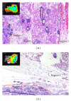
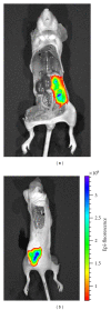
Positive margins have been a critical issue that hinders the success of breast- conserving surgery. The incidence of positive margins is estimated to range from 20% to as high as 60%. Currently, there is no effective intraoperative method for margin assessment. It would be desirable if there is a rapid and reliable breast cancer margin assessment tool in the operating room so that further surgery can be continued if necessary to reduce re-excision rate. In this study, we seek to develop a sensitive and specific molecular probe to help surgeons assess if the surgical margin is clean. The molecular probe consists of the unique aqueous quantum dots developed in our laboratory conjugated with antibodies specific to breast cancer markers such as Tn-antigen. Excised tumors from tumor-bearing nude mice were used to demonstrate the method. AQD-Tn mAb probe proved to be sensitive and specific to identify cancer area quantitatively without being affected by the heterogeneity of the tissue. The integrity of the surgical specimen was not affected by the AQD treatment. Furthermore, AQD-Tn mAb method could determine margin status within 30 minutes of tumor excision, indicating its potential as an accurate intraoperative margin assessment method.
Figures


Similar articles
-
Quantitative assessment of Tn antigen in breast tissue micro-arrays using CdSe aqueous quantum dots.Biomaterials. 2014 Mar;35(9):2971-80. doi: 10.1016/j.biomaterials.2013.12.034. Epub 2014 Jan 8. Biomaterials. 2014. PMID: 24411673
-
Close/positive margins after breast-conserving therapy: additional resection or no resection?Breast. 2013 Aug;22 Suppl 2:S115-7. doi: 10.1016/j.breast.2013.07.022. Breast. 2013. PMID: 24074771 Review.
-
Intraoperative margin assessment and re-excision rate in breast conserving surgery.Eur J Surg Oncol. 2004 Apr;30(3):233-7. doi: 10.1016/j.ejso.2003.11.008. Eur J Surg Oncol. 2004. PMID: 15028301
-
Low re-excision rate for positive margins in patients treated with ultrasound-guided breast-conserving surgery.Breast. 2013 Oct;22(5):698-702. doi: 10.1016/j.breast.2012.12.019. Epub 2013 Jan 17. Breast. 2013. PMID: 23333255
-
Local failure and margin status in early-stage breast carcinoma treated with conservation surgery and radiation therapy.Ann Surg. 1993 Jul;218(1):22-8. doi: 10.1097/00000658-199307000-00005. Ann Surg. 1993. PMID: 8328825 Free PMC article. Review.
Cited by
-
The status of contemporary image-guided modalities in oncologic surgery.Ann Surg. 2015 Jan;261(1):46-55. doi: 10.1097/SLA.0000000000000622. Ann Surg. 2015. PMID: 25599326 Free PMC article. Review.
-
Application of functional quantum dot nanoparticles as fluorescence probes in cell labeling and tumor diagnostic imaging.Nanoscale Res Lett. 2015 Apr 10;10:171. doi: 10.1186/s11671-015-0873-8. eCollection 2015. Nanoscale Res Lett. 2015. PMID: 25897311 Free PMC article.
-
Topical dual-stain difference imaging for rapid intra-operative tumor identification in fresh specimens.Opt Lett. 2013 Dec 1;38(23):5184-7. doi: 10.1364/OL.38.005184. Opt Lett. 2013. PMID: 24281541 Free PMC article.
-
Topical dual-probe staining using quantum dot-labeled antibodies for identifying tumor biomarkers in fresh specimens.PLoS One. 2020 Mar 11;15(3):e0230267. doi: 10.1371/journal.pone.0230267. eCollection 2020. PLoS One. 2020. PMID: 32160634 Free PMC article.
-
Use of monoclonal antibody-IRDye800CW bioconjugates in the resection of breast cancer.J Surg Res. 2014 May 1;188(1):119-28. doi: 10.1016/j.jss.2013.11.1089. Epub 2013 Nov 22. J Surg Res. 2014. PMID: 24360117 Free PMC article.
References
-
- Breast Cancer Overview. American Cancer Society; 2012 http://www.cancer.org/Cancer/BreastCancer/OverviewGuide/breast-cancer-ov....
-
- Balch GC, Mithani SK, Simpson JF, Kelley MC, Gwin J, Bland K. Accuracy of intraoperative gross examination of surgical margin status in women undergoing partial mastectomy for breast malignancy. American Surgeon. 2005;71(1):22–28. - PubMed
-
- Cabioglu N, Hunt KK, Sahin AA, et al. Role for intraoperative margin assessment in patients undergoing breast-conserving surgery. Annals of Surgical Oncology. 2007;14(4):1458–1471. - PubMed
-
- Kurniawan ED, Wong MH, Windle I, et al. Predictors of surgical margin status in breast-conserving surgery within a breast screening program. Annals of Surgical Oncology. 2008;15(9):2542–2549. - PubMed
LinkOut - more resources
Full Text Sources
Other Literature Sources

