

3D Pathology Volumetric Technique: A Method for Calculating Breast Tumour Volume from Whole-Mount Serial Section Images
- PMID: 23320179
- PMCID: PMC3540737
- DOI: 10.1155/2012/691205
3D Pathology Volumetric Technique: A Method for Calculating Breast Tumour Volume from Whole-Mount Serial Section Images
Abstract
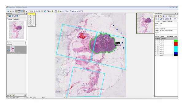
Tumour size, most commonly measured by maximum linear extent, remains a strong predictor of survival in breast cancer. Tumour volume, proportional to the number of tumour cells, may be a more accurate surrogate for size. We describe a novel "3D pathology volumetric technique" for lumpectomies and compare it with 2D measurements. Volume renderings and total tumour volume are computed from digitized whole-mount serial sections using custom software tools. Results are presented for two lumpectomy specimens selected for tumour features which may challenge accurate measurement of tumour burden with conventional, sampling-based pathology: (1) an infiltrative pattern admixed with normal breast elements; (2) a localized invasive mass separated from the in situ component by benign tissue. Spatial relationships between key features (tumour foci, close or involved margins) are clearly visualized in volume renderings. Invasive tumour burden can be underestimated using conventional pathology, compared to the volumetric technique (infiltrative pattern: 30% underestimation; localized mass: 3% underestimation for invasive tumour, 44% for in situ component). Tumour volume approximated from 2D measurements (i.e., maximum linear extent), assuming elliptical geometry, was seen to overestimate volume compared to the 3D volumetric calculation (by a factor of 7x for the infiltrative pattern; 1.5x for the localized invasive mass).
Figures


References
-
- Tabár L, Vitak B, Chen HH, et al. The Swedish two-county trial twenty years later: updated mortality results and new insights from long-term follow-up. Radiologic Clinics of North America. 2000;38(4):625–651. - PubMed
-
- Fisher B, Slack NH, Bross ID. Cancer of the breast: size of neoplasm and prognosis. Cancer. 1969;24(5):1071–1080. - PubMed
-
- Carter CL, Allen C, Henson DE. Relation of tumor size, lymph node status, and survival in 24,740 breast cancer cases. Cancer. 1989;63(1):181–187. - PubMed
-
- Smart CR, Myers MH, Gloeckler LN. Implications from SEER data on breast cancer management. Cancer. 1978;41(3):787–789. - PubMed
LinkOut - more resources
Full Text Sources
