Review
. 2013 Apr;13(2):139-51.
doi: 10.2174/1566523211313020008.

Translating induced pluripotent stem cells from bench to bedside: application to retinal diseases

Affiliations
Review

Translating induced pluripotent stem cells from bench to bedside: application to retinal diseases

Alona O Cramer et al. Curr Gene Ther. 2013 Apr.

Abstract

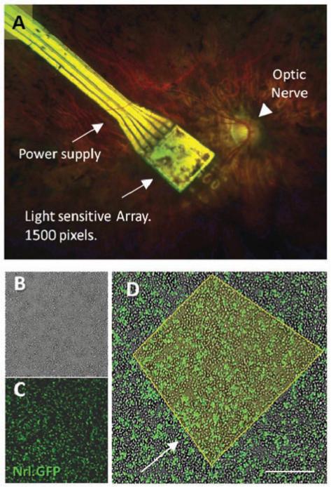
Induced pluripotent stem cells (iPSc) are a scientific and medical frontier. Application of reprogrammed somatic cells for clinical trials is in its dawn period; advances in research with animal and human iPSc are paving the way for retinal therapies with the ongoing development of safe animal cell transplantation studies and characterization of patient- specific and disease-specific human iPSc. The retina is an optimal model for investigation of neural regeneration; amongst other advantageous attributes, it is the most accessible part of the CNS for surgery and outcome monitoring. A recent clinical trial showing a degree of visual restoration via a subretinal electronic prosthesis implies that even a severely degenerate retina may have the capacity for repair after cell replacement through potential plasticity of the visual system. Successful differentiation of neural retina from iPSc and the recent generation of an optic cup from human ESc invitro increase the feasibility of generating an expandable and clinically suitable source of cells for human clinical trials. In this review we shall present recent studies that have propelled the field forward and discuss challenges in utilizing iPS cell derived retinal cells as reliable models for clinical therapies and as a source for clinical cell transplantation treatment for patients suffering from genetic retinal disease.


Figures

Fig. (1)
Fig. (1)
The layered structure of the retina. The first order sensory neurons of the visual system, cone and rod photoreceptors, reside in the outer nuclear layer bordering on RPE cells. Light signals are transferred to the bipolar cells of the inner nuclear layer and pass through ganglion cells and the optic nerve to reach the brain.
Fig. (2)
Fig. (2)
1. Somatic cells, such as skin fibroblasts are obtained from a patient with a genetic retinal degeneration. 2. Cells are reprogrammed to an ES-like pluripotent state. 3. Cells are expanded in-vitro and differentiated to reach an appropriate developmental state. 4. Developing cells may provide an in-vitro model of disease development and treatment or may be subjected to gene therapy with the aim of correcting the genetic source of degeneration ex-vivo. 5. In order to prevent adverse consequences of therapy, cell-colonies are to be screened and purified of proliferative and potentially malignant cells. 6. Patient receives a subretinal injection to replace degenerate cells with reprogrammed, suitably differentiated, corrected and purified autologous cells.
Fig. (3)
Fig. (3)
There is extensive genetic heterogeneity and variability in the phenotypes of outer retinal disease. The four examples of photoreceptor degeneration shown here represent different degeneration mechanisms with divergent phenotypes. A. Retinal degeneration due to rod specific gene- neural retina leucine zipper (NRL). B. Retinal degeneration due to a gene expressed outside the eye- ornithine amino-transferase (OAT). C. Retinal degeneration due to an RPE specific gene- RPE65. D. Retinal degeneration due to acquired infection - rubella. These different phenotypes would require different treatments based on the underlying genetic background and cell types needing to be replaced.
Fig. (4)
Fig. (4)
A. Subretinal electronic implant in a human degenerate retina. The retinal implant device (Retina Implant AG, Reutlingen, Germany) is a light-sensitive electronic chip with 1,500 pixel-generating elements located under the retina in contact with bipolar cells and powered via a subretinal cable. B. Dissociated retinal cells from early postnatal (PN3) Nrl.GFP mouse retina in culture. C. florescence microscopy image of retinal dissociation, florescent cells are rod photoreceptor precursors expressing Nrl. D. Merged image; Nrl.GFP cells in the in vitro cultured retina. Yellow outline represents an area of light sensitive cells, corresponding to the 1,500 pixels transmitted by the electronic chip. Scale bar 30μm. Observations of restored vision in patients with the electronic retinal implant are proof of concept for how an iPS cell-generated photoreceptor monolayer might similarly reverse blindness in retinitis pigmentosa (RP). The cell-therapy approach however has the advantage of generating energy from the oxidation of glucose and would not therefore require an external power supply.

References

    1. Gurdon JB. Adult frogs derived from the nuclei of single somatic cells. Dev Biol. 1962;4:256–73. - PubMed
    1. Takahashi K, Yamanaka S. Induction of pluripotent stem cells from mouse embryonic and adult fibroblast cultures by defined factors. Cell. 2006;126:663–76. - PubMed
    1. MacLaren RE. Re-establishment of visual circuitry after optic nerve regeneration. Eye. 1999;13:277–284. - PubMed
    1. Sung CH, Chuang JZ. The cell biology of vision. J Cell Biol. 2010;190:953–63. - PMC - PubMed
    1. MacLaren RE, Taylor JS. Regeneration in the developing optic nerve: correlating observations in the opossum to other mammalian systems. Prog Neurobiol. 1997;53(3):381–98. - PubMed

Publication types

MeSH terms