. 2013 Jan 15:10:9.
doi: 10.1186/1742-2094-10-9.

TWEAK/Fn14 pathway modulates properties of a human microvascular endothelial cell model of blood brain barrier

Affiliations

TWEAK/Fn14 pathway modulates properties of a human microvascular endothelial cell model of blood brain barrier

Delphine Stephan et al. J Neuroinflammation. .

Abstract

Background: The TNF ligand family member TWEAK exists as membrane and soluble forms and is involved in the regulation of various human inflammatory pathologies, through binding to its main receptor, Fn14. We have shown that the soluble form of TWEAK has a pro-neuroinflammatory effect in an animal model of multiple sclerosis and we further demonstrated that blocking TWEAK activity during the recruitment phase of immune cells across the blood brain barrier (BBB) was protective in this model. It is now well established that endothelial cells in the periphery and astrocytes in the central nervous system (CNS) are targets of TWEAK. Moreover, it has been shown by others that, when injected into mice brains, TWEAK disrupts the architecture of the BBB and induces expression of matrix metalloproteinase-9 (MMP-9) in the brain. Nevertheless, the mechanisms involved in such conditions are complex and remain to be explored, especially because there is a lack of data concerning the TWEAK/Fn14 pathway in microvascular cerebral endothelial cells.

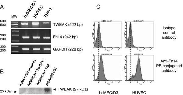
Methods: In this study, we used human cerebral microvascular endothelial cell (HCMEC) cultures as an in vitro model of the BBB to study the effects of soluble TWEAK on the properties and the integrity of the BBB model.

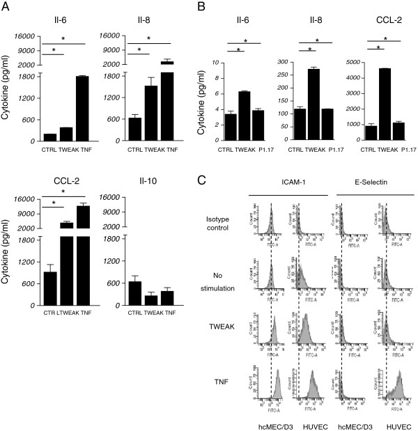
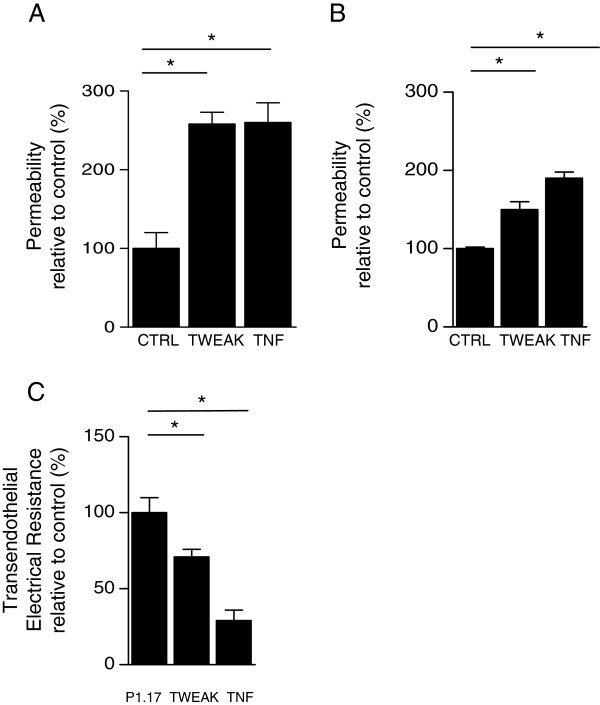
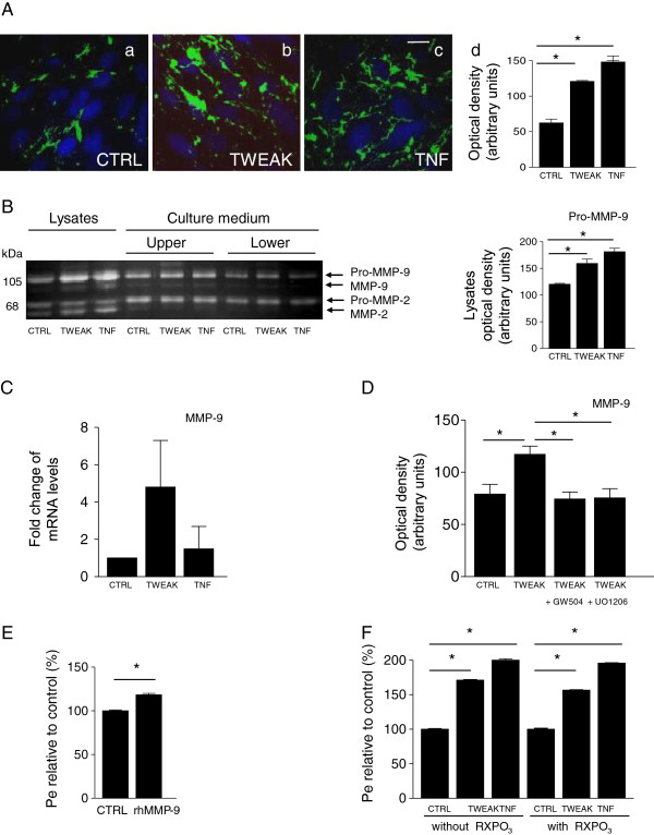
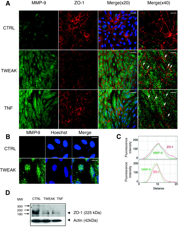
Results: We showed that soluble TWEAK induces an inflammatory profile on HCMECs, especially by promoting secretion of cytokines, by modulating production and activation of MMP-9, and by expression of cell adhesion molecules. We also demonstrated that these effects of TWEAK are associated with increased permeability of the HCMEC monolayer in the in vitro BBB model.

Conclusions: Taken together, the data suggest a role for soluble TWEAK in BBB inflammation and in the promotion of BBB interactions with immune cells. These results support the contention that the TWEAK/Fn14 pathway could contribute at least to the endothelial steps of neuroinflammation.


Figures

Figure 1
Figure 1
Expression of TWEAK and its receptor, Fn 14 by hCMEC/D3 cells. (A) Steady-state levels of TWEAK, Fn14, and GAPDH mRNAs were assessed by semi-quantitative RT-PCR in cultures of hCMEC/D3, HUVECs, and THP-1 cells. PCR products were analyzed by electrophoresis on a 2% agarose gel containing ethidium bromide and PCR products of the expected sizes were obtained for TWEAK (522 bp), Fn14 (242 bp), and GAPDH (226 bp) indicating expression of all three genes at the mRNA level. (B) Western blot analysis of protein extracts from hCMEC/D3 or MDA-MB-231 cells (positive control) following separation by SDS PAGE and transfer onto nitrocellulose membranes. Membranes were probed with a primary antibody against TWEAK. Note the absence of TWEAK protein expression by the hCMEC/D3 cells as compared with the MDA-MB-231 cells. (C) FACS analysis of membrane bound Fn14 on differentiated hCMEC/D3 and HUVECs using anti-human Fn14 or isotype control phycoerythrin-conjugated antibodies. Note the presence of Fn14 at the plasma membrane of the hCMEC/D3 cells.
Figure 2
Figure 2
Proliferative effects of TWEAK on hCMEC/D3. Unstimulated (CTRL) or stimulated (TWEAK, TNF) hCMEC/D3 cell proliferation was determined by measurement of BrdU incorporation during DNA synthesis by chemiluminescence detection. Results indicate increased hCMEC/D3 cell proliferation on TNF and TWEAK stimulation, the latter being more prominent. * p < 0.05 according to Student’s t test.
Figure 3
Figure 3
hCMEC/D3 cells produced chemokines and membrane ICAM-1 after TWEAK exposure. (A) ELISA analysis of CCL-2, IL-8, Il-6, and IL-10 levels in the supernatants of hCMEC/D3 differentiated cells stimulated with TWEAK or TNF for 24 h or not (CTRL); all samples were analyzed in triplicates. The detection threshold was 16 pg/ml of cytokine. Note the increased secretion of CCL-2, Il-8, and Il-6. (B) Primary HCMECs were stimulated with Fc-TWEAK or its isotype control P1.17 for 24 h or not (CTRL). Supernatants were collected and Il-6, IL-8, and CCL-2; levels were evaluated by ELISA. All samples were analyzed in triplicate. The detection threshold was 16 pg/ml of cytokine. (C) FACS analysis of membrane expression of ICAM-1 and E-selectin in differentiated hCMEC/D3 and HUVEC cells stimulated with TWEAK or TNF for 24 h or not (CTRL). Cells were incubated with anti-human ICAM-1 and E-selectin or isotype control fluorescein-conjugated antibodies. TWEAK induces ICAM-1 labeling at the membrane of hCMEC/D3 cells. In (A) and (B), * p < 0.05 according to Student’s t test.
Figure 4
Figure 4
WEAK increased Lucifer yellow permeability in an in vitro model of the BBB. (A) hCMEC/D3 cells were differentiated on coated Transwell® filters and stimulated (TWEAK, TNF) or not (CTRL) during 24 hours. At time t = 0, Lucifer yellow (LY) was applied in the apical compartment. After 60 min, LY fluorescence was assessed in the lower compartment and the permeability coefficient (Pe) was calculated taking into account the relation between the permeability of the monolayer and the permeability of empty filters (pre-coated, without cells). Each condition was run in triplicate in three independent experiments. TWEAK induces increased passage of LY across the hCMEC/D3 cell monolayer. (B) The passage of BSA-FITC was used to assess transport through primary HCMEC seeded and differentiated on coated filters. Primary HCMEC were stimulated with Fc-TWEAK or TNF for 24 h or not (CTRL). At time t = 0, BSA-FITC was applied in the apical compartment. After 60 min, the fluorescence levels were assessed in the basal compartment. Each condition was run in triplicate. (C) The assay setup of HCMEC for TEER was the same as for the BBB permeability assay described in (B). HCMEC TEER was measured by the Electrical Resistance System Millicell-ERS-2. Coated inserts without cells were used as a blank (minimum resistance). The electrical resistance of each insert following treatment with TWEAK, P1.17, or TNF was calculated by subtracting the blank from each reading. Each condition was run in duplicate, and the resistance measured twice for each well. In (A), (B) and (C) * p < 0.05 according to Student’s t test.
Figure 5
Figure 5
Gelatinase activity and expression in hCMEC/D3 cells treated with TWEAK and effects of MMP inhibition. (A) Epifluorescence photomicrographs in live hCMEC/D3 cells with gelatin-quenched fluorescent substrate (green), and nuclear intercalant Hoechst (blue). Net gelatinolytic activity (a) increased with TWEAK (b) and TNF (c), shown by densitometric analysis (d). Scale bar, 10 μm. (B) Gel zymography showing expression and secretion of MMP-2 and MMP-9 in cell lysates and culture media after treatment of hCMEC/D3 with TWEAK or TNF. Nonstimulated endothelial cells express pro-MMP-9, pro-MMP-2, and active MMP-2. Active MMP-9 is barely detected. Control cells secrete pro-MMP-9 and pro-MMP-2 in both compartments, and low levels of their active forms. In cell lysates, TWEAK or TNF treatment has no significant effect on the expression of pro- and active MMP-2 but significantly induces pro-MMP-9 and active MMP-9 expression; there is no effect on the secreted forms of MMP-2 and MMP-9. (Densitometric analysis of pro-MMP-9 zymograms of the lysates.) (C) MMP-9 mRNA quantification by qPCR, expressed as fold change ratios of treated versus control samples after normalization with Abelsson mRNA. (D) Inhibition of TWEAK-induced MMP-9 activity by ERK and JUNK inhibitors in hCMEC/D3 cells. After serum-starvation for 24 h, cells were pretreated with ERK and JUNK inhibitors (5 μM GW5074 and 0.5 μM U0126, respectively) for 1 h and then treated with TWEAK. TWEAK readily induces MMP-9 expression. Both inhibitors significantly inhibit TWEAK-induction of MMP-9 in the cell lysate (densitometric analysis of zymograms). (E) Exogenous recombinant human MMP-9 (rhMMP-9, 250 ng/ml for 24 h) enhanced the permeability (Pe) of the endothelial cell monolayer by 20%. (F) Inhibition of MMP activity with the broad-spectrum MMP inhibitor RXPO3 for 24 h has no effect on TWEAK- or TNFα-increased Pe and barrier impairment. In (A,B,D-F) * P < 0.05 according to Student’s t test.
Figure 6
Figure 6
MMP-9 and Zo-1 expression and distribution in hCMEC/D3 cells treated with TWEAK. (A) Colabeling immunocytochemistry showing expression and distribution of MMP-9 and ZO-1 in control (CTRL), TWEAK-, and TNFα-treated hCMEC/D3 cells. MMP-9 showed a punctuate, vesicular-like pattern distributed throughout the cytoplasm that was increased in the TWEAK- and TNFα-treated cells. Superimposition of MMP-9 and ZO-1 images shows colocalization of both proteins (Merge (×20), scale bar 10 μm) in yellow-orange along the plasma membrane, visible as distinct puncta (arrows in Merge (×40), scale bar 2.5 μm). Note the global down-regulation of ZO-1 expression in TWEAK and TNF-treated hCMEC/D3 cells as compared with CTRL. (B) Confocal analysis of a single z-axis plan of the TWEAK-treated cells shows nuclear accumulation of MMP-9. Scale bar 2 μm. (C) Kymographs were constructed and analyzed with ImageJ software from single-pixel width lines taken from each channel of the confocal images in A (Merge (×40)). Profiles of the signal intensities of MMP-9 (green line) and ZO-1 (red line) measured along the single-pixel width lines drawn in Merge (×40) indicate colocalization. (D) Western blot analysis of protein extracts from TWEAK-treated hCMEC/D3 cells following separation by SDS PAGE and transfer onto nitrocellulose membranes, which were probed with primary antibodies against ZO-1 and actin. Both TWEAK and TNF treatments diminish ZO-1 expression in the hCMEC/D3 cells. Representative Western blot of three independent experiments.

Similar articles

Cited by

References

    1. Chicheportiche Y, Bourdon PR, Xu H, Hsu YM, Scott H, Hession C, Garcia I, Browning JL. TWEAK, a new secreted ligand in the tumor necrosis factor family that weakly induces apoptosis. J Biol Chem. 1997;272:32401–32410. doi: 10.1074/jbc.272.51.32401. - DOI - PubMed
    1. Zhao Z, Burkly LC, Campbell S, Schwartz N, Molano A, Choudhury A, Eisenberg RA, Michaelson JS, Putterman C. TWEAK/Fn14 interactions are instrumental in the pathogenesis of nephritis in the chronic graft-versus-host model of systemic lupus erythematosus. J Immunol. 2007;179:7949–7958. - PubMed
    1. van Kuijk AWR, Wijbrandts CA, Vinkenoog M, Zheng TS, Reedquist KA, Tak PP. TWEAK and its receptor Fn14 in the synovium of patients with rheumatoid arthritis compared to psoriatic arthritis and its response to tumour necrosis factor blockade. Ann Rheum Dis. 2010;69:301–304. doi: 10.1136/ard.2008.090548. - DOI - PMC - PubMed
    1. Dohi T, Burkly LC. The TWEAK/Fn14 pathway as an aggravating and perpetuating factor in inflammatory diseases: focus on inflammatory bowel diseases. J Leukoc Biol. 2012;92:265–279. doi: 10.1189/jlb.0112042. - DOI - PubMed
    1. Michaelson JS, Wisniacki N, Burkly LC, Putterman C. Role of TWEAK in lupus nephritis: a bench-to-bedside review. J Autoimmun. 2012;39:130–142. doi: 10.1016/j.jaut.2012.05.003. - DOI - PMC - PubMed

Publication types

MeSH terms

LinkOut - more resources