

Mir143 expression inversely correlates with nuclear ERK5 immunoreactivity in clinical prostate cancer
- PMID: 23321517
- PMCID: PMC3553517
- DOI: 10.1038/bjc.2012.510
Mir143 expression inversely correlates with nuclear ERK5 immunoreactivity in clinical prostate cancer
Abstract
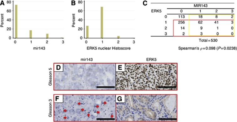
Background: Aberrant mitogen/extracellular signal-regulated kinase 5 (MEK5)-extracellular signal-regulated protein kinase 5 (ERK5)-mediated signalling has been implicated in a number of tumour types including prostate cancer (CaP). The mechanism for ERK5 activation in CaP remains to be fully elucidated. Studies have recently implicated the role of microRNA (miRNA) mir143 expression in the regulation of ERK5 expression.
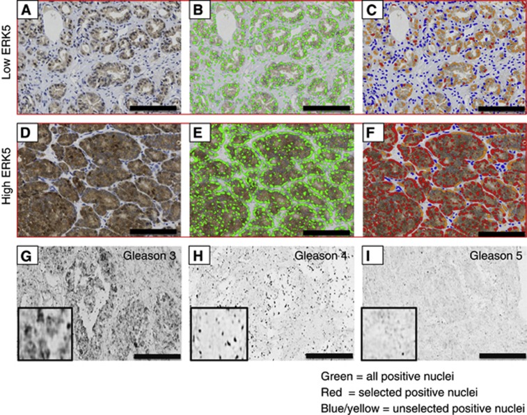
Methods: We utilised a tissue microarray (TMA) of 530 CaP cores from 168 individual patients and stained for both mir143 and ERK5. These TMAs were scored by a combination of observer and automated methods.
Results: We observed a strong inverse relation between ERK5 and mir143, which manifested itself most strongly in the subgroup of 417 cores with non-zero mir143 and ERK5 immunoreactivity, or with only one of mir143 or ERK5 being zero (cc=0.2558 and P<0.0001). Mir143 neither correlate with Gleason scores or prostate-specific antigen levels, nor was it a predictor of disease-specific survival on univariate analysis.
Conclusion: Although the mechanism for ERK5 activation in CaP remains to be fully elucidated, we have further validated the potential role of mir143 in regulating ERK5 levels in the clinical context. In addition, we demonstrate that the automated counting method for nuclear ERK5 is a clinically useful alterative to observer counting method in patient stratification in the context of ERK5 targeting therapy.
Figures
References
-
- Abate-Shen C, Shen MM (2000) Molecular genetics of prostate cancer. Genes Dev 14(19): 2410–2434 - PubMed
-
- Akao Y, Nakagawa Y, Hirata I, Iio A, Itoh T, Kojima K, Nakashima R, Kitade Y, Naoe T (2010) Role of anti-oncomirs miR-143 and -145 in human colorectal tumors. Cancer Gene Ther 17(6): 398–408 - PubMed
-
- Andriole GL, Crawford ED, Grubb RL, Buys SS, Chia D, Church TR, Fouad MN, Gelmann EP, Kvale PA, Reding DJ, Weissfeld JL, Yokochi LA, O’Brien B, Clapp JD, Rathmell JM, Riley TL, Hayes RB, Kramer BS, Izmirlian G, Miller AB, Pinsky PF, Prorok PC, Gohagan JK, Berg CD (2009) Mortality results from a randomized prostate-cancer screening trial. N Engl J Med 360(13): 1310–1319 - PMC - PubMed
-
- Bartel DP (2004) MicroRNAs: genomics, biogenesis, mechanism, and function. Cell 116(2): 281–297 - PubMed
-
- Berney DM, Gopalan A, Kudahetti S, Fisher G, Ambroisine L, Foster CS, Reuter V, Eastham J, Moller H, Kattan MW, Gerald W, Cooper C, Scardino P, Cuzick J (2009) Ki-67 and outcome in clinically localised prostate cancer: analysis of conservatively treated prostate cancer patients from the Trans-Atlantic Prostate Group study. Br J Cancer 100(6): 888–893 - PMC - PubMed
Publication types
MeSH terms
Substances
Grants and funding
LinkOut - more resources
Full Text Sources
Other Literature Sources
Medical
Miscellaneous
