. 2013 Mar 1;126(Pt 5):1122-33.
doi: 10.1242/jcs.112607. Epub 2013 Jan 15.

PAK-PIX interactions regulate adhesion dynamics and membrane protrusion to control neurite outgrowth

Affiliations

PAK-PIX interactions regulate adhesion dynamics and membrane protrusion to control neurite outgrowth

Miguel Santiago-Medina et al. J Cell Sci. .

Abstract

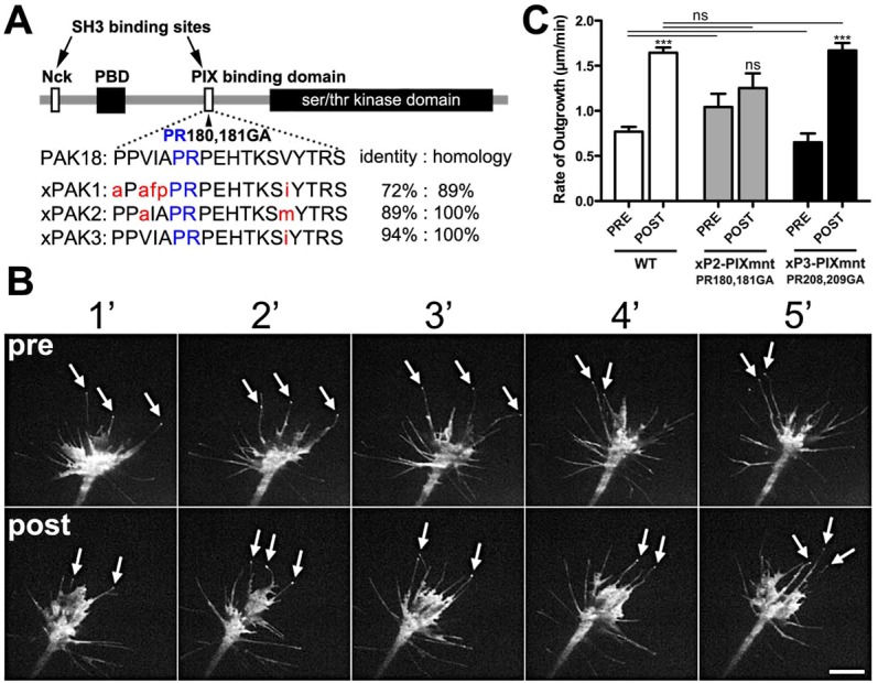
The roles of P21-activated kinase (PAK) in the regulation of axon outgrowth downstream of extracellular matrix (ECM) proteins are poorly understood. Here we show that PAK1-3 and PIX are expressed in the developing spinal cord and differentially localize to point contacts and filopodial tips within motile growth cones. Using a specific interfering peptide called PAK18, we found that axon outgrowth is robustly stimulated on laminin by partial inhibition of PAK-PIX interactions and PAK function, whereas complete inhibition of PAK function stalls axon outgrowth. Furthermore, modest inhibition of PAK-PIX stimulates the assembly and turnover of growth cone point contacts, whereas strong inhibition over-stabilizes adhesions. Point mutations within PAK confirm the importance of PIX binding. Together our data suggest that regulation of PAK-PIX interactions in growth cones controls neurite outgrowth by influencing the activity of several important mediators of actin filament polymerization and retrograde flow, as well as integrin-dependent adhesion to laminin.


Figures

Fig. 1.
Fig. 1.
PAK and PIX isoforms are expressed in the embryonic spinal cord and fusion proteins localize to distinct sites within live growth cones. (A) RT-PCR amplification of PAK1, 2, 3 and α-, β-PIX from stage 24 Xenopus spinal cord shows that PAK2 and PAK3, as well as α-PIX are most highly expressed. (B) Western blot of PAK1, 2 and 3 from stage 24 Xenopus spinal cord confirms that PAK2 and PAK3 are highly expressed. (C–F) TIRF images of representative live spinal neuron growth cones on LN expressing PXN–GFP or PXN–mCh together with different isoforms of fluorescent PAK (C–E) or α-PIX (F). (C) xPAK1 does not concentrate at any distinct location within this growth cone and does not colocalize with PXN-containing point contacts (PCs). (D) xPAK2 localizes to PXN-containing PCs (arrows) and to filopodial tips that contain little or no PXN (arrowheads). (E) xPAK3 localizes to only PXN-containing PCs (arrows). (F) α-PIX localizes to only PXN-containing PCs (arrows). (G,H) Montages of merged images of the growth cones shown in D and E, respectively, expressing mCh–xPAK2 and PXN–GFP (G) or mCh–xPAK3 and PXN–GFP (H); images were taken at 15 second intervals. Note in G that xPAK2 is present at the tips of extending filopodia (arrowheads) and colocalizes with PXN at stable point contacts (dashed box). In H xPAK3 is not present in the filopodia tips (arrowheads), but does colocalize with PXN at stable point contacts (dashed box). Scale bar: 10 µm.
Fig. 2.
Fig. 2.
Dose-dependent effects of acute inhibition of PAK–PIX interactions on growth cone motility and morphology. (A–C) Phase-contrast images at 5 minute intervals of growth cones on LN during stimulation with the indicated concentrations of PAK18 (at 0 minutes). (D) The rate of neurite outgrowth of neurons on LN and PDL after stimulation with increasing concentrations of PAK18 normalized to the pretreatment rate of outgrowth. Note that PAK18 maximally stimulates axon outgrowth on LN at 1 µM, but has no effect on neurite outgrowth on PDL at any concentration. Kruskal–Wallis test with Dunn’s post-hoc analysis, n≥60. (E) Kymographs generated from the leading edge of fluorescent growth cones on LN (above) and PDL (below) during stimulation with 1 µM PAK18 (at black arrowhead). Note differences in scale bars. (F) DIC images of a growth cone on LN at 5 minute intervals during stimulation with 1 µM PAK18. Note an increase in growth cone area, as well as filopodia number and length after PAK18 stimulation. (G) Growth cone area was measured on fixed neurons after 5 minutes stimulation with different concentrations of PAK18. Kruskal–Wallis test with Dunn’s post-hoc analysis, n≥148. (H) Number of filopodia and their length in response to 1 µM PAK18. The y-axis represents quantity. Student’s t-test, n≥27. **P<0.01, ***P<0.001. Scale bar: 20 µm (A–C) and 10 µm (F).
Fig. 3.
Fig. 3.
Acute inhibition of PAK with PAK18 has dose-dependent effects on PAK targets and regulates actin polymerization. (A–D) Representative growth cones treated for 5 minutes with control medium or increasing concentrations of PAK18, and immunolabeled for p-XAC (Ser3). (A′–D′) Merged images of p-XAC (green) and F-actin labeling (red). (E) Fluorescence intensity measurements, normalized to control, of p-XAC labeling of growth cones treated with increasing concentrations of PAK18. (F–I). Representative growth cones treated for 5 minutes with control medium or increasing concentrations of PAK18 and immunolabeled for p-MLC. (F′–I′) Merged images of p-MLC (green) and F-actin labeling (red). (J) Normalized fluorescence intensity measurements of p-MLC-labeled growth cones. (K–N) Representative growth cones treated for 5 minutes with control medium or increasing concentrations of PAK18 and labeled for G-actin (Alexa-Fluor-488–DNase1). (K′–N′) The same growth cones as in K″–N″ labeled for F-actin with Alexa-Fluor-546–phalloidin. (K″–N″) Merged images of G-actin (green) and F-actin labeling (red). (O) Fluorescence intensity measurements of G-actin and F-actin labeling in growth cones treated with PAK18. (P) G-/F-actin ratiometric measurements of growth cones treated with PAK18. **P<0.01, ***P<0.001, Kruskal–Wallis test with Dunn’s post-hoc analysis, n≥35. Scale bars: 10 µm.
Fig. 4.
Fig. 4.
Acute inhibition of PAK–PIX interactions decelerates F-actin retrograde flow. (A) A live growth cone labeled with the F-actin barbed-end binding probe, TMR–KabC. The red box denotes the region used to generate a kymograph (see Materials and Methods). (B) Kymograph from the boxed region of the growth cone in A indicating the rearward flow of KabC-capped actin filaments (red arrow). Note that the angle of flow lines indicates the rate of retrograde flow. (C) Growth cone in A, 5 minutes after stimulation with 1 µM PAK18. (D) Kymograph from the boxed region of the growth cone in C. Note the retrograde flow lines appear more shallow (red arrow), indicating the rate of actin rearward flow is reduced by 1 µM PAK18. (E) The average rate of retrograde flow is significantly reduced by both 1 and 10 µM PAK18. ***P<0.001, Kruskal–Wallis test with Dunn’s post-hoc analysis, n = 10. Scale bar: 10 µm (A,C).
Fig. 5.
Fig. 5.
Acute inhibition of PAK–PIX interactions regulates point contact formation and turnover. (A–D) Representative growth cones treated for 5 minutes with control medium or increasing concentrations of PAK18 and immunolabeled for p-PXN (Tyr118). (A′–D′) Merged images of p-PXN (green) and F-actin labeling (red). (E–G) Quantification of adhesions using particle analysis (see Materials and Methods) of p-PXN-labeled growth cones normalized to untreated control growth cones. **P<0.01, ***P<0.001, Kruskal–Wallis test with Dunn’s post-hoc analysis, n≥29. (H,I) Time-lapse TIRF images of a live growth cone expressing PXN–GFP shown 10 minutes before (H) and after (I) stimulation with 1 µM PAK18. (H′–I′) Pseudocolored heat maps (see Materials and Methods), which illustrate point contact lifetimes over the 15 minute periods before and after stimulation with 1 µM PAK18. Note that many adhesions with short lifetimes form after 1 µM PAK18. (J,K) Time-lapse TIRF images of a live growth cone expressing PXN–GFP shown 10 minutes before (J) and after (K) stimulation with 10 µM PAK18. (J′–K′) Pseudocolored heat maps, which illustrates point contact lifetimes over the 15 minute periods before and after stimulation with 10 µM PAK18. Note that many adhesions with long lifetimes form after 10 µM PAK18. (L–M) Point contact adhesion lifetimes measured before and after stimulation with 1 µM PAK18 (L) and 10 µM PAK18 (M). ***P<0.001, Kruskal–Wallis test with Dunn’s post-hoc analysis, n = 10. Scale bar: 10 µm.
Fig. 6.
Fig. 6.
Acute inhibition of PAK–PIX interactions displaces PAK from paxillin-based adhesions. (A,B) TIRF images of a live growth cone expressing both PXN–GFP and mCh–xPAK3 before (A) and after (B) stimulation with 10 µM PAK18. The white boxes indicate point contacts. (C,D) TIRF images of point contacts from boxed regions in A and B presented at 30 second intervals. Before PAK18 addition at 0 seconds, PXN targets to stable point contacts, which colocalize, with some delay, with PAK3 (arrowheads). However, upon addition of PAK18 at 0 seconds, PAK3 is lost from point contacts. In the continued presence of PAK18 (D), new point contacts recruit little PAK3 and are long lived. (E) Measurement of mCh–xPAK3 fluorescence at PXN–GFP point contacts shows reduced PAK3 after PAK18 addition. ***P<0.001, Student’s t-test, n>134 point contacts in seven growth cones. (F) Measurement of point contact adhesion lifetimes shows that PXN and PAK3 remain within point contacts longer after PAK18. *P<0.05, ***P<0.001, Kruskal–Wallis test with Dunn’s post-hoc analysis, n = 72 point contacts in seven growth cones. Scale bars: 5 µm (A,B); 2 µm (C).
Fig. 7.
Fig. 7.
S273 paxillin regulates point contact adhesion dynamics and growth cone motility. (A) Schematic diagram of paxillin showing the position of the S273 residue that is phosphorylated by PAK and was mutated to generate phosphomimetic (S273D) and nonphosphorylatable (S273A) variants of paxillin. (B) From time-lapse TIRF images of S273D/A-PXN–GFP, the lifetime of point contacts was measured before and after stimulation with 1 µM PAK18 in growth cones expressing wild-type and mutant variants of PXN. n = 10. (C) The rate of neurite outgrowth 15 minutes before and after stimulation with 1 µM PAK18 in growth cones expressing wild-type and mutant variants of PXN. *P<0.05, **P<0.01, ***P<0.001, Kruskal–Wallis test with Dunn’s post-hoc analysis, n≥38.
Fig. 8.
Fig. 8.
A PIX-binding PAK2 mutant blocks the effects of PAK18. (A) Schematic diagram of PAK showing the PIX-binding domain and amino acid mutations that disrupt PAK binding to PIX. The PIX-binding domain of all three PAK isoforms is compared with the sequence of PAK18. Note strong sequence homology between PAK18 and xPAK2 and xPAK3, but less homology with xPAK1. Red indicate sequence divergence; blue indicates the amino acids necessary for PAK–PIX binding. (B) Time-lapse TIRF images of a growth cone expressing GFP–xPAK2-PR180,181GA (GFP–xP2-PIXm) shown at 1 minute intervals for 5 minutes before and after stimulation with 1 µM PAK18. Note that GFP–xP2-PIXm does not localize to point contacts, but does localize to filopodia tips (arrows), even after PAK18 treatment (lower panel). Also, note little morphological effect on GFP–xP2-PIXm-expressing growth cone after 1 µM PAK18. (C) Rate of neurite outgrowth measured 15 minutes before and after stimulation with 1 µM PAK18. ***P<0.001, Kruskal–Wallis test with Dunn’s post-hoc analysis, n≥22. Scale bar: 10 µm.

Similar articles

Cited by

References

    1. Aizawa H., Wakatsuki S., Ishii A., Moriyama K., Sasaki Y., Ohashi K., Sekine–Aizawa Y., Sehara–Fujisawa A., Mizuno K., Goshima Y.et al.2001). Phosphorylation of cofilin by LIM-kinase is necessary for semaphorin 3A-induced growth cone collapse. Nat. Neurosci. 4367–373 10.1038/86011 - DOI - PubMed
    1. Ang L. H., Kim J., Stepensky V., Hing H. (2003). Dock and Pak regulate olfactory axon pathfinding in Drosophila. Development 130, 1307–1316 10.1242/dev.00356 - DOI - PubMed
    1. Arias–Romero L. E., Chernoff J. (2008). A tale of two Paks. Biol. Cell 100, 97–108 10.1042/BC20070109 - DOI - PubMed
    1. Bagrodia S., Taylor S. J., Jordon K. A., Van Aelst L., Cerione R. A. (1998). A novel regulator of p21-activated kinases. J. Biol. Chem. 273, 23633–23636 10.1074/jbc.273.37.23633 - DOI - PubMed
    1. Bisson N., Islam N., Poitras L., Jean S., Bresnick A., Moss T. (2003). The catalytic domain of xPAK1 is sufficient to induce myosin II dependent in vivo cell fragmentation independently of other apoptotic events. Dev. Biol. 263, 264–281 10.1016/j.ydbio.2003.07.002 - DOI - PubMed

Publication types

Substances