. 2013 Jan 15;3(1):e211.
doi: 10.1038/tp.2012.135.

RNAi-mediated serotonin transporter suppression rapidly increases serotonergic neurotransmission and hippocampal neurogenesis

Affiliations

RNAi-mediated serotonin transporter suppression rapidly increases serotonergic neurotransmission and hippocampal neurogenesis

A Ferrés-Coy et al. Transl Psychiatry. .

Abstract

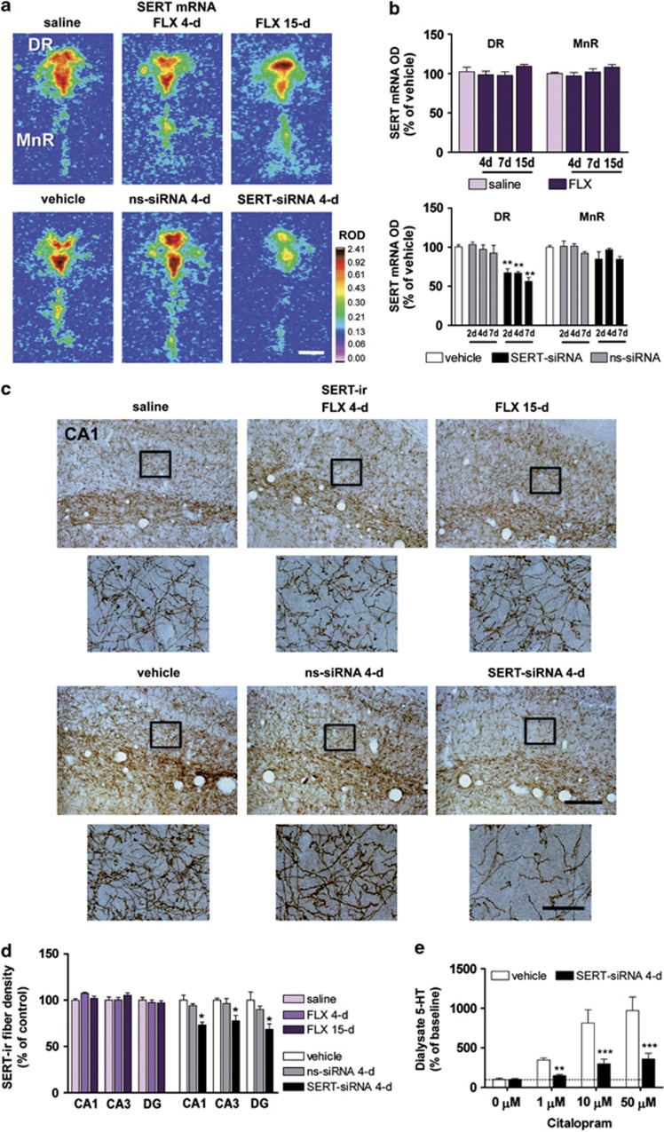
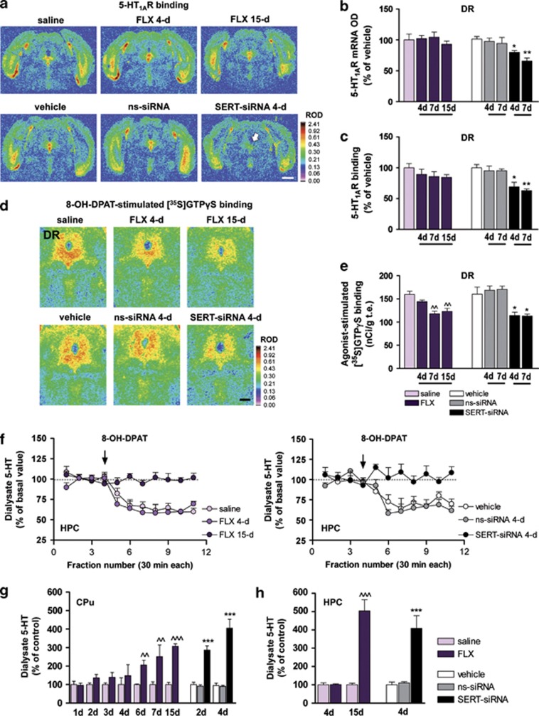
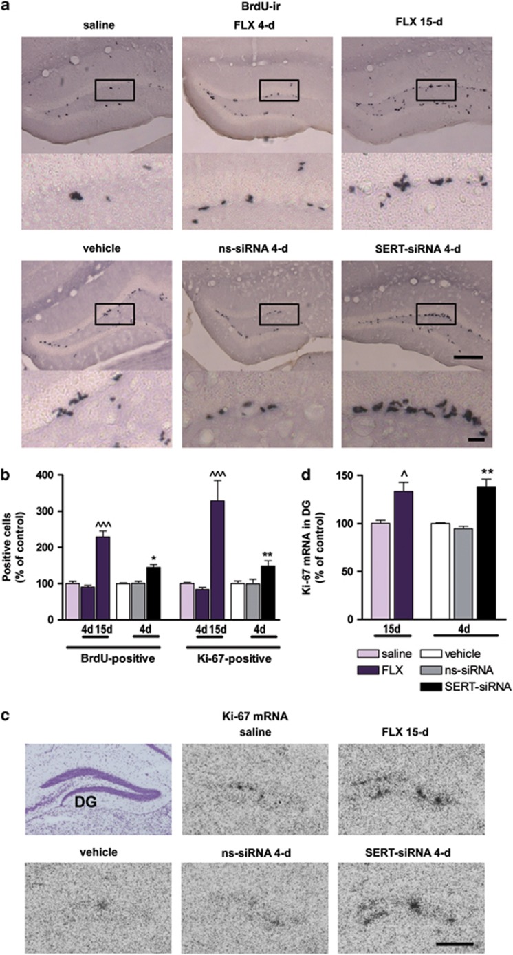
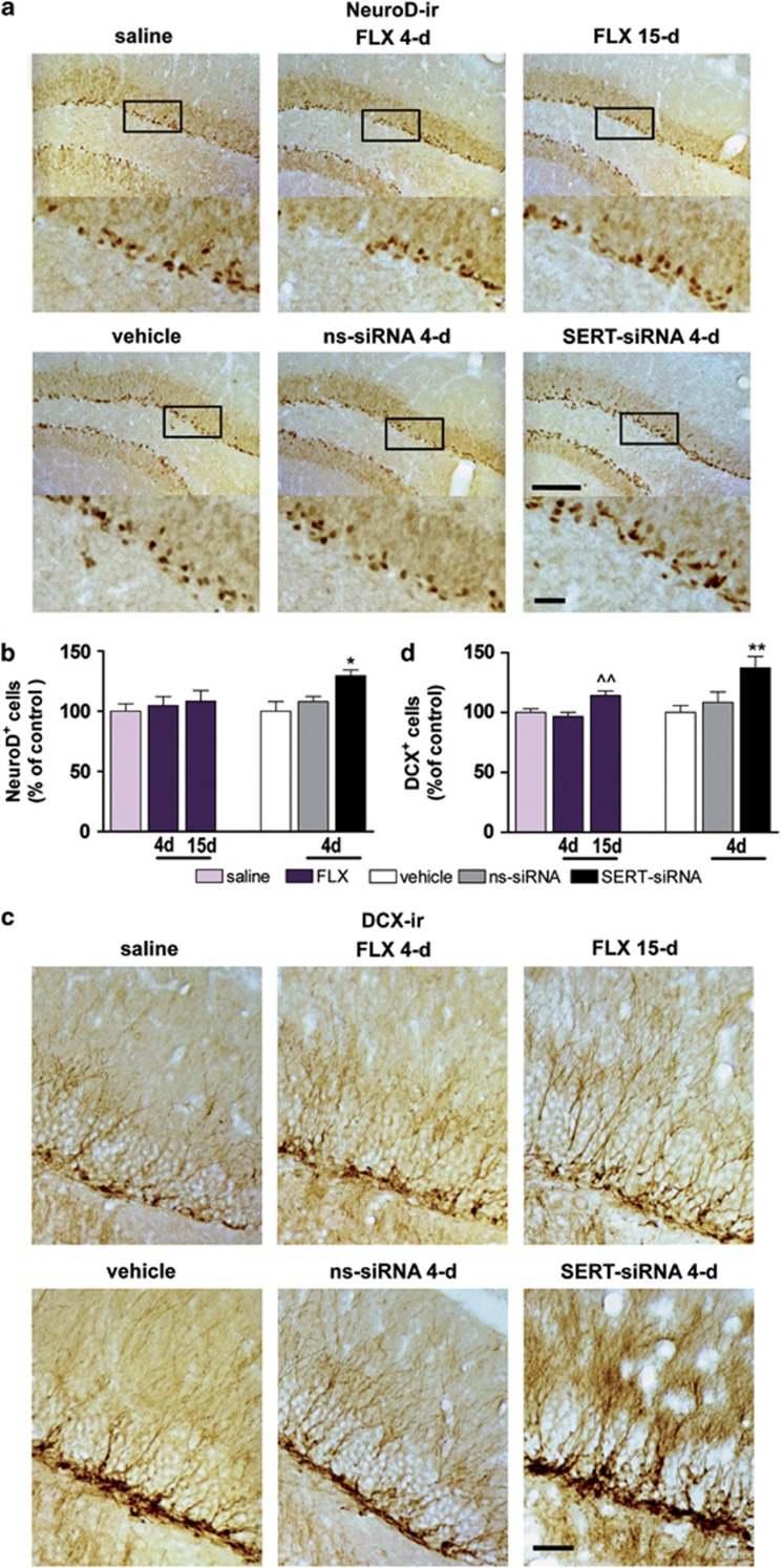
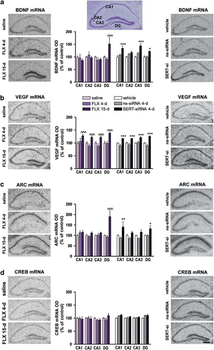
Current antidepressants, which inhibit the serotonin transporter (SERT), display limited efficacy and slow onset of action. Here, we show that partial reduction of SERT expression by small interference RNA (SERT-siRNA) decreased immobility in the tail suspension test, displaying an antidepressant potential. Moreover, short-term SERT-siRNA treatment modified mouse brain variables considered to be key markers of antidepressant action: reduced expression and function of 5-HT(1A)-autoreceptors, elevated extracellular serotonin in forebrain and increased neurogenesis and expression of plasticity-related genes (BDNF, VEGF, Arc) in hippocampus. Remarkably, these effects occurred much earlier and were of greater magnitude than those evoked by long-term fluoxetine treatment. These findings highlight the critical role of SERT in serotonergic function and show that the reduction of SERT expression regulates serotonergic neurotransmission more potently than pharmacological blockade of SERT. The use of siRNA-targeting genes in serotonin neurons (SERT, 5-HT(1A)-autoreceptor) may be a novel therapeutic strategy to develop fast-acting antidepressants.


Figures

Figure 1
Figure 1
Serotonin transporter-small interference RNA (SERT-siRNA) treatment, but not fluoxetine (FLX), downregulates SERT mRNA and protein levels. Mice were infused with two-, four- or seven-dose SERT- or nonsense siRNA (ns-siRNA) (10 μg–0.7 nmol per day) or vehicle into the dorsal raphe nucleus (DR). Other groups of mice were treated with 4, 7 or 15-day FLX (20 mg kg−1 per day, intraperitoneally) or saline. (a) Representative coronal brain sections showing reduced SERT expression in the DR of mice treated with SERT-siRNA (four dose), but not with FLX, as assessed by in situ hybridization. Scale bar=500 μm. (b) Bar graphs showing no differences in SERT mRNA level in DR and median raphe nucleus (MnR) of FLX-treated mice. However, SERT-siRNA at different doses significantly reduced SERT mRNA level exclusively in DR (n=5–10 mice per group). Two-way analysis of variance revealed a significant effect of group (F2,33=59.32, P<0.001). (c) Representative images of SERT-immunoreactive (SERT-ir) axons in the hippocampal CA1 region. Short-term SERT-siRNA treatment (four dose) significantly decreased the target SERT protein density as compared with vehicle and ns-siRNA-treated mice. In contrast, FLX treatment did not alter SERT-ir axon density. Box insets represent regions of high-magnification photomicrographs. Scale bars: lower magnification=100 μm and high magnification=20 μm. (d) SERT-ir fiber density in different hippocampal subfields including CA1, CA2, CA3 and dentate gyrus (DG) was measured and expressed as the percentage of the density in the respective vehicle-treated mice (n=4–6 mice per group). Two-way ANOVA showed an effect of group (F2,11=11.16, P<0.01). (e) Local citalopram (SSRI) infusion by reverse dialysis induced a concentration-dependent increase of extracellular 5-HT in caudate putamen (CPu) of vehicle-treated mice (n=7). However, this effect was lesser marked in SERT-siRNA-treated mice (n=9). Two-way ANOVA showed an effect of group (F1,14=17.17, P<0.0001), concentration (F3,42=27.06, P<0.0001) and group-by-concentration interaction (F3,42=7.72, P<0.0001). *P<0.05, **P<0.01, ***P<0.001 compared with vehicle and ns-siRNA-treated mice. Values are mean±s.e.m.
Figure 2
Figure 2
(af) Short-term serotonin transporter-small interference RNA (SERT-siRNA) treatment, but not fluoxetine (FLX), reduces 5-HT1A-autoreceptor expression and function. Mice were infused with four or seven dose SERT- or nonsense siRNA (ns-siRNA; 10 μg per day) or vehicle into dorsal raphe nucleus (DR). Other groups of mice were treated with 4, 7 or 15-day FLX (20 mg kg−1 per day, intraperitoneally (i.p.) or saline. (a) Representative coronal midbrain sections showing [3H]-8-OH-DPAT binding to 5-HT1AR in DR. The arrow indicates the decreased DR 5-HT1AR density in SERT-siRNA-treated mice (four dose). Scale bar=2 mm. (b) Bar graphs showing no differences in DR 5-HT1AR mRNA levels of vehicle- and FLX-treated mice. However, SERT-siRNA significantly reduced 5-HT1AR mRNA level in DR compared with vehicle and ns-siRNA groups (n=3–5 mice per group). Two-way analysis of variance (ANOVA) revealed an effect of group (F2,15=22.59, P<0.001). (c) Quantitative [3H]-8-OH-DPAT binding showed a decreased DR 5-HT1AR density after SERT-siRNA treatment, but not with FLX (n=4–7). Two-way ANOVA showed a significant effect of group (F2,15=20.69, P<0.001). (d) Autoradiograms of coronal midbrain sections of mice showing 5-HT1AR 8-OH-DPAT agonist-stimulated [35S]GTPγS binding. Scale bar=500 μm. (e) FLX induced a decreased DR 8-OH-DPAT-stimulated [35S]GTPγS binding from days 7 to 15 of treatment (n=5–8). Two-way ANOVA showed an effect of group (F3,25=11.40, P<0.001). However, SERT-siRNA produced a fast 5-HT1A-autoreceptor desensitization detected after four dose treatment (n=4–8). Two-way ANOVA revealed an effect of group (F2,31=10.47, P<0.001). (f) The 5-HT1AR agonist 8-OH-DPAT (0.5 mg kg−1, i.p.) decreased extracellular 5-HT concentration in ventral hippocampus (HPC) of saline- and FLX-treated mice (4-day), but not after 15-day FLX treatment (n=5–9). Two-way ANOVA showed an effect of group (F2,18=41.32, P<0.0001), time (F10,180=13.96, P<0.001) and interaction (F20,180=4.41, P<0.001). Similarly, 8-OH-DPAT had no effect on ventral HPC 5-HT release in SERT-siRNA-treated mice (four-dose), unlike to control groups (n=4). Two-way ANOVA showed an effect of group (F2,8=23.13, P<0.0001), time (F10,80=6.38, P<0.001) and interaction (F20,80=4.41, P<0.001). (gh) Time course of the increase in extracellular 5-HT levels in forebrain. Mice received an intra-DR infusion of two or four dose siRNA or vehicle. Microdialysis experiments were performed on the 3rd or 6th day, respectively. In addition, groups of mice were treated with FLX (20 mg kg−1 per day, i.p.) or saline and microdialysis experiments were performed 24 h after the last administration on 2nd, 3rd, 4th, 5th, 7th, 8th and 16th day. (g) FLX treatment significantly increased extracellular 5-HT levels in caudate putamen (CPu) from six dose onwards, compared with saline-treated mice (F13,80=7.43, P<0.0001; n=7–10 mice per group). In contrast, SERT-siRNA-treated mice displayed a larger and faster increase in CPu 5-HT levels, which was significantly different from control already after two-dose treatment (F5,60=32.52, P<0.0001; n=7–12 mice per group). (h) Long-term FLX treatment (15 dose) also increased 5-HT levels in ventral HPC compared with saline-treated mice (F3,20=39.93, P<0.0001; n=4–9). SERT-siRNA-treated mice (four dose) also showed a rapid and significant increase of HPC 5-HT levels relative to vehicle and ns-siRNA-treated mice (F2,18=14.51, P<0.001; n=4–9 mice). ^^P<0.01, ^^^P<0.001 versus saline-treated mice; *P<0.05, **P<0.01, ***P<0.001 versus vehicle and ns-siRNA-treated mice. Values are mean±s.e.m.
Figure 3
Figure 3
Serotonin transporter (SERT) silencing accelerates neural proliferation in the adult hippocampus compared with fluoxetine (FLX). Mice were infused with four dose SERT- or nonsense siRNA (ns-siRNA; 10 μg per day) or vehicle into dorsal raphe nucleus (DR). Other groups of mice were treated with 4 or 15-day FLX (20 mg kg−1 per day, intraperitoneally) or saline. (a) Representative images showing an increased number of 5-Bromo-2′-deoxyuridine (BdrU)-positive cells in the dentate gyrus (DG) of FLX-treated mice (15-day) or serotonin transporter-small interference RNA (SERT-siRNA)-treated mice (four dose) compared with their respective control mice. Box insets frame regions of high-magnification photomicrographs shown below. Scale bars: lower magnification=100 μm and high magnification=20 μm. (b) Cell proliferation was assessed by the number of BdrU-positive cells (n=5–8 mice) and Ki-67-positive cells (n=5–9 mice) in the granule cell layer. Quantitative analysis indicated a significant increase in both BdrU- and Ki-67-positive cell number for longer treatment with FLX (BdrU: F2,20=53.16; Ki-67: F2,17=20.72) or SERT-siRNA (BdrU: F2,13=14.10; Ki-67: F2,18=6.26). (c) Representative coronal brain sections showing Ki-67 expression in DG assessed by in situ hybridization. Scale bar=100 μm. (d) Bar graph showing increased Ki-67 mRNA levels in DG following FLX (15-day) and SERT-siRNA (four dose) treatment as compared with respective control groups (n=3–4 mice). One-way analysis of variance revealed a significant effect of group (F4,11=9.73, P<0.01). ^P<0.05, ^^^P<0.001 versus saline-treated mice; *P<0.05, **P<0.01 versus vehicle and ns-siRNA-treated mice. Values are mean±s.e.m.
Figure 4
Figure 4
Serotonin transporter (SERT) silencing rapidly increases the number of NeuroD- and DCX-positive cells in hippocampus. Mice were infused with four dose SERT- or nonsense siRNA (ns-siRNA; 10 μg per day) or vehicle into dorsal raphe nucleus (DR). Other groups of mice were treated with 4 or 15-day fluoxetine (FLX; 20 mg kg−1 per day, intraperitoneally) or saline. (a) Immunohistochemical images showing NeuroD-positive progenitors in the dentate gyrus (DG) of mice. Box insets represent regions of high-magnification photomicrographs. Scale bars: lower magnification=100 μm and high magnification=20 μm. (b) Quantitative analysis indicated a significant increase in the number of NeuroD-positive cells in serotonin transporter-small interference RNA (SERT-siRNA)-treated mice (four dose) compared with vehicle and ns-siRNA-treated mice (n=5–9 mice). One-way analysis of variance (ANOVA) showed an effect of group (F2,11=5.87, P<0.05). (c) Representative photomicrographs showing DCX-positive cells, bearing a complex dendritic morphology in the DG of mice. Scale bar=20 μm. (d) Quantitative analysis revealed a significant increase in the number of DCX-positive cells in both 15-day FLX (F2,29=7.71, P<0.01) and four dose SERT-siRNA-treated mice (F2,18=5.94, P<0.05) (n=6–10). ^^P<0.01 versus saline-treated mice; *P<0.05, **P<0.01 versus vehicle and ns-siRNA-treated mice. Values are mean±s.e.m.
Figure 5
Figure 5
Serotonin transporter (SERT) silencing enhances hippocampal plasticity-associated gene expression. Mice were infused with four dose SERT- or nonsense siRNA (ns-siRNA; 10 μg per day) or vehicle into dorsal raphe nucleus (DR). Other groups of mice were treated with 4 or 15-day fluoxetine (FLX; 20 mg kg−1 per day, intraperitoneally) or saline. (ad) Representative autoradiograms of hippocampal sections of mice are shown for (a) brain-derived neurotrophic factor (BDNF), (b) vascular endothelial growth factor (VEGF), (c) activity-regulated cytoskeletal protein (Arc) and (d) cAMP response element binding protein (CREB) mRNA expression. Scale bar=100 μm. Densitometric analyses were performed in different hippocampal regions: CA1, CA2, CA3 and dentate gyrus (DG), shown in the cresyl violet-stained section (top). Levels of mRNA for each gene are shown in the bar graphs next to the representative autoradiograms (n=4–5 mice). (a) BDNF mRNA levels in DG were significantly increased after 15-day FLX treatment compared with saline-treated mice (two-way analysis of variance (ANOVA): effect of region F3,33=5.76 and interaction group-by-region F6,33=7.86). However, serotonin transporter-small interference RNA (SERT-siRNA) treatment (four dose) increased BDNF expression in CA1, CA3 and DG compared with respective control groups (significant effect of group F2,8=18.03 and region F3,24=4.33). (b) VEGF mRNA levels were augmented in all hippocampal subfields after FLX (15-day) or SERT-siRNA (four dose) treatments compared with respective control groups (two-way ANOVA; FLX: effect of group F2,10=10.11, region F3,30=12.80 and interaction F6,30=4.54; SERT-siRNA: effect of group F2,8=17.32, region F3,24=5.00 and interaction F6,24=5.42). (c) Arc mRNA levels in DG were significantly increased following FLX (15-day) compared with saline-treated mice (two-way ANOVA: effect of group F2,11=9.30, region F3,33=4.93 and interaction group-by-region F6,33=7.75). SERT-siRNA treatment (four dose) increased Arc expression in CA1 and DG compared with respective control groups (significant effect of group F2,13=11.53, region F3,39=6.77 and interaction F6,39=4.05). (d) CREB mRNA levels were unchanged following any treatment. ^^^P<0.001 versus saline-treated mice; *P<0.05, **P<0.01, ***P<0.001 versus vehicle and ns-siRNA-treated mice. Values are mean±s.e.m.

Similar articles

Cited by

References

    1. Kessler RC, Chiu WT, Demler O, Merikangas KR, Walters EE. Prevalence, severity, and comorbidity of 12-month DSM-IV disorders in the National Comorbidity Survey Replication. Arch Gen Psychiatry. 2005;62:617–627. - PMC - PubMed
    1. Krishnan V, Nestler EJ. The molecular neurobiology of depression. Nature. 2008;455:894–902. - PMC - PubMed
    1. Smith K. Trillion-dollar brain drain. Nature. 2011;478:15. - PubMed
    1. Wong ML, Licinio J. Research and treatment approaches to depression. Nature Rev Neurosci. 2001;2:343–351. - PubMed
    1. Rush AJ, Trivedi MH, Wisniewski SR, Nierenberg AA, Stewart JW, Warden D, et al. Acute and longer-term outcomes in depressed outpatients requiring one or several treatment steps: a STAR*D report. Am J Psychiatry. 2006;163:1905–1917. - PubMed

Publication types

MeSH terms

Substances