. 2013 Jun;133(6):1533-40.
doi: 10.1038/jid.2013.4. Epub 2013 Jan 15.

CD8(+) granzyme B(+)-mediated tissue injury vs. CD4(+)IFNγ(+)-mediated parasite killing in human cutaneous leishmaniasis

Affiliations
Free PMC article

CD8(+) granzyme B(+)-mediated tissue injury vs. CD4(+)IFNγ(+)-mediated parasite killing in human cutaneous leishmaniasis

Claire da Silva Santos et al. J Invest Dermatol. 2013 Jun.
Free PMC article

Erratum in

  • J Invest Dermatol. 2014 Nov;134(11):2850

Abstract

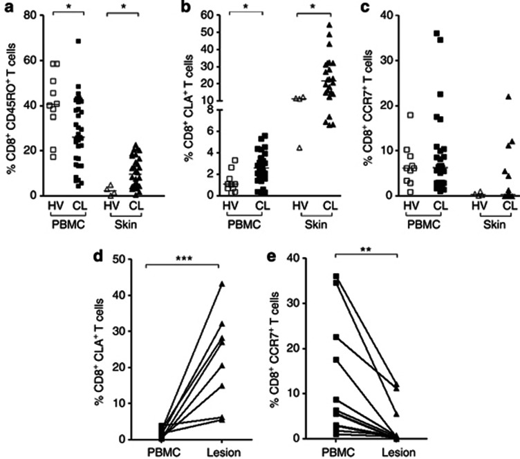
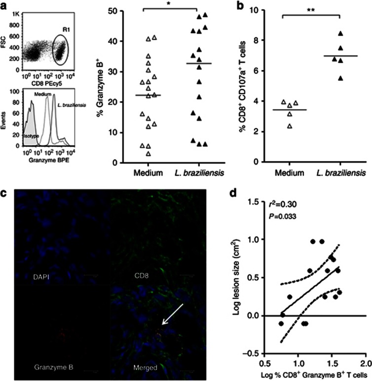
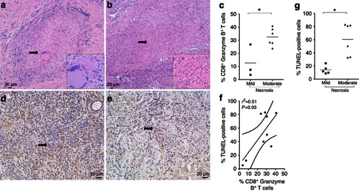
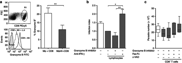
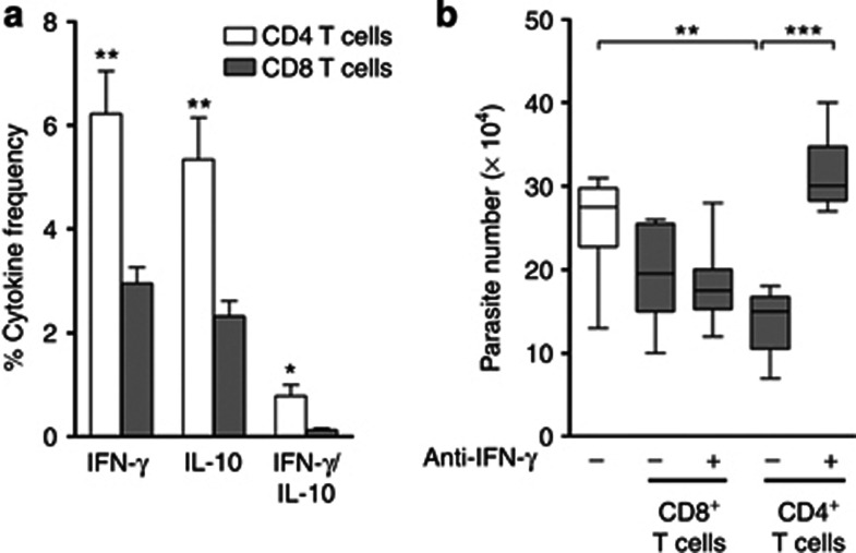
A protective or deleterious role of CD8(+)T cells in human cutaneous leishmaniasis (CL) has been debated. The present report explores the participation of CD8(+)T cells in disease pathogenesis as well as in parasite killing. CD8(+)T cells accumulated in CL lesions as suggested by a higher frequency of CD8(+)CD45RO(+)T cells and CD8(+)CLA(+)T cells compared with peripheral blood mononuclear cells. Upon Leishmania braziliensis restimulation, most of the CD8(+)T cells from the lesion expressed cytolytic markers, CD107a and granzyme B. Granzyme B expression in CL lesions positively correlated with lesion size and percentage of TUNEL-positive cells. We also observed a significantly higher percentage of TUNEL-positive cells and granzyme B expression in the biopsies of patients showing a more intense necrotic process. Furthermore, coculture of infected macrophages and CD8(+)T lymphocytes resulted in the release of granzyme B, and the use of granzyme B inhibitor, as well as z-VAD, Fas:Fc, or anti-IFN-γ, had no effect upon parasite killing. However, coculture of infected macrophages with CD4(+)T cells strongly increased parasite killing, which was completely reversed by anti-IFN-γ. Our results reveal a dichotomy in human CL: CD8(+) granzyme B(+)T cells mediate tissue injury, whereas CD4(+)IFN-γ(+)T cells mediate parasite killing.


Figures

Figure 1
Figure 1
Recruitment and expression of homing markers in CD8+ T cells from cutaneous leishmaniasis (CL) lesions. (a) Ex vivo analysis of CD8+CD45RO+ T cells, (b) CD8+CLA+ T cells, and (c) CD8+CCR7+ T cells in peripheral blood mononuclear cells (PBMC) from healthy volunteers (HV) (n=10, open squares), from CL patients (n=35, closed squares), and in skin biopsies from healthy controls (n=4, open triangles) and lesions (n=30, closed triangles) from CL patients. (d, e) Ex vivo analysis of homing markers in PBMC (closed squares) and lesion cells (closed triangles) from CL patients (n=9). Each symbol represents one individual and the lines represent results obtained in the same patient. *P<0.05. Statistical comparisons were done using the Mann–Whitney U-test. **P<0.01, ***P<0.001.
Figure 2
Figure 2
Expression of cytolytic markers by lesion-derived CD8+ T cells from cutaneous leishmaniasis (CL) patients after stimulation. (a) R1 was analyzed from the CD8 versus forward scatter (FSC) dot plot (top). Histograms are representative of 18 CL patients and the graph displays the frequency of CD8+ T cells expressing granzyme B. (b) Expression of CD107a in CD8 T cells. (c) Representative images from confocal microscopy. 4',6-Diamidino-2-phenylindole (DAPI) counterstained (top left panel); CD8 staining (top right panel); granzyme B staining (bottom left panel); overlay showing expression of granzyme B in CD8+ T cells (white arrow; bottom right panel), bar=10 μm. (d) Correlation analysis between the frequency of CD8+Granzyme B+ T cells and lesion size. *P<0.05, **P<0.01. Statistical comparisons were done using the Mann–Whitney U-test and the Spearman's (r2) rank test. PE, phycoerythrin.
Figure 3
Figure 3
Histopathology and TUNEL-positive cells in cutaneous leishmaniasis (CL) lesions. (a, b) Necrosis scores 1 and 2 showed by arrowhead and inset. (c) Analysis between intensity of necrosis and percentage of CD8+Granzyme B+ T cells. DNA damage in biopsies with (d) higher and (e) lower percentages of TUNEL-positive cells, which are indicated by black arrows. (f) Correlation analysis between the percentage of CD8+Granzyme B+ T cells in the tissue and the percentage of TUNEL+ cells. (g) Analysis between the intensity of necrosis and the number of TUNEL+ cells. Bar=20 μm. *P<0.05. Statistical comparisons were done using the Mann–Whitney U-test and the Spearman's (r2) rank test. Pictures represented a magnification of × 200, insets × 1,000, and TUNEL+ cells × 400.
Figure 4
Figure 4
Cytotoxic CD8+ T cell does not contribute to parasite killing in cutaneous leishmaniasis (CL) patients. Macrophages from peripheral blood mononuclear cells (PBMC) of CL patients were infected or not infected with parasites and cocultured with autologous lymphocytes or peripheral CD8+ T cells for 48 hours. (a) R1 was analyzed from the CD8 versus forward scatter (FSC) dot plot (top). Histograms are representative of six CL patients. The graph shows the expression of granzyme B in CD8+ T cells. (b, c) Infected macrophages cocultured (closed bars) or not (open bar) with peripheral lymphocytes (b) or with purified CD8+ T cells (closed bars) (c) in the absence or presence of inhibitors. *P<0.05; **P<0.01. Statistical comparisons were done using the Mann–Whitney U-test, the Kruskal–Wallis test, or the Friedman test, followed by the Dunn's multiple comparison test.
Figure 5
Figure 5
CD4+, but not CD8+, T cells are the principal source of cytokines in cutaneous leishmaniasis (CL) lesions and are responsible for the killing of parasites. (a) The frequency of lesional CD4+ T cells (open bars) and CD8+ T cells (closed bars) expressing IFN-γ and IL-10 or coexpressing these cytokines, after restimulation in vitro with L. braziliensis. (b) Parasite load in infected macrophages (open bar) and in coculture of infected macrophages with peripheral CD8+ or CD4+ T cells (closed bars) in the absence or presence of anti-IFN-γ. *P<0.05; **P<0.01; ***P<0.001. Statistical comparisons were done using the Mann–Whitney U-test or the Friedman test, followed by the Dunn's multiple comparison test.

References

    1. Akdis M, Simon HU, Weigl L, et al. Skin homing (cutaneous lymphocyte-associated antigen-positive) CD8+ T cells respond to superantigen and contribute to eosinophilia and IgE production in atopic dermatitis. J Immunol. 1999;163:466–475. - PubMed
    1. Antonelli LR, Dutra WO, Almeida RP, et al. Activated inflammatory T cells correlate with lesion size in human cutaneous leishmaniasis. Immunol Lett. 2005;101:226–230. - PubMed
    1. Bacellar O, Lessa H, Schriefer A, et al. Up-regulation of Th1-type responses in mucosal leishmaniasis patients. Infect Immun. 2002;70:6734–6740. - PMC - PubMed
    1. Barral-Netto M, Barral A, Brodskyn C, et al. Cytotoxicity in human mucosal and cutaneous leishmaniasis. Parasite Immunol. 1995;17:21–28. - PubMed
    1. Betts MR, Brenchley JM, Price DA, et al. Sensitive and viable identification of antigen-specific CD8+ T cells by a flow cytometric assay for degranulation. J Immunol Methods. 2003;281:65–78. - PubMed

Publication types

MeSH terms