. 2013:4:1355.
doi: 10.1038/ncomms2346.

A structural basis for streptomycin-induced misreading of the genetic code

Affiliations

A structural basis for streptomycin-induced misreading of the genetic code

Hasan Demirci et al. Nat Commun. 2013.

Abstract

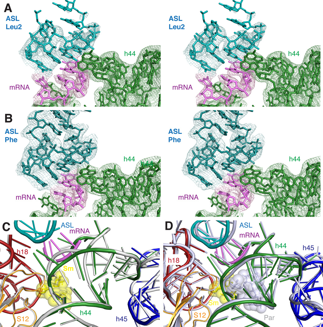
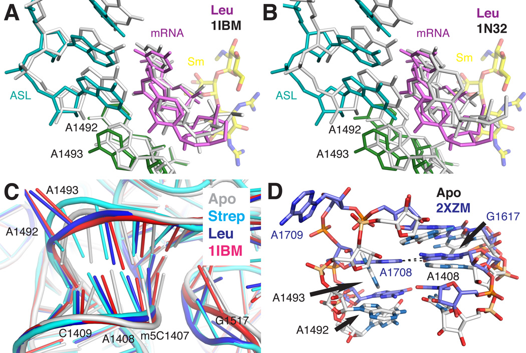
During protein synthesis, the ribosome selects aminoacyl-transfer RNAs with anticodons matching the messenger RNA codon present in the A site of the small ribosomal subunit. The aminoglycoside antibiotic streptomycin disrupts decoding by binding close to the site of codon recognition. Here we use X-ray crystallography to define the impact of streptomycin on the decoding site of the Thermus thermophilus 30S ribosomal subunit in complexes with cognate or near-cognate anticodon stem-loop analogues and messenger RNA. Our crystal structures display a significant local distortion of 16S ribosomal RNA induced by streptomycin, including the crucial bases A1492 and A1493 that participate directly in codon recognition. Consistent with kinetic data, we observe that streptomycin stabilizes the near-cognate anticodon stem-loop analogue complex, while destabilizing the cognate anticodon stem-loop analogue complex. These data reveal how streptomycin disrupts the recognition of cognate anticodon stem-loop analogues and yet improves recognition of a near-cognate anticodon stem-loop analogue.


Conflict of interest statement

Conflict of Interest

The authors declare no conflict of interest.

Figures

Figure 1
Figure 1. Conformational changes upon binding of streptomycin to the 30S subunit
a. Cartoon representation of the 16S rRNA seen from the subunit interface. The region shown in (b) is indicated with a black rectangle. b. Streptomycin binding site in the 30S subunit. Streptomycin is shown as yellow sticks and spheres. Helix 44 is shown as dark green cartoon, helix 18 is colored in red, helix 27 in cyan, helix 45 in blue, and protein S12 in orange. c. Final σA-weighted m2Fo-dFc electron density map of the streptomycin-binding site contoured at 1 σ. d. Secondary structure diagram of the decoding site region in helix 44 and of the helix 45 tetraloop (indicated with a box). e. Conformational changes in the decoding site upon streptomycin binding. The streptomycin-bound 30S structure is shown superimposed onto the apo 30S structure shown in light grey. f. Comparison of the two helix 45 tetraloop conformations induced by streptomycin (data set Strep, PDB accession code 4DR3) and paromomycin (data set Par, PDB accession code 4DR2, colored in light orange).
Figure 2
Figure 2. Helix 45 conformation in four 30S crystal structures
Final σA-weighted 2mFo-DFc electron density contoured at 1σ is shown for helix 44 colored in green and helix 45 colored in blue with A1518 and A1519 colored in cyan. (a) streptomycin-bound, (b) streptomycin, mRNA and ASLGAGLeu2, (c) paromomycin, and (d) paromomycin, mRNA and ASLGAAPhe.
Figure 3
Figure 3. Decoding of near-cognate ASLGAGLeu2 in the presence of streptomycin
a. σA-weighted 2mFo-DFc electron density (contoured at 1σ) for the ASLGAGLeu2, mRNA, and helix 44 in the presence of streptomycin. ASL, mRNA and helix 44 are colored in blue, violet, and green, respectively. b. σA-weighted 2mFo-DFc electron density (contoured at 1σ) for the ASLGAAPhe, mRNA, and helix 44 in the presence of paromomycin. c. Conformational changes (and compensatory shift of the phosphate backbone) of bases A1492 and A1493 in the presence of the near-cognate ASLGAGLeu2, mRNA, and streptomycin (colored as in Fig. 1) compared to the streptomycin-bound 30S subunit (colored in light grey). d. Comparison of near-cognate ASLGAGLeu2 position in the presence of streptomycin (colored as before) with cognate ASLGAAPhe in the presence of paromomycin (MAP data, PDB accession code 4DR4, colored in grey).
Figure 4
Figure 4. Conformational flexibility in the 30S decoding site
a. Comparison of decoding interactions for near-cognate ASLGAGLeu2 (data set Leu, PDB accession code 4DR6, colored as before) with cognate decoding interactions (PDB accession code 1IBM colored in grey) b. Data set Leu colored as before compared to PDB accession code 1N32 (30S subunit in complex with near-cognate ASLGAGLeu2, mRNA, and paromomycin, colored in grey). Paromomycin-induced domain closure shifts the position of the near-cognate ASL, mRNA, and A1492/A1493 when compared to the open, streptomycin stabilized interaction shown in (a). c. Restructuring of the helix 44 region by streptomycin. The position of bases 1407 to 1409 opposite of A1492 and A1493 is shifted upon streptomycin binding both in the absence (cyan) and presence (blue) of ASL and mRNA when compared with the apo-30S structure (grey) or the cognate decoding structure PDB accession code 1IBM (red). d. Comparison of the helix 44 decoding region (colored in white) with the eukaryotic 40S subunit from Tetrahymena thermophila (colored in blue, PDB accession code 2XZM). A1708 (equivalent to A1492) forms a non-Watson-Crick base pair with G1617 (equivalent to A1408), while A1709 is flipped out of the helix.

References

    1. Poehlsgaard J, Douthwaite S. The bacterial ribosome as a target for antibiotics. Nature Reviews: Microbiology. 2005;3:870–881. - PubMed
    1. Gale EF, Cundliffe E, Reynolds PE, Richmond MH, Waring MJ. The molecular basis of antibiotic action. London: Wiley & Sons; 1981. pp. 402–547.
    1. Gorini L, Gundersen W, Burger M. Genetics of regulation of enzyme synthesis in the arginine biosynthetic pathway of Escherichia coli. Cold Spring Harbor Symposia on Quantitative Biology. 1961;26:173–182. - PubMed
    1. Davies J, Gilbert W, Gorini L. Streptomycin, Suppression, and the Code. Proceeding of the National Academy of Sciences of the United States of America. 1964;51:883–890. - PMC - PubMed
    1. Gorini L, Jacoby GA, Breckenridge L. Ribosomal ambiguity. Cold Spring Harbor Symposia on Quantitative Biology. 1966;31:657–664. - PubMed

Publication types

MeSH terms

LinkOut - more resources