

Carcinoembryonic antigen is the preferred biomarker for in vivo colorectal cancer targeting
- PMID: 23322207
- PMCID: PMC3593555
- DOI: 10.1038/bjc.2012.605
Carcinoembryonic antigen is the preferred biomarker for in vivo colorectal cancer targeting
Abstract
Background: Colorectal cancer-specific biomarkers have been used as molecular targets for fluorescent intra-operative imaging, targeted PET/MRI, and selective cytotoxic drug delivery yet the selection of biomarkers used is rarely evidence-based. We evaluated sensitivities and specificites of four of the most commonly used markers: carcinoembryonic antigen (CEA), tumour-associated glycoprotein-72 (TAG-72), folate receptor-α (FRα) and Epithelial growth factor receptor (EGFR).
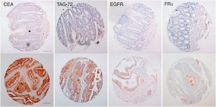
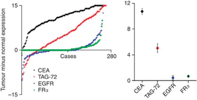
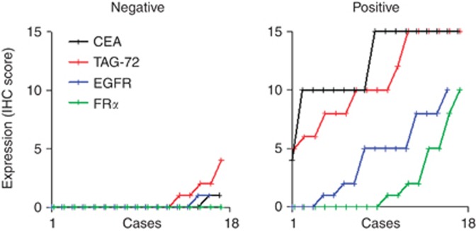
Methods: Marker expression was evaluated semi-quantitatively in matched mucosal and colorectal cancer tissues from 280 patients using immunohistochemistry (scores of 0-15). Matched positive and negative lymph nodes from 18 patients were also examined.
Results: Markers were more highly expressed in tumour tissue than in matched normal tissue in 98.8%, 79.0%, 37.1% and 32.8% of cases for CEA, TAG-72, FRα and EGFR, respectively. Carcinoembryonic antigen showed the greatest differential expression, with tumours scoring a mean of 10.8 points higher than normal tissues (95% CI 10.31-11.21, P<0.001). Similarly, CEA showed the greatest differential expression between positive and negative lymph nodes. Receiver operating characteristic analyses showed CEA to have the best sensitivity (93.7%) and specificity (96.1%) for colorectal cancer detection.
Conclusion: Carcinoembryonic antigen has the greatest potential to allow highly specific tumour imaging and drug delivery; future translational research should aim to exploit this.
Figures

References
-
- Barbet J, Bardies M, Bourgeois M, Chatal J-F, Cherel M, Davodeau F, Faivre-Chauvet A, Gestin J-F, Kraeber-Bodere F. Radiolabeled antibodies for cancer imaging and therapy. Methods Mol Biol. 2012;907:681–697. - PubMed
-
- Bhargava R, Chen B, Klimstra DS, Saltz LB, Hedvat C, Tang LH, Gerald W, Teruya-Feldstein J, Paty PB, Qin J, Shia J. Comparison of two antibodies for immunohistochemical evaluation of epidermal growth factor receptor expression in colorectal carcinomas, adenomas, and normal mucosa. Cancer. 2006;106 (8:1857–1862. - PubMed
-
- Boerman OC, Oyen WJG. Immuno-PET of cancer: a revival of antibody imaging. J Nucl Med. 2011;52 (8:1171–1172. - PubMed
-
- Buckley AF, Kakar S. Comparison of the dako EGFR pharmDx kit and zymed EGFR antibody for assessment of EGFR status in colorectal adenocarcinoma. Appl Immunohistochem. 2007;15 (3:305–309. - PubMed
-
- Camp RL, Charette LA, Rimm DL. Validation of tissue microarray technology in breast carcinoma. Lab Invest. 2000;80 (12:1943–1949. - PubMed
Publication types
MeSH terms
Substances
Grants and funding
LinkOut - more resources
Full Text Sources
Other Literature Sources
Medical
Research Materials
Miscellaneous
