. 2013 Jun;28(6):1412-21.
doi: 10.1002/jbmr.1871.

Compensatory regulation of the Snai1 and Snai2 genes during chondrogenesis

Affiliations

Compensatory regulation of the Snai1 and Snai2 genes during chondrogenesis

Ying Chen et al. J Bone Miner Res. 2013 Jun.

Abstract

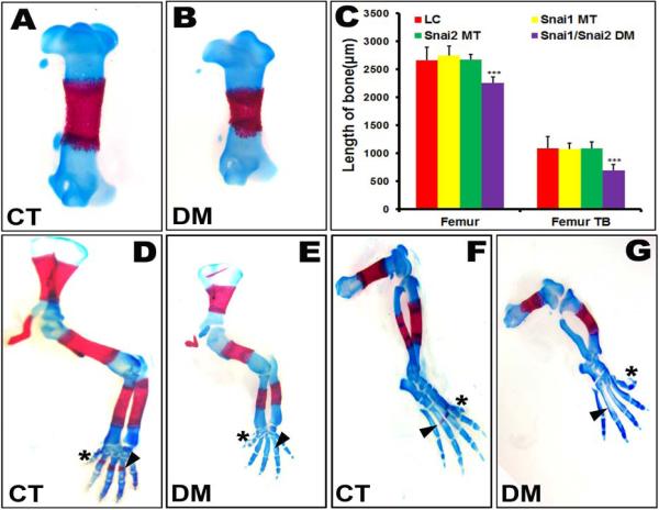
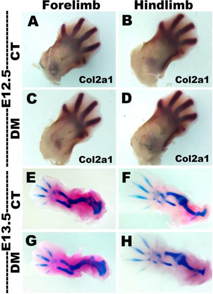
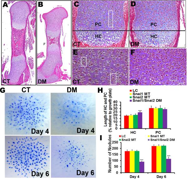
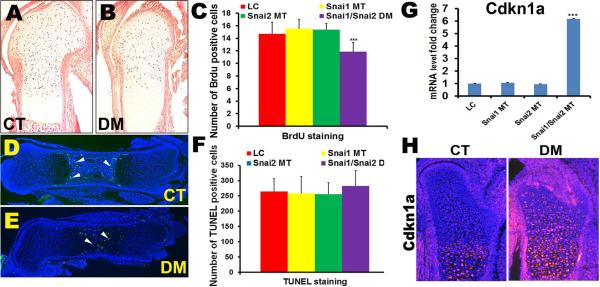
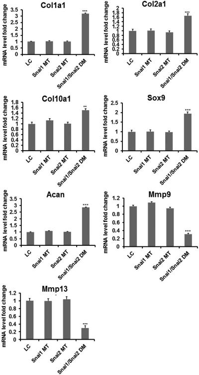
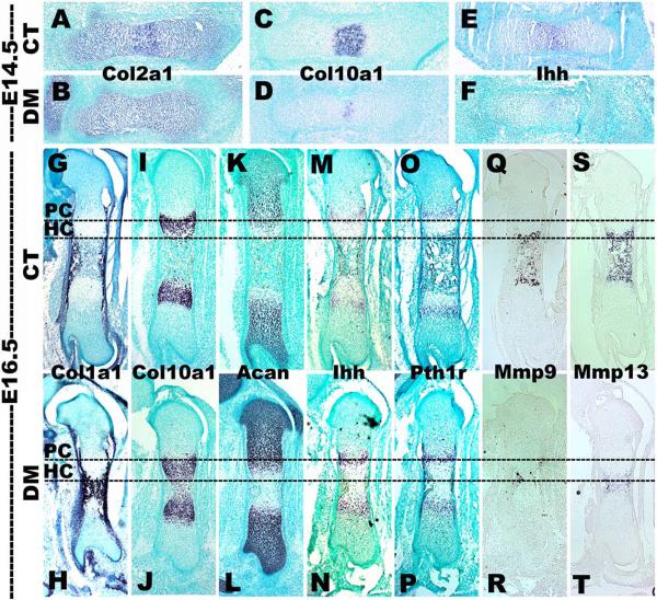
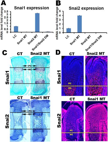
Endochondral bone formation is a multistep process during which a cartilage primordium is replaced by mineralized bone. Several genes involved in cartilage and bone development have been identified as target genes for the Snail family of zinc finger transcriptional repressors, and a gain-of-function study has demonstrated that upregulation of Snai1 activity in mouse long bones caused a reduction in bone length. However, no in vivo loss-of-function studies have been performed to establish whether Snail family genes have an essential, physiological role during normal bone development. We demonstrate here that the Snai1 and Snai2 genes function redundantly during embryonic long bone development in mice. Deletion of the Snai2 gene, or limb bud-specific conditional deletion of the Snai1 gene, did not result in obvious defects in the skeleton. However, limb bud-specific Snai1 deletion on a Snai2 null genetic background resulted in substantial defects in the long bones of the limbs. Long bones of the Snai1/Snai2 double mutants exhibited defects in chondrocyte morphology and organization, inhibited trabecular bone formation, and delayed ossification. Chondrocyte proliferation was markedly reduced, and transcript levels of genes encoding cell cycle regulators, such as p21(Waf1/Cip1) , were strikingly upregulated in the Snai1/Snai2 double mutants, suggesting that during chondrogenesis Snail family proteins act to control cell proliferation by mediating expression of cell-cycle regulators. Snai2 transcript levels were increased in Snai1 mutant femurs, whereas Snai1 transcript levels were increased in Snai2 mutant femurs. In addition, in the mutant femurs the Snai1 and Snai2 genes compensated for each other's loss not only quantitatively, but also by expanding their expression into the other genes' normal expression domains. These results demonstrate that the Snai1 and Snai2 genes transcriptionally compensate temporally, spatially, and quantitatively for each other's loss, and demonstrate an essential role for Snail family genes during chondrogenesis in mice.


Figures

Fig. 1
Fig. 1
Snai1/Snai2 double mutant embryos exhibit shortened long bones and delayed ossification. (A, B) Alcian blue-Alizarin red staining of E16.5 control (CT) and Snai1/Snai2 double mutant (DM) embryos reveals shortened long bones, such as the femurs shown here, with a reduction of their trabecular bone. *** p < 0.001. (C) Length of the femur and its trabecular bone (TB) at E16.5. The femur and its trabecular bone were significantly shorter in Snai1/Snai2 DM mice. (D–G) Snai1/Snai2 DM embryos exhibited altered orientation in digit 5 of the hindlimbs (asterisks in D, E) and delayed ossification in digits of both forelimbs (arrowheads in D, E) and hindlimbs (arrowheads in F, G), as well as in the tibia (arrows in F, G).
Fig. 2
Fig. 2
Early limb bud patterning is normal in Snai1/Snai2 DM limbs. (A–D) In situ hybridization for Col2a1 expression at E12.5. (E–F) Alcian blue-Alizarin red staining of control and DM embryos at E13.5. Both forelimbs and hindlimbs of Snai1/Snai2 DM embryos exhibit normal patterning and are similar in size and stage to limbs of control littermates.
Fig. 3
Fig. 3
Defects in chondrocyte morphology and growth in Snai1/Snai2 DM mutant embryos. (A–F) Hematoxylin-eosin-stained sections of E16.5 control and DM femurs. Snai1/Snai2 DM growth plates lost the columnar arrangement (areas in rectangles) and the flattened lens shape exhibited by control chondrocytes. PC: proliferating chondrocytes; HC: hypertrophic chondrocytes. (G) Snai1/Snai2 DM micromass cultures generated fewer Alcian blue-staining nodules than cultures set up from control limb buds. (H) Quantification of the lengths of the hypertrophic chondrocyte and proliferating chondrocyte zones in growth plates of Snai1/Snai2 DM and control and single mutant littermate femurs at E16.5. The Snai1/Snai2 DM growth plates exhibited an increased length of the hypertrophic chondrocyte zone, but no significant difference in the length of the proliferating chondrocyte zone. *** p < 0.001. (I) Quantification of nodule formation in limb bud micromass cultures revealed that Snai1/Snai2 DM cultures generated approximately half as many Alcian blue-staining nodules as cultures set up from control or single mutant limb buds on both days 4 and 6 of culture. *** p < 0.001.
Fig. 4
Fig. 4
Defects in chondrocyte proliferation in Snai1/Snai2 DM growth plates. (A–C) BrdU incorporation into proliferating chondrocytes was significantly reduced in Snai1/Snai2 DM growth plates, but not in the other genotypes (C). *** p < 0.001. (D–F) Detection of apoptotic cells by TUNEL staining. (D) In the control (CT) group, apoptotic cells were mainly located at regions of terminal differentiation adjacent to the trabecular region. (E) In DM femurs, apoptotic cells were found in throughout the central region of the femur. (F) However, no significant differences in the numbers of TUNEL-positive cells were observed in any genotype group. (G) Quantitative RT-PCR revealed that the Cdkn1a gene (encoding the cyclin-dependent kinase inhibitor p21Waf1/Cip1) was upregulated in DM femurs, but not in control or single mutant femurs. *** p < 0.001. (H) Immunofluorescent staining with an antibody to the p21Waf1/Cip1 protein revealed increased protein expression in DM femurs.
Fig. 5
Fig. 5
Quantitative RT-PCR of chondrocyte differentiation markers in femurs at E16.5. mRNAs encoding Col1a1, Col2a1, Col10a1, Acan and Sox9 were significantly increased in Snai1/Snai2 DM femurs compared to the other genotypes, whereas Mmp9 and Mmp13 mRNA levels were significantly decreased in DM femurs. ** p<0.05; *** p < 0.001.
Fig. 6
Fig. 6
Altered chondrocyte gene expression in Snai1/Snai2 DM femurs. In situ hybridization of femurs isolated at E14.5 (A–F) and E16.5 (G–T). At E14.5, Snai1/Snai2 DM femurs exhibited reduced expression of Col10a1 (a hypertrophic chondrocyte marker) (C, D) and Indian hedgehog (Ihh) (a prehypertrophic chondrocyte marker) (E, F). Expression of the Col10a1 (I, J) and Ihh (M, N) genes had recovered in DM femurs at E16.5. Expression of the Mmp9 (Q, R) and Mmp13 (S, T) genes (encoding matrix metalloproteases) was strongly decreased in DM femurs at E16.5. PC: proliferating chondrocytes; HC: hypertrophic chondrocytes.
Fig. 7
Fig. 7
Compensatory regulation of the Snai1 and Snai2 genes during chondrogenesis. (A) qRT-PCR demonstrates that Snai1 transcript levels were increased 3.3-fold in Snai2 MT femurs. As expected, no Snai1 transcripts were detected in Snai1 MT or Snai1/Snai2 DM femurs. (B) Snai2 transcript levels were increased 3.9-fold in Snai1 MT femurs. No Snai2 transcripts were detected in Snai2 MT or Snai1/Snai2 DM femurs. Both in situ hybridization (C) and immunofluorescence (D) demonstrate that in control (CT) growth plates, Snai1 is expressed in hypertrophic chondrocytes (HC), whereas Snai2 is highly expressed in proliferating chondrocytes (PC). However, in Snai2 MT growth plates, Snai1 expression expanded into proliferating chondrocytes, while in Snai1 MT growth plates Snai2 expression expanded into hypertrophic chondrocytes.

Similar articles

Cited by

References

    1. Hartmann C. Transcriptional networks controlling skeletal development. Curr Opin Genet Dev. 2009;19:437–43. - PubMed
    1. Karsenty G, Kronenberg HM, Settembre C. Genetic control of bone formation. Annu Rev Cell Dev Biol. 2009;25:629–48. - PubMed
    1. Barrallo-Gimeno A, Nieto MA. The Snail genes as inducers of cell movement and survival: implications in development and cancer. Development. 2005;132:3151–61. - PubMed
    1. Haraguchi M. The role of the transcriptional regulator snail in cell detachment, reattachment and migration. Cell Adh Migr. 2009;3:259–63. - PMC - PubMed
    1. Wu Y, Zhou BP. Snail: More than EMT. Cell Adh Migr. 2010;4:199–203. - PMC - PubMed

Publication types

MeSH terms

Substances