

Discrimination of dementia with Lewy bodies from Alzheimer's disease using voxel-based morphometry of white matter by statistical parametric mapping 8 plus diffeomorphic anatomic registration through exponentiated Lie algebra
- PMID: 23322456
- PMCID: PMC3659278
- DOI: 10.1007/s00234-013-1138-9
Discrimination of dementia with Lewy bodies from Alzheimer's disease using voxel-based morphometry of white matter by statistical parametric mapping 8 plus diffeomorphic anatomic registration through exponentiated Lie algebra
Abstract
Introduction: The purpose of this study was to identify brain atrophy specific for dementia with Lewy bodies (DLB) and to evaluate the discriminatory performance of this specific atrophy between DLB and Alzheimer's disease (AD).
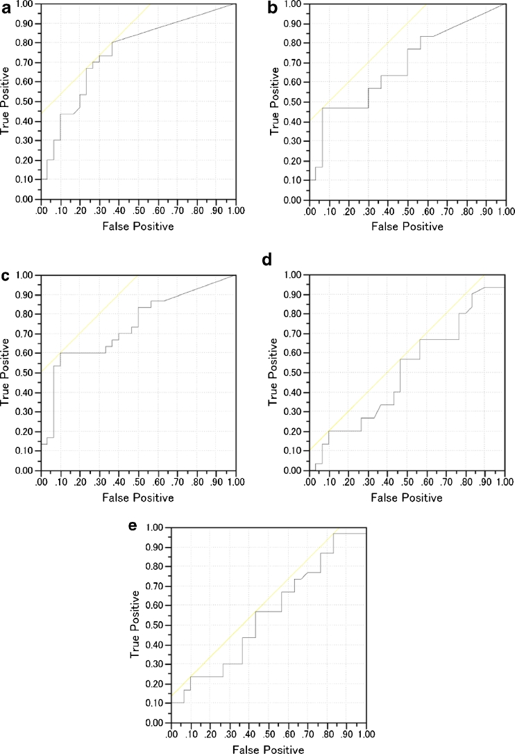
Methods: We retrospectively reviewed 60 DLB and 30 AD patients who had undergone 3D T1-weighted MRI. We randomly divided the DLB patients into two equal groups (A and B). First, we obtained a target volume of interest (VOI) for DLB-specific atrophy using correlation analysis of the percentage rate of significant whole white matter (WM) atrophy calculated using the Voxel-based Specific Regional Analysis System for Alzheimer's Disease (VSRAD) based on statistical parametric mapping 8 (SPM8) plus diffeomorphic anatomic registration through exponentiated Lie algebra, with segmented WM images in group A. We then evaluated the usefulness of this target VOI for discriminating the remaining 30 DLB patients in group B from the 30 AD patients. Z score values in this target VOI obtained from VSRAD were used as the determinant in receiver operating characteristic (ROC) analysis.
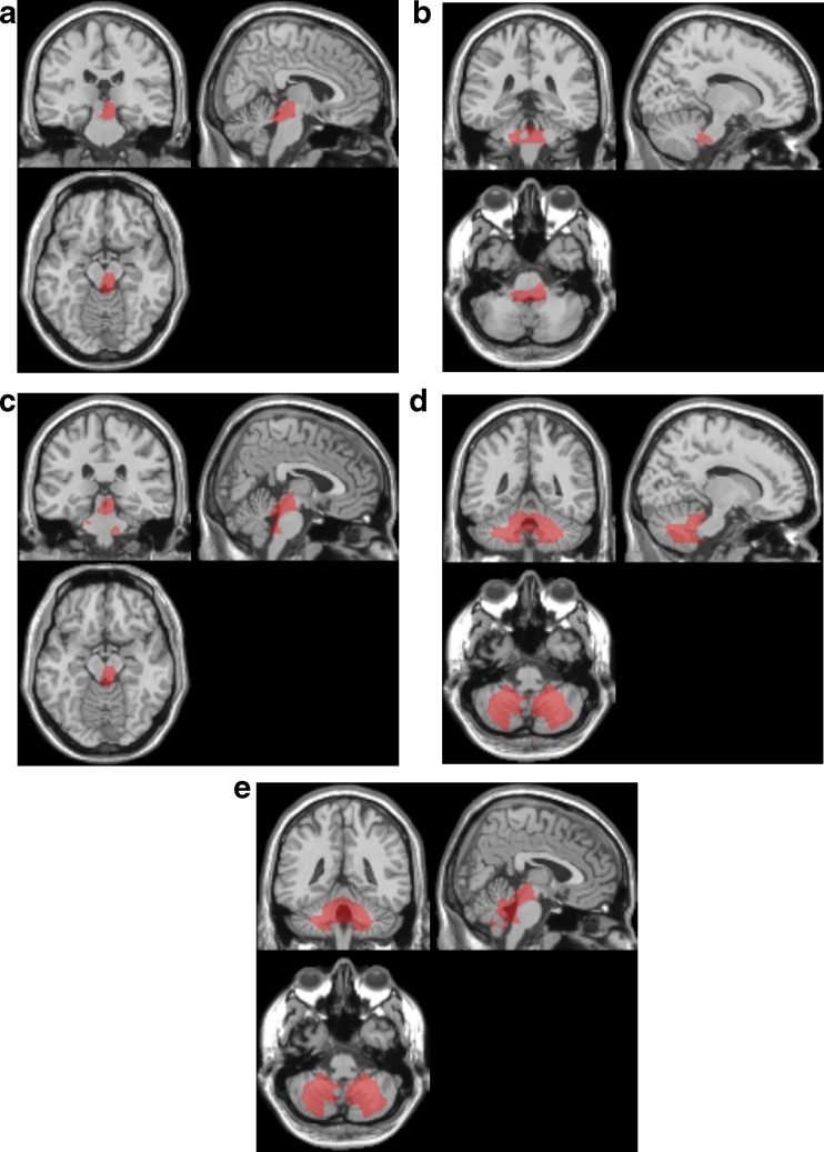
Results: Specific target VOIs for DLB were determined in the right-side dominant dorsal midbrain, right-side dominant dorsal pons, and bilateral cerebellum. ROC analysis revealed that the target VOI limited to the midbrain exhibited the highest area under the ROC curves of 0.75.
Conclusions: DLB patients showed specific atrophy in the midbrain, pons, and cerebellum. Midbrain atrophy demonstrated the highest power for discriminating DLB and AD. This approach may be useful for determining the contributions of DLB and AD pathologies to the dementia syndrome.
Figures

Similar articles
-
The feasibility of white matter volume reduction analysis using SPM8 plus DARTEL for the diagnosis of patients with clinically diagnosed corticobasal syndrome and Richardson's syndrome.Neuroimage Clin. 2014 Feb 27;7:605-10. doi: 10.1016/j.nicl.2014.02.009. eCollection 2015. Neuroimage Clin. 2014. PMID: 26082887 Free PMC article.
-
Measurement of gray and white matter atrophy in dementia with Lewy bodies using diffeomorphic anatomic registration through exponentiated lie algebra: A comparison with conventional voxel-based morphometry.AJNR Am J Neuroradiol. 2010 Nov;31(10):1873-8. doi: 10.3174/ajnr.A2200. Epub 2010 Jul 15. AJNR Am J Neuroradiol. 2010. PMID: 20634303 Free PMC article. Clinical Trial.
-
Automatic voxel-based morphometry of structural MRI by SPM8 plus diffeomorphic anatomic registration through exponentiated lie algebra improves the diagnosis of probable Alzheimer Disease.AJNR Am J Neuroradiol. 2012 Jun;33(6):1109-14. doi: 10.3174/ajnr.A2935. Epub 2012 Feb 2. AJNR Am J Neuroradiol. 2012. PMID: 22300935 Free PMC article.
-
Structural imaging in dementia with Lewy bodies: the potential of multivariate data analysis.Psychiatry Res Neuroimaging. 2020 Dec 30;306:111180. doi: 10.1016/j.pscychresns.2020.111180. Epub 2020 Sep 5. Psychiatry Res Neuroimaging. 2020. PMID: 32948404
-
Imaging in Dementia With Lewy Bodies: An Overview.J Geriatr Psychiatry Neurol. 2016 Sep;29(5):254-60. doi: 10.1177/0891988716654984. J Geriatr Psychiatry Neurol. 2016. PMID: 27502300 Review.
Cited by
-
Voxel-based morphometry analysis of double inversion-recovery magnetic resonance imaging for detecting microscopic lesions: a simulation study.Radiol Phys Technol. 2019 Jun;12(2):149-155. doi: 10.1007/s12194-019-00501-1. Epub 2019 Feb 22. Radiol Phys Technol. 2019. PMID: 30796738
-
Atypical Parkinsonian Syndromes: Structural, Functional, and Molecular Imaging Features.AJNR Am J Neuroradiol. 2024 Dec 9;45(12):1865-1877. doi: 10.3174/ajnr.A8313. AJNR Am J Neuroradiol. 2024. PMID: 39209485 Review.
-
Structural and Functional Deficits in Patients with Poststroke Dementia: A Multimodal MRI Study.Neural Plast. 2021 Nov 3;2021:3536234. doi: 10.1155/2021/3536234. eCollection 2021. Neural Plast. 2021. PMID: 34777496 Free PMC article.
-
Symplectomorphic registration with phase space regularization by entropy spectrum pathways.Magn Reson Med. 2019 Feb;81(2):1335-1352. doi: 10.1002/mrm.27402. Epub 2018 Sep 19. Magn Reson Med. 2019. PMID: 30230014 Free PMC article.
-
Neurodegeneration and inflammation crosstalk: Therapeutic targets and perspectives.IBRO Neurosci Rep. 2022 Dec 16;14:95-110. doi: 10.1016/j.ibneur.2022.12.003. eCollection 2023 Jun. IBRO Neurosci Rep. 2022. PMID: 37388502 Free PMC article.
References
MeSH terms
LinkOut - more resources
Full Text Sources
Other Literature Sources
Medical
