. 2013 Feb 1;54(2):1268-79.
doi: 10.1167/iovs.12-11172.

Oxidized porous silicon particles covalently grafted with daunorubicin as a sustained intraocular drug delivery system

Affiliations

Oxidized porous silicon particles covalently grafted with daunorubicin as a sustained intraocular drug delivery system

Jay Chhablani et al. Invest Ophthalmol Vis Sci. .

Abstract

Purpose: To test the feasibility of covalent loading of daunorubicin into oxidized porous silicon (OPS) and to evaluate the ocular properties of sustained delivery of daunorubicin in this system.

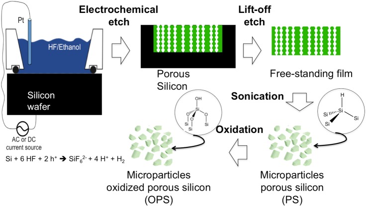
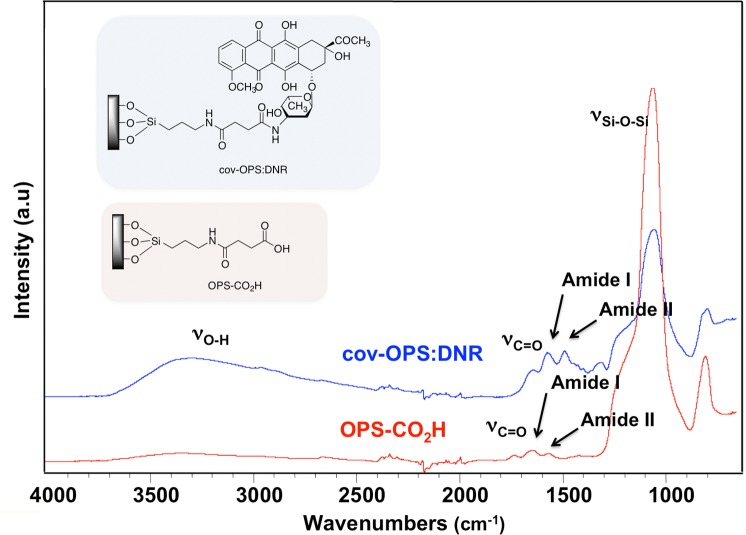
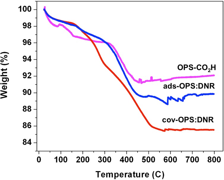
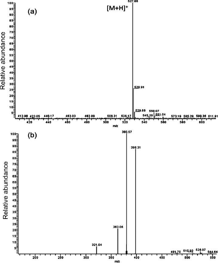
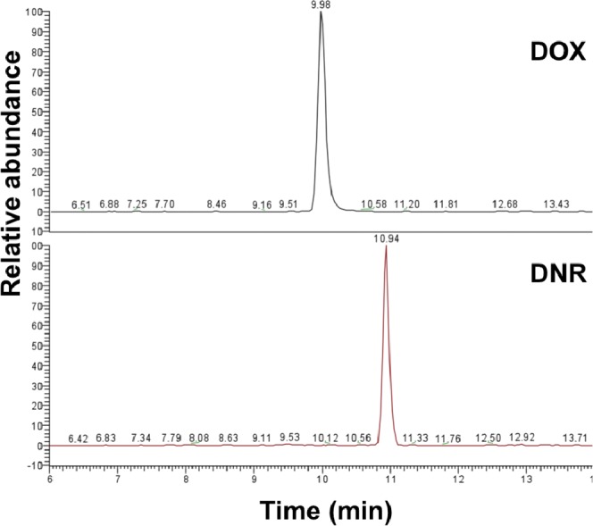
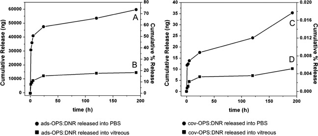
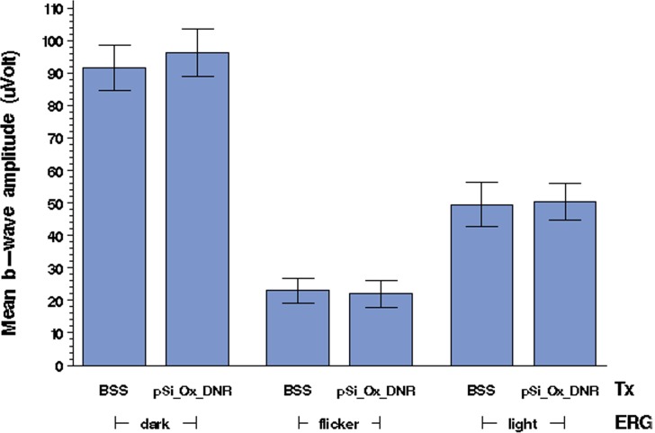
Methods: Porous silicon was heat oxidized and chemically functionalized so that the functional linker on the surface was covalently bonded with daunorubicin. The drug loading rate was determined by thermogravimetric analysis. Release of daunorubicin was confirmed in PBS and excised rabbit vitreous by mass spectrometry. Daunorubicin-loaded OPS particles (3 mg) were intravitreally injected into six rabbits, and ocular properties were evaluated through ophthalmic examinations and histology during a 3-month study. The same OPS was loaded with daunorubicin using physical adsorption and was evaluated similarly as a control for the covalent loading.

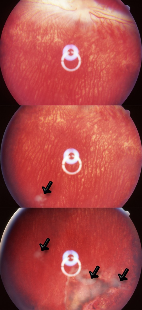
Results: In the case of covalent loading, 67 ± 10 μg daunorubicin was loaded into each milligram of the particles while 27 ± 10 μg/mg particles were loaded by physical adsorption. Rapid release of daunorubicin was observed in both PBS and excised vitreous (~75% and ~18%) from the physical adsorption loading, while less than 1% was released from the covalently loaded particles. Following intravitreal injection, the covalently loaded particles demonstrated a sustained degradation of OPS with drug release for 3 months without evidence of toxicity; physical adsorption loading revealed a complete release within 2 weeks and localized retinal toxicity due to high daunorubicin concentration.

Conclusions: OPS with covalently loaded daunorubicin demonstrated sustained intravitreal drug release without ocular toxicity, which may be useful to inhibit unwanted intraocular proliferation.


Conflict of interest statement

Disclosure: J. Chhablani, None; A. Nieto, None; H. Hou, None; E.C. Wu, None; W.R. Freeman, Spinnaker Biosciences (C); M.J. Sailor, Spinnaker Biosciences (I); L. Cheng, None

Figures

Figure 1.
Figure 1.
Schematic diagram of the synthesis of porous silicon microparticles. The electrochemical etch cell and apparatus used, as well as the Si electro corrosion half-reaction responsible for porous Si formation, is shown. The synthesis of porous Si involves the electrochemical corrosion of a single crystal wafer. In the presence of an electrolyte containing hydrofluoric acid, electrochemical corrosion removes Si in the form of SiF62−, generating myriads of nanometer-scale pores. Pores nucleate uniformly across the surface of the Si wafer, and they propagate preferentially in the <100> crystallographic direction.
Figure 2.
Figure 2.
Fourier transform infrared (FTIR) spectra of oxidized porous silicon microparticles (OPS-CO2H) before daunorubicin loading and after daunorubicin loading by covalent attachment (cov-OPS:DNR).
Figure 3.
Figure 3.
(A) Schematic illustration of loading of daunorubicin into oxidized porous silicon microparticles by covalent attachment. (B) Oxidized porous silicon microparticles before (B1) and after (B2) daunorubicin loading.
Figure 4.
Figure 4.
Thermogravimetric analysis (TGA) curves of OPS-CO2H (oxidized porous silicon with carboxylic acid functional surface), ads-OPS:DNR (oxidized porous silicon particles containing physically adsorbed daunorubicin), and cov-OPS:DNR (oxidized porous silicon particles containing daunorubicin covalently attached to the pore walls as described in the text). Weight percent is reported relative to initial weight of sample, prior to heating. The initial decrease in weight at 120°C is attributed to the loss of water from the sample. The decrease in weight observed from 120°C to 600°C is attributed to the organic functionalization and the drug loaded into the particles. The weight loss difference obtained by subtraction between particles before (OPS-CO2H) and after drug loading (ads-OPS:DNR or cov-OPS:DNR) accounts exclusively for the organic matter corresponding to the drug loaded into the particles.
Figure 5.
Figure 5.
Typical positive ion mode ESI-MS and ESI-MS/MS spectra of daunorubicin: (a) full scan mode; (b) MS/MS analysis on m/z 528 ([M+H]+, where M = the molecular mass of daunorubicin).
Figure 6.
Figure 6.
Selected reaction monitoring (SRM) LC-ESI-MS/MS chromatograms of daunorubicin released at 8 days from oxidized porous silicon microparticles loaded with daunorubicin by covalent attachment (cov-OPS:DNR). DOX was used as an internal standard.
Figure 7.
Figure 7.
Left panel: Daunorubicin released ex vivo from ads-OPS:DNR (oxidized porous silicon particles containing physically adsorbed daunorubicin) into PBS (A) and into vitreous (B). Right panel: Daunorubicin released ex vivo from cov-OPS:DNR (oxidized porous silicon particles containing daunorubicin covalently attached to the pore walls) into PBS (C) and into vitreous (D). In each graph, the left y-axis represents cumulative amount of drug released over time, and the right y-axis represents cumulative percentage of drug released.
Figure 8.
Figure 8.
Fundus photographs obtained on day 14 of three rabbits injected with ads-OPS:DNR particles (oxidized porous silicon particles containing physically adsorbed daunorubicin). Localized pigmentation changes and atrophy are apparent in small regions of the inferior retina. The white material was the aggregates of empty porous silicon particles after daunorubicin leached out (arrows).
Figure 9.
Figure 9.
Fundus photographs of rabbit eye at 3 days (3d), 2 weeks (2wk), 5 weeks (5wk), and 9 weeks (9wk) postinjection of cov-OPS:DNR particles. The oxidized porous silicon particles contained daunorubicin covalently attached to the inner pore walls as described in the text. Initially, the OPS particles appear red due to the presence of daunorubicin. The image obtained at 9 weeks shows significantly fewer particles with a less intense red color, indicative of particle degradation and drug release.
Figure 10.
Figure 10.
Mean (n = 3) IOP observed in the cov-OPS:DNR group (pSi_Ox_D) and control group (BSS) as a function of day postinjection. pSi_Ox_D: Oxidized porous silicon particles containing daunorubicin covalently attached to the pore walls as described in the text. No significant difference in IOP between study and control eyes was observed at any of the measured time points.
Figure 11.
Figure 11.
Mean (n = 3) b-wave amplitude values, in microvolts, obtained by ERG measurements on study (pSi_Ox_D, cov-OPS:DNR) and control (BSS) rabbit eyes. Dark-adapted, flicker, and light-adapted ERG measurements, as indicated, were performed on days 14 and 90 postinjection. BSS, control eyes injected with balanced salt solution; cov-OPS:DNR, eyes injected with oxidized porous silicon particles containing daunorubicin, covalently attached to the pore walls as described in the text. No significant difference in b-wave amplitude was observed between the treatment group and the control group.
Figure 12.
Figure 12.
Detection of apoptotic cells by TUNEL staining from the treated and control eyes harvested 2 weeks following intravitreal injection. (A) A section from a study control eye showing normal retinal structures and no apoptotic cells. (B) A section from a cov-OPS:DNR-injected eye, showing normal retinal structures similar to those in the section in (A) from its contralateral control eye. No apoptotic activity was detected. (C) Negative control section. (D) Positive control section, showing brownish positive staining of cells in all three layers of the retina. The sections were counterstained with hematoxylin. All four images are at ×62.5 magnification.

Similar articles

Cited by

References

    1. Pastor JC. Proliferative vitreoretinopathy: an overview. Surv Ophthalmol. 1998; 43: 3–18 - PubMed
    1. Cheng L, Hostetler K, Valiaeva N, et al. Intravitreal crystalline drug delivery for intraocular proliferation diseases. Invest Ophthalmol Vis Sci. 2010; 51: 474–481 - PMC - PubMed
    1. Turgut B, Uyar F, Ustundag B, Celiker U, Akpolat N, Demir T. The impact of tacrolimus on growth factors in experimental proliferative vitreoretinopathy. Retina. 2012; 32: 232–241 - PubMed
    1. Hou J, Li Y, Zhou Z, et al. Antiproliferative property of hexadecyloxypropyl 9-[2-(phosphonomethoxy) ethyl] guanine (HDP-PMEG) for unwanted ocular proliferation. Mol Vis. 2011; 17: 627–637 - PMC - PubMed
    1. Wiedemann P, Sorgente N, Bekhor C, Patterson R, Tran T, Ryan SJ. Daunomycin in the treatment of experimental proliferative vitreoretinopathy. Effective doses in vitro and in vivo. Invest Ophthalmol Vis Sci. 1985; 26: 719–725 - PubMed

Publication types

MeSH terms