

Longitudinal 1H MRS of rat forebrain from infancy to adulthood reveals adolescence as a distinctive phase of neurometabolite development
- PMID: 23322706
- PMCID: PMC3634877
- DOI: 10.1002/nbm.2913
Longitudinal 1H MRS of rat forebrain from infancy to adulthood reveals adolescence as a distinctive phase of neurometabolite development
Abstract
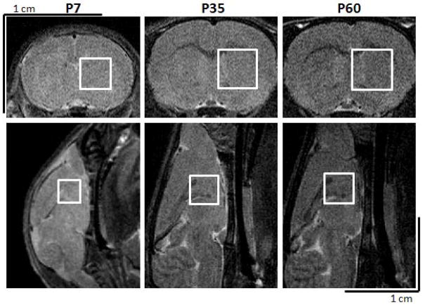
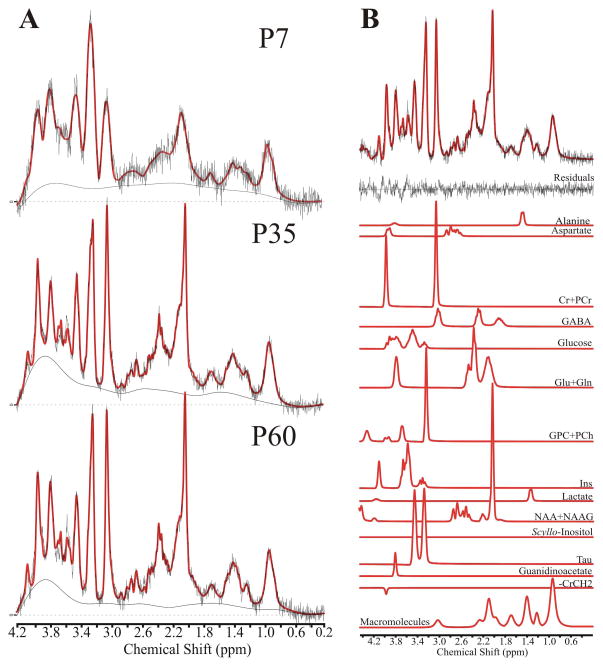
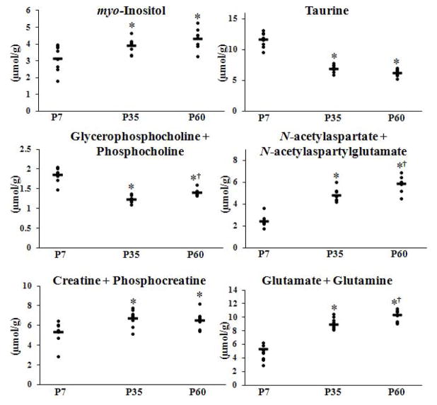
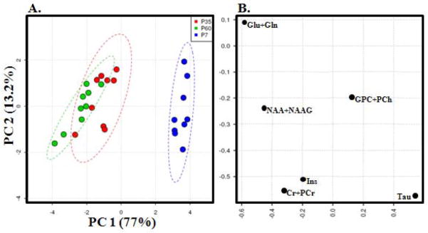
This study represents the first longitudinal, within-subject (1) H MRS investigation of the developing rat brain spanning infancy, adolescence and early adulthood. We obtained neurometabolite profiles from a voxel located in a central location of the forebrain, centered on the striatum, with smaller contributions for the cortex, thalamus and hypothalamus, on postnatal days 7, 35 and 60. Water-scaled metabolite signals were corrected for T1 effects and quantified using the automated processing software LCModel, yielding molal concentrations. Our findings indicate age-related concentration changes in N-acetylaspartate + N-acetylaspartylglutamate, myo-inositol, glutamate + glutamine, taurine, creatine + phosphocreatine and glycerophosphocholine + phosphocholine. Using a repeated measures design and analysis, we identified significant neurodevelopment changes across all three developmental ages and identified adolescence as a distinctive phase in normative neurometabolic brain development. Between postnatal days 35 and 60, changes were observed in the concentrations of N-acetylaspartate + N-acetylaspartylglutamate, glutamate + glutamine and glycerophosphocholine + phosphocholine. Our data replicate past studies of early neurometabolite development and, for the first time, link maturational profiles in the same subjects across infancy, adolescence and adulthood.
Copyright © 2013 John Wiley & Sons, Ltd.
Figures
Similar articles
-
Validation of in vivo MRS measures of metabolite concentrations in the human brain.NMR Biomed. 2019 Mar;32(3):e4058. doi: 10.1002/nbm.4058. Epub 2019 Jan 21. NMR Biomed. 2019. PMID: 30663818
-
Magnetic resonance spectroscopic analysis of neurometabolite changes in the developing rat brain at 7T.Brain Res. 2016 Nov 15;1651:114-120. doi: 10.1016/j.brainres.2016.09.028. Epub 2016 Sep 20. Brain Res. 2016. PMID: 27663970
-
Regional age dependence of human brain metabolites from infancy to adulthood as detected by quantitative localized proton MRS.Pediatr Res. 1999 Oct;46(4):474-85. doi: 10.1203/00006450-199910000-00019. Pediatr Res. 1999. PMID: 10509371
-
Proton magnetic resonance spectroscopy as a probe into the pathophysiology of autism spectrum disorders (ASD): a review.Autism Res. 2013 Apr;6(2):119-33. doi: 10.1002/aur.1273. Epub 2013 Feb 21. Autism Res. 2013. PMID: 23436782 Review.
-
Proton magnetic resonance spectroscopic evaluation of brain tumor metabolism.Semin Oncol. 2004 Oct;31(5):605-17. doi: 10.1053/j.seminoncol.2004.07.003. Semin Oncol. 2004. PMID: 15497114 Review.
Cited by
-
Concentrations of glutamate and N-acetylaspartate detected by magnetic resonance spectroscopy in the rat hippocampus correlate with hippocampal-dependent spatial memory performance.Front Mol Neurosci. 2024 Aug 16;17:1458070. doi: 10.3389/fnmol.2024.1458070. eCollection 2024. Front Mol Neurosci. 2024. PMID: 39219740 Free PMC article.
-
Continuous Ingestion of Lacticaseibacillus rhamnosus JB-1 during Chronic Stress Ensures Neurometabolic and Behavioural Stability in Rats.Int J Mol Sci. 2022 May 5;23(9):5173. doi: 10.3390/ijms23095173. Int J Mol Sci. 2022. PMID: 35563564 Free PMC article.
-
Longitudinal Biochemical and Behavioral Alterations in a Gyrencephalic Model of Blast-Related Mild Traumatic Brain Injury.Neurotrauma Rep. 2024 Mar 14;5(1):254-266. doi: 10.1089/neur.2024.0002. eCollection 2024. Neurotrauma Rep. 2024. PMID: 38515547 Free PMC article.
-
Altered Forebrain Functional Connectivity and Neurotransmission in a Kinase-Inactive Met Mouse Model of Autism.Mol Imaging. 2019 Jan-Dec;18:1536012118821034. doi: 10.1177/1536012118821034. Mol Imaging. 2019. PMID: 30799683 Free PMC article.
-
Effect of diet on brain metabolites and behavior in spontaneously hypertensive rats.Behav Brain Res. 2014 Aug 15;270:240-7. doi: 10.1016/j.bbr.2014.05.013. Epub 2014 May 19. Behav Brain Res. 2014. PMID: 24855038 Free PMC article.
References
-
- Dobbing J, Sands J. Comparative aspects of the brain growth spurt. Early Hum Dev. 1979;3:79–83. - PubMed
-
- Chung SM. Safety issues in magnetic resonance imaging. J Neuroophthalmol. 2002;22:35–39. - PubMed
-
- Clancy B, Kersh B, Hyde J, Darlington RB, Anand KJ, Finlay BL. Web-based method for translating neurodevelopment from laboratory species to humans. Neuroinformatics. 2007;5:79–94. - PubMed
-
- Tkác I, Rao R, Georgieff MK, Gruetter R. Developmental and regional changes in the neurochemical profile of the rat brain determined by in vivo 1H NMR spectroscopy. Magn Reson Med. 2003;50 :24–32. - PubMed
Publication types
MeSH terms
Substances
Grants and funding
LinkOut - more resources
Full Text Sources
Other Literature Sources
Miscellaneous
